KLAUS RUTZL B re din-Arc hb old-
Smithsonian Biological Survey of Dominica:
r Burrowing Sponges, Genus Siphonodictyon hrgquist, From
The Caribbean
SMITHSONIAN CONTRIBUTIONS TO ZOOLOGY NUMBER 77
SERIAL P U B L I C A T I O N S OF THE SMITHSONIAN INSTITUTION The emphasis upon publications as a means of diffusing knowledge was expressed by the first Secretary of the Smithsonian Institution. In his formal plan for the Insti- tution, Joseph Henry articulated a program that included the following statement:
"It is proposed to publish a series of reports, giving an account of the new discoveries in science, and of the changes made from year to year in all branches of knowledge."
This keynote of basic research has been adhered to over the years in the issuance of thousands of titles in serial publications under the Smithsonian imprint, com- mencing with Smithsonian Contributions to Knowledge in 1848 and continuing with the following active series:
Smithsonian Annals of Flight Smithsonian Contributions to Anthropology
Smithsonian Contributions to Astrophysics Smithsonian Contributions to Botany Smithsonian Contributions to the Earth Sciences
Smithsonian Contributions to Paleobiology Smithsonian Contributions to Zoology Smithsonian Studies in History and Technology
In these series, the Institution publishes original articles and monographs dealing with the research and collections of its several museums and offices and of profes- sional colleagues at other institutions of learning. These papers report newly acquired facts, synoptic interpretations of data, or original theory in specialized fields. These publications are distributed by subscription to libraries, laboratories, and other in- terested institutions and specialists throughout the world. Individual copies may be obtained from the Smithsonian Institution Press as long as stocks are available.
S. DILLON RIPLEY
Secretary
Smithsonian Institution
SMITHSONIAN CONTRIBUTIONS TO ZOOLOGY
NUMBER 77
Klaus Kutzier Bredin-Archbold-
Smithsonian Biological Survey of Dominica:
Burrowing Sponges, Genus Siphonodictyon Bergquist, From
The Caribbean
SMITHSONIAN INSTITUTION PRESS
CITY OF WASHINGTON
ABSTRACT
Riitzler, Klaus. Bredin—Archbold—Smithsonian Biological Survey of Dominica:
Burrowing Sponges, Genus Siphonodictyon Bergquist, from the Caribbean. Smith- sonian Contributions to Zoology, number 77, 37 pages. 1971.—Siphonodictyon (Adociidae, Porifera) was hitherto known only from one species (Siphonodictyon mucosum Bergquist) from the Pacific Ocean. In the present paper the species is recorded also from the Indian Ocean. Two new species (Siphonodictyon cachacrouense, new species and Siphonodictyon coralliphagum, new species) are described from the Caribbean Sea. Based on morphological differences Siphono- dictyon coralliphagum is divided into four forms (forma typica, forma obruta, forma tubulosa and forma incrustans). All species excavate burrows in coral skeletons. Another Caribbean species with sand-burrowing habit (Siphonochalina siphona de Laubenfels) is here transferred to Siphonodictyon. Based on the new material the definition of the genus is revised. Histological features and ecological data are discussed for all species.
Official publication date is handstamped in a limited number of initial copies and is recorded in the Institution's annual report, Smithsonian Year.
UNITED STATES GOVERNMENT PRINTING OFFICE WASHINGTON : 1971
For sale by the Superintendent of Documents, U.S. Government Printing Office Washington, D.C. 20402 - Price 50 cents (paper cover)
Klaus
Bredin-Archbold-
Smithsonian Biological Survey of Dominica:
Burrowing Sponges, Genus Siphonodictyon Bergquist, From
The Caribbean
Introduction
Marine sponges which excavate calcium carbonate substrata have been known for well over a century and have since been found within the full horizontal and vertical range of benthic habitats. They are commonly called "boring sponges" and, with more or less justification, are lumped in the family Clionidae (order Clavaxinellida). They burrow in limestone and in all kinds of calcareous algal and invertebrate skeletons. Clionids usually excavate characteristic tunnels and chambers, with only orifice-bearing papillae showing on the substratum surface. Some species can reach a massive epilithic stage (gamma stage) after their original substratum fragment has been completely destroyed.
During several diving descents to Caribbean coral reefs I frequently observed deep-yellow sponge chimneys protruding from living coral heads in depths below 20 m. It was striking to note that these processes were merely vents for larger mucous masses of sponge enclosed deep in the coral skeleton. The regular shape of the excavations and the immaculate white contact areas between substratum and sponge
Klaus Rutzier, Department of Invertebrate Zoology, National Museum of Natural History, Smithsonian Institution, Wash- ington, D.C. 20560.
suggested that the sponges were actively excavating in the coral rather than occupying deserted burrows of other organisms. Micro-anatomical preparation of the yellow burrowers revealed that they were congeneric with Siphonodictyon mucosum described by Bergquist (1965) from the Pacific Ocean. Further search in the same environment in Jamaica, Domi- nica, and Barbados and additional study of pre- served material collected by colleagues in Jamaica and Puerto Rico produced what are here considered to be four distinct morphological forms of the same new species (Siphonodictyon coraliiphagum, new species). A second species was discovered in a Dominican coral reef. Specimens of a sand-dwelling species were collected in Bimini and were found to be conspecific with Siphonochalina siphona Lau- benfels (1949). Based on this new material the species is here transferred to Siphonodictyon. Study of burrowing sponges collected during the Inter- national Indian Ocean Expedition revealed one specimen of the type species from Indonesia.
Methods
After the specimens were photographed under water they were collected, using rock hammer and chisel.
Fragments of the substratum were kept for later 1
identification. Hexamethylenetetramine-buffered 4% fonnalin-seawater was used as fixative (up to two weeks); subsequently the specimens were trans- ferred into 80% ethyl alcohol. Anatomical prepara- tions consisted of hand sections stained in basic fuchsin and spicule slides produced after separate boiling of ectosomal and choanosomal tissue in con- centrated nitric acid. Paraffin and frozen sections were used for histological observations. They were routinely stained with toluidin blue, Mallory's triple stain, and Ehrlich's hematoxylin-eosin.
For each specimen studied 200 spicule measure- ments were taken by scanning the slides under the microscope and measuring maximum length and width of 100 spicules which entered the field of view and were unbroken and lying flat. Two size cate- gories of oxea were more or less distinct in the choanosome. The thinner and usually shorter oxea are probably younger growth stages but were still measured separately. In case of doubt the shape of the ends was used as a criterion; they are smooth and gradually pointed in the smaller category, and rough, stepped, mucronate, or mammiform in the larger. An additional rapid scan of the slides in- dicated whether the complete size range had been included in the random measuring technique. The Smithsonian time-share computer was used to evalu- ate the data.
The terminology here used follows Borojevic' et al (1968). Abbreviations: USNM: United States Na- tional Museum (National Museum of Natural His- tory, Smithsonian Institution, Washington, D.C.);
YPM: Peabody Museum of Natural History (Yale University, New Haven, Connecticut); AMNH:
American Museum of Natural History (New York, N.Y.).
Acknowledgements
This report is based on work carried out during the Bredin-Archbold-Smithsonian Biological Survey of Dominica. I thank Dr. Horton H. Hobbs, Jr., Na- tional Museum of Natural History, for making my participation in the survey possible. Mr. P. Brand and Mr. B. Robinson of Dominica gave valuable help. A large number of Jamaican specimens and microscope slides were made available to me for study through the generosity of Dr. W. D. Hartman, Peabody Museum of Natural History, Yale Uni- versity. Dr. T. F. Goreau was my kind host in
Jamaica and first introduced me to Caribbean deep- water reefs. The laboratory facilities of The Bellairs Research Institute, Barbados were generously made available by acting director Dr. T. H. Carefoot.
Drs. E. Kirsteuer, American Museum of Natural History and H. Pulpan, University of Alaska were patient diving companions in Dominica and Bar- bados. I also acknowledge financial support during the International Indian Ocean Expedition (Te Vega cruise A), during my stay at the Lerner Marine Laboratory, Bimini (Office of Naval Research con- tract NONR 552 [027]) and during laboratory work (Smithsonian Office of Systematics). Dr. J. A. Peters, National Museum of Natural History, introduced me to the use of the Smithsonian time-share com- puter. Miss M. Dwyer prepared Figure 4. Mrs. C.
Sterrer, Bermuda, prepared all other drawings from original photographs and photomicrographs.
Family ADOCIIDAE Laubenfels, 1934 Genus Siphonodictyon Bergquist, 1965
Gender: neuter. Type-species: Siphonodictyon mu- cosum Bergquist, 1965
Definition (revised).—Adociidae lacking micro- scleres. Ectosomal spicule tracts ending in perpendic- ular brushes supporting surface net. Spongin strongly reduced. Sponginous spicule reinforced membrane sheaths lining exhalant system. Pulpy mucus-secreting choanosome with oogonia cysts and spicules in confusion. Spicules are oxea with mu- cronate, stepped or rough tips. Smaller size category with gradually pointed tips also present in choano- some (possibly younger growth stages). Habit of burrowing in limestone substrata, only apertures bearing ectosomal structures protruding.
Siphonodictyon mucosum Bergquist, 1965
FIGURES 1, 10a; PLATE 1
Siphonodictyon mucosa Bergquist, 1965, pp. 158-161, figures 20a, b, table 4.
HOLOTYPE.-USMN 23697; Iwayama Bay, Palau Archipelago; 0.9-6 m; 14 August 1955; "Project Coral Fish," col.
MATERIAL.-USNM 24105; Te Vega Station 98, Poeloe Melila, Delapan Group (2°15'N, 97°25'E), Indonesia; 0.6 m (low tide); 23 November 1963.
NUMBER 77
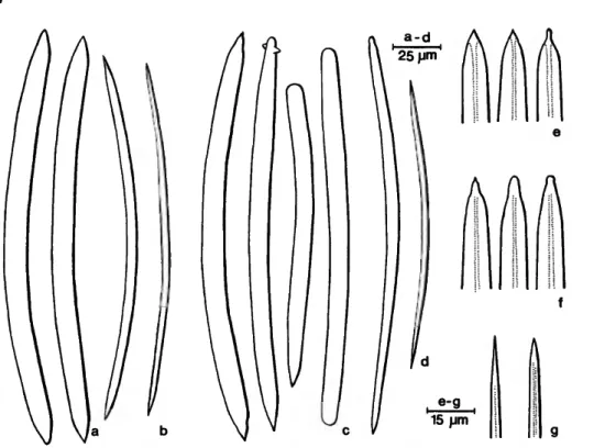
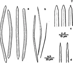
FIGURE 1 .—Siphonodictyon mucosum Bergquist: large (a,e) and small (b) oxea of the Indian Ocean specimen; large oxea and derivatives (c,f) and small oxea (d,g) of the holotype.
DESCRIPTION.—Shape and size: From a substrate area of 150 cm2 26 hollow tubes were protruding.
They are 5-10 mm in diameter and attain 88 mm from base to tip. Several of them are bifurcated, two or even three adjoining ones can be fused together sideways in their upper half. Seven of the tubes terminate in oscula, the remaining ones in sieve areas. The choanosome fills a cavity in the coral substrate, approximately 250 cm3 in volume.
Color: The tubes are blackish brown in life, the choanosome greyish tan. The color fades slightly in alcohol.
Consistency: The tubes are stiff and brittle, the choanosome soft, compressible and mucous.
Surface: Microhispid; the bases of the processes are incrusted with various sediment particles.
Apertures: The oscula are restricted to the term- inal opening of the tubes. They are circular and measure 8-5.5 mm in diameter. The ostia (23-150 fim) * are distributed over the entire tube surface.
Skeleton: Bundles of stout longitudinal fibers run parallel to the lumina of the ectosomal tubes and radiate toward the surface. They are interconnected
by less well defined secondary strands. The primary fibers fan out at the surface where they support a dense tangential reticulation. The main fibers meas- ure 100-375 fim,* the secondaries 100-150 fim, the meshes of the reticulum 100-450 fim. The spicula- tion of the choanosome is in confusion.
Spicules: Rubust oxea with little developed cen- tral canal. Two size categories are distinct in the choanosome. The larger oxea have acute to rau- cronate, sometimes mammiform tips, the smaller ones are gradually and sharply pointed. Modifica- tions to styles and strongyles are present.
Measurements (of the Indian Ocean specimen) : Given in Table 2, and can be compared with the holotype.
REMARKS.—This record extends the range of this species from the Pacific to the Indo-Pacific region.
•Resolution 7 of the Thirteenth General Conference on Weights and Measures, 1968, substitutes for the micron (p) — 10-» meter the micrometer (^m), the symbol ^ being reserved for the prefix micro- in this and other metric uses. See also the recent (1970) "Conversion to the Metric System," by Lord Ritchie-Calder (Scientific American 223(1): 17-25).
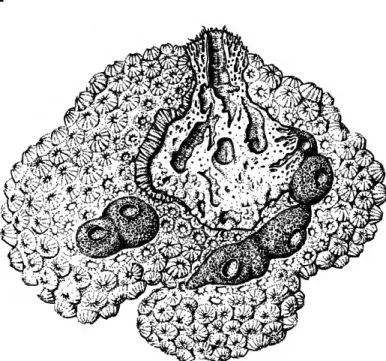
FIGURE 2.—Montastrea annularis infested by Siphonodictyon cachacrouense, new species. Part of the coral is removed to show the internal structure of the sponge (0.6 X)-
Probably a second Pacific species is Siphonodictyon aberrans (Dendy) from Three Kings Islands, New Zealand, transferred to this genus by Bergquist
(1965).
Siphonodictyon cachacrouense, new species
FIGURES 2, 3,106; PLATES 2, 8a, b, e
HOLOTYPE.-USNM 24094 (Figures 2, 3; Plate 2; Table 2). Scotts Head Bay, Dominica, West Indies; 35 m; 19 June 1966 (only specimen).
DIAGNOSIS.—Epilithic sponge portion forming a tough elastic, irregular pillow. Color: dark greyish brown. Surface strongly hispid. Oscula circular, elevated with brush-like collar. Choanosome endoli- thic, irregularly massive, greyish tan. Oxea, large:
203.9 x 6.9 ^m; oxea, small: 181.0 x 3.0 /an (means of 50 measurements each category).
DESCRIPTION.—Shape and size: The exposed ecto- somal portion of the sponge forms an irregular pillow extending over an area of 120 cm2. There are approximately 25 conical mounds reaching 25 mm in height, each terminating in an osculum. In some places coral fragments are showing between the elevations; these are remnants of the coral sub- strate. The massive choanosome extends 60 mm into the coral, filling a cavity of approximately 500 cm3.
A fh
fh
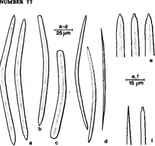
FIGURE 3.—Siphonodictyon cachacrouense, new species: large (a,c) and small (b,d) oxea of the holotype.
NUMBER 77
Color: The ectosome and the walls of the aquifer- ous system are dark greyish brown. The choanosome is light greyish tan with large dirty yellow embryos (June) contrasting from it. In alcohol the color remains the same although it fades slightly.
Consistency: The ectosome is very tough and elastic. In the transition zone to the choanosome it is ligneous due to the spicule-reinforced walls of the aquiferous canals. The choanosome is crumbly soft and compressible, with large amounts of mucus.
Surface: It is smooth to microhispid on the rising sides of the sponge, hispid, like a brush on top.
Fiber bristles are slightly higher around the oscula.
Apertures: The elevated circular oscula are 4-8 mm in diameter. Ostia in the side portions of the sponge where they are not obscured by the bristle structure, measure 100-140 /an.
Skeleton: Stout spicule bundles radiate from the choanosome towards the surface. They branch fre- quently along their way and are interconnected by short secondary tracts. In the last 4-5 mm before the primary fibers reach the top surface of the sponge pillow the connecting secondaries are absent, thus causing the brush-like appearance of the surface.
At the side portions of the sponge, however, the primaries are also interconnected near the surface, where they support a tangential fiber reticulation.
The primary bundles measure 120-400 fim in diam- eter, the interconnecting secondary ones 50-150 fim. The meshes of the surface net are 80-200 fim in diameter, its fibers 50-80 fxm. In the choanosome spicules are abundant but show no orientation.
Spicules: Bent oxea, quite regular, with thin cen- tral canal. The tips are sharply pointed, usually smooth but sometimes rough, seldom stepped. A few modifications to styles or strongyles occur. In the choanosome a second (smaller and thinner) size category is quite distinct.
Measurements: Given in Table 2.
REMARKS.—This species is named after Pointe Cachacrou (Patois for Scotts Head) where it was collected. It is so distinct that it is described as new without hesitation although only one specimen was available for study.
Siphonodictyon coralliphagum, new species HOLOTYPE.-USNM 24095 (Figure 5; Plates 3 e,f, 4 a-d; Table 2). Discovery Bay, Jamaica; 25 m;
24 March 1966.
DIAGNOSIS.—Epilithic ectosomal portions, if pres- ent, are brittle crusts, hollow cones or tubes. They extend over 2 to 600 cm2. Color deep yellow, lemon yellow to whitish yellow. Circular oscula on top of elevations, if such are existing. Surface microhispid.
Choanosome usually endolithic, ovoid to irregular massive. Color: beige yellow to yellow tan. Oxea, large: 135.9-169.3 x 4.7-6.9 fim; oxea, small: 118.9- 159.6 x 2.1-3.2 fim (range of means of all 22 speci- mens attributed to the species).
Based on morphological and spicule-dimension differences, four forms can be distinguished which might be different ecophenotypes and or growth stages:
Forma typica: Ectosomal structures consisting of single conical chimneys or tubes protruding from the substratum. Oxea, large: 142.1-156.3 x 5.0-6.4 /an; oxea, small: 129.0-142.9 x 2.2-2.7 fim (range of means).
Forma obrnta: No ectosomal structures present.
Oxea, large: 135.9-144.0 x 4.9-6.2 fim; oxea, small: 118.9-129.4 x 2.1-2.7 fim (range of means).
Forma tubulosa: Clusters of ectosomal tubes, trum- pets or finger-shaped hollow processes. Oxea, large: 165.9-169.3 x 6.1-6.9 fim; oxea, small:
144.5-152.2 x 2.2-3.2 /xm (range of means).
Forma incrustans: Extensive flat ectosomal crusts or pillows. Oxea, large: 150.0-164.9 x 4.7-5.7 fim, oxea, small: 140.5-154.7 x 2.3-2.8 /an (range of means).
Siphonodictyon coralliphagum forma typica
FIGURES 4, 5, 9b, c, lOr; PLATES 3, Aa-d, 8c, d, f, 9a-c, e, f
MATERIAL.—USNM 24095; Discovery Bay, Jama- ica; 25 m; 24 March 1966 (holotype of the species).
USNM 24096; Scotts Head Bay, Dominica, West Indies; 1.5 m; 4 April 1966. USNM 24097; Scotts Head Bay, Dominica, West Indies; 15 m; 11 July 1969. YPM 6487A; Discovery Bay, Jamaica; 45 m;
19 July 1966; J. Lang, col. YPM 6507; Runaway Bay, Pear Tree Bottom, Jamaica; 30-33 m; 22 July 1966; y. Lang, col. (paratypes of the species) .
DESCRIPTION.—Shape and size: Tuberculate chim- neys with thick walls are protruding from the live coral substrate. The osculum on top is half to one- third of the chimney diameter. This typical ap- pearance can be modified if no osculum is present;
one can find irregular papillae of all sizes, sometimes
FIGURE 4.—Stephanocoenia michelinii infested by Siphonodic- tyon coralliphagum, new species (forma typica). Part of the coral is removed and one tubular process is cut open to dem- onstrate the internal structure of the sponge (0.6 x ) -
only a tuberculate pillow. These can also be an outgrowth from the chimney base. Occasional thin- walled tubes are never vase-shaped. On top of some papillae the usually dense network of spicule strands is loosened up in various degree suggesting the break-through of a new osculum. The various epilithic structures are 5-35 mm in diameter, 6-40 mm high. They stand 2-10 cm apart, and 1 to a maximum of 4 were counted for each sponge speci- men. The choanosome is endolithic; it may also reach the coral surface or be 20-30 mm removed and connected with the epilithic structures through tunnels of 10-15 mm diameter. The choanosome completely fills the ovoid to irregular cavities in the coral-rock. It has a volume of 20-130 cm3.
Color: The exposed ectosome is of consistent deep yellow (comparable to egg-yolk) in the live speci- mens. If left in contact with air for some minutes the color changes to pinkish yellow. The choano- some is yellowish beige; it stains the fingers yellow
when handled. Numerous large embryos (April and July) are conspicuous by their bright yellow color.
In alcohol the color changes to greyish-tan, light in the ectosome, dark in the choanosome.
Consistency: The ectosome is friable but the chimneys as a whole are quite elastic, due to spicule reinforcement in the walls of the large exhalant canals. The choanosome is soft and mucous and un- elastically compressible.
Surface: It is finely porous and microhispid.
Apertures: The oscula are always elevated, i.e., on top of the chimneys or tubes. They are circular, 2- 10 mm in diameter. The ostia are distributed over the entire surface and measure 100-250 [im.
Skeleton: A delicate, more or less regular network of polygonal meshes overlays most of the surface parts. The spicule tracts forming the meshes are 2-6 parallel spicules wide. The mesh diameter is 75-125 jim.
The aquiferous canals of the choanosome, as well as the lumina of the oscular chimneys are lined with multiple layers of detachable membranes. These contain loose two-dimensional spicule strands em- bedded in a spongin matrix. This seems to be the
NUMBER 77
place of origin for massive longitudinal spicule bundles which start deep in the choanosome and run parallel to the canal lumina to the top surface of the sponge. They are supporters of the exposed sponge processes. They also radiate toward all other parts of the sponge surface, subdivide and are interconnected by secondary tracts to form a solid network. This becomes looser and more del- icate the closer it approaches the surface, where it finally supports the tangential surface net.
The primary strands are 100-175 fiva across but are frequently fused together to fascicles. The secondary strands measure 40-100 fiva, the meshes they form average 120-300 /urn. In the choanosome spicules are in confusion.
Spicules: Oxea, usually slightly bent, with more or less distinct central canal. A smaller and thinner size category is distinct in the choanosome. The tips are typically mucronate but many stepped, rounded, mammiform, and tylote modifications are present.
Measurements: Given in table 2.
FIGURE 5.—Siphonodictyon coralliphagum, new species (forma typica); large (a/:) and small (b,d) oxea (and derivatives) of the holotype.
Siphonodictyon coralliphagum forma obruta
FIGURES 6, 9a, 10c; PLATE 4e-g
MATERIAL.-USNM 24098; Southwest of Hole- town, Barbados, West Indies; 25 m; 5 July 1969
A A A
FIGURE 6.—Siphonodictyon coralliphagum, new species, forma obruta: large (a/:) and small (b,d) oxea (and derivatives).
(specimen representative for the form). USNM 24099; South of Holetown, Barbados, West Indies;
20 m; 7 July 1969. USNM 24100; off Littlegood Harbour, Barbados, West Indies; 23 m; 7 July 1969.
YPM 4398; Cayo de Caballo Ahogao, near La Par- guera, Puerto Rico; 18 June 1959; W.D. Hartman and J. Rivero, col.
DESCRIPTION.—Shape and size: The entire sponge is endolithic. Its choanosome fills cavities in coral rock, 5-10 mm below the coral surface. The cavities are spherical in small specimens (5-10 mm in diameter), ovoid in larger ones (10-20 mm), the larger axis being parallel to the substrate surface.
One or two tissue-lined tunnels (0.5-1.8 mm diam- eter) lead to an opening on the coral rock surface.
These tunnels always run at an angle of 20-40 degrees to the perpendicular coral structure, at least in the specimens available.
Color: Uniformly yellowish beige.
Consistency: Soft compressible and slightly mucous.
Surface: No epilithic parts are present.
Apertures: The only apertures detectable are the circular vents of the tunnels mentioned above. They have the same size as the tunnel diameter.
Skeleton: The oxea are abundant but without orientation, like in the choanosome of the typical form. Only near the coral substratum they have a tendency to be placed tangentially.
8
Spicules: Two size categories of more or less distinctly bent oxeas. Styles occur occasionally. The tips are stepped, mucronate to mammiform.
Measurements: Given in Table 2.
Siphonodictyon coralliphagum forma tubulosa
FIGURES 7, 9e, 10c; PLATES 5a, b, 6
MATERIAL.-USNM 24101; Scotts Head Bay, Dominica, West Indies; 20 m; 30 May 1966 (speci- men representative for the form). USNM 24102;
Scotts Head Bay, Dominica, West Indies; 35 m; 18 June 1966. USNM 24103; Scotts Head Bay, Domi- nica, West Indies, 35 m; 18 June 1966. USNM 24104; Scotts Head Bay, Dominica, West Indies;
15 m; 11 July 1969.
DESCRIPTION.—Shape and size: The epilithic part of single specimens extends over areas of 70-950 cm2 between live coral heads. It consists of dense clusters of thin-walled tubes (0.8-1.2 mm wall thickness) frequently vase- or funnel-shaped, alter- nating with hollow finger-shaped processes which can be fused together along their sides, forming humpy pillows. The ectosome between these aqui- ferous processes (their distance from each other:
5-25 mm) is encrusted with coralline algae which again provide substrate for secondary settlers (algae, sponges, bryozoans, tunicates). The coralline algal crusts can reach up to 5 mm below the oscular rim of the larger tubes (e.g., specimen USNM 24101). The following counts and measurements were taken on the fresh specimens (Table 1):
The choanosome extends horizontally as far as the ectosome. The upper portion lies level with the TABLE I.—Specimen Measurement of Siphonodictyon coralliphagum
forma tubulosa.
Specimen Number
USNM 24101
(not preserved)
USNM 24102
USNM 24103
Area Measured
600 cm2
300 cm2
200 cm2
70 cm2
Type open tubes
closed fistules, single closed fistules, double
and triple open tubes
closed fistules and low papillae (some double or triple)
open tubes, vase-shaped closed fistules, single
or double
low papillae, all fused together, some also fused with tubes open tubes papillae, single papillae, fused
Process Number
9 43 8
26 68
6 14
70 (approx.
2 4 -
Diameter 15-25 mm 8-12 mm 12-39 mm
7-16 mm 5-15 mm
9-20 mm 5-10 mm
100 mm )
18, 20 mm 6-12 mm
60 mm
Height 50-65 mm
6-30 mm 25-30 mm
10-55 mm 8—26 mm
15-48 mm 8-15 mm
6-14 mm
20, 25 mm 6™o mm 11-15 mm
NUMBER 77 9
A A A
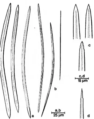
FIGURE 7.—Siphonodictyon coralliphagum, new species, forma tubulosa: large oxea (a,e), style (b), strongyle (c) and small oxea (d, /).
coral substrate, most of which has been dissolved;
only some coral fragments are still embedded; the lower portion is endolithic. It attains approximately 2 liters in volume in a large specimen.
Color: The ectosome is a light yolk yellow; one specimen (USNM 24102) was whitish yellow in life. During exposure to air the same color change to pinkish yellow occurs as in the typical form. The color of the choanosome is yellowish beige with bright yellow embryos, as in the typical form. In alcohol the color changes to dark beige or greyish- tan.
Consistency: All ectosomal processes are very fragile. In the transition zone to the choanosome, numerous exhalant canals with spicule-reinforced walls cause a ligneous consistency. The choanosome is soft, compressible, and very mucous.
Surface: The surface, where not covered by cor- alline algae, is microhispid.
Apertures: The oscula on top of the thin-walled tubes measure 7-19 mm in diameter. The ostia situated on the surface of all processes are 120-280 /u.m in diameter.
Skeleton: Structure and measurements of the spicule bundles fall in the range of the typical form.
Spicules: Bent oxea with fine central canal. The two size categories of the choanosome are distinct.
The tips are rather blunt to mammiform, usually
smooth, sometimes rough to stepped. Some styles and strongyles occur.
Siphonodictyon coralliphagum forma incrustans
FIGURES 8,9d, 10c; PLATES 5C, d.
MATERIAL.—YPM 4757; Runaway Bay, Jamaica;
30-33 m; 16 July 1961; D. Fraser, E. Graham, col.
(specimen representative for the form). YPM 4816;
Runaway Bay, Jamaica; 24 m; 21 July 1961; E.
Graham, col. YPM 4831; Runaway Bay, Jamaica;
15-27 m; 21 July 1961; I. Goodbody, R. L. Walker, col. YPM 4930; Runaway Bay, Jamaica; 15 m; 23 July 1961; T. F. Goreau, E. Graham, R. Dalzeel, col. YPM 5346, YPM 5359, YPM 5388, YPM 5395;
Runaway Bay, Jamaica; 33-39 m; 21 May 1961;
T. F. Goreau, D. Fraser, L. M. Passano, col. YPM 6496, YPM 6896; Discovery Bay, Jamaica; 57 m;
21 July 1966; J. Lang, col.
DESCRIPTION.-Shape and size: Flat crusts, irregu- lar pillows, or flat cones show above substrate level.
Oscula, if present are not, or only slightly, elevated.
The maximum diameter of these structures varies between 2 and 26 cm, surface area is approximately 3.5 to 350 cm2. The choanosome is buried in the substrate; its thickness is about one-third the diameter of the whole sponge.
Color: The ectosome varies from deep to lemon yellow, the choanosome is mustard yellow to yellow- tan. Preserved specimens are ochre to dark brown;
the preservation alcohol of larger specimens is also stained brown.
Consistency: the ectosome is firm but brittle; the choanosome soft, compressible, and very mucous.
Surface: Microhispid and porous. Oscula are distinct only in larger specimens.
Apertures: The oscula are usually level with the sponge surface, are sometimes slightly elevated, and are surrounded by a slight rim; their shape is typi- cally circular. Occasionally, however, two or more adjoining ones become fused together presenting an oval to irregular outline (very distinct in specimen YPM 6496). The interior of the oscula appears as dark in color as the choanosome (in preserved state). The ostia measure 80-300 fxm.
Skeleton: Structure of the skeleton and size of the spicule bundles is not different from the typical form.
A
A15pm
FIGURE 8.—Siphonodictyon coralliphagum, new species, forma incrustans: large (a,c) and small (b) oxea (and derivatives).
Spicules: The oxea are usually slightly, sometimes sharply bent. A central canal is distinct only in the ectosomal spicules. Separation into two size cate- gories in the choanosome is only slight since there are many transitional forms. Some styles are present.
Tips are blunt, mammiform or mucronate, degen- erated where there is a wide central canal developed.
Measurements: Given in table 2.
DISCUSSION.—It can be expected that further and more directed studies in the Caribbean area will shed light on the proper systematic status of the various forms of Siphonodictyon coralliphagum.
The species is certainly more widely distributed than the available specimens would suggest. Accord- ing to the author's field notes—which unfortunately, are not documented by specimens—formae typica and obruta were encountered also around Bimini.
Particularly, during earlier collecting forma obruta has been frequently discarded because it was con- sidered a secondary settler in clionid burrows and the material was too sparse to be properly recog- nized. Forma typica could on several occasions be clearly identified from amateur underwater photo- graphs taken in Caribbean deep-water reefs. There is no indication, however, that forma tubulosa occurs outside of Dominica and that forma incrus-
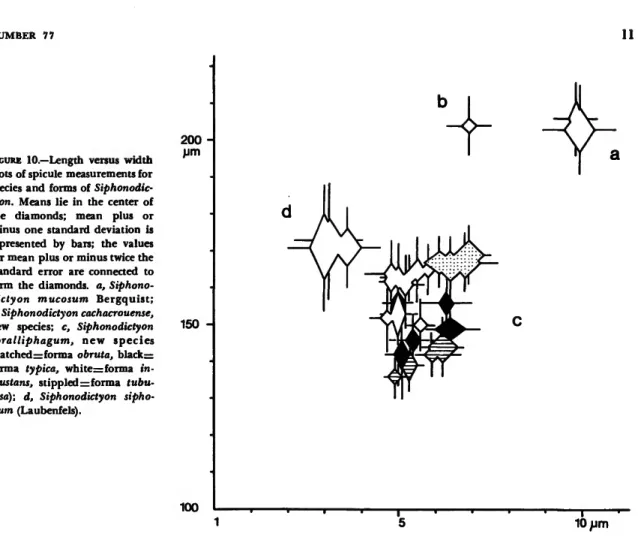
tans has been found outside of Jamaica. Transi- tional stages between forma typica and forma tubu- losa on the one hand and forma incrustans on the other have been observed at Dominica and Jamaica, respectively. No transitional forms between obruta and typica are evident, although it can be antici- pated with little speculation that the latter derives from the former (Figure 9). After plotting means, standard deviation, and double standard error of length and width of the oxea for each species and form (Figure 10) one also arrives at the conclusion that, because of the overlap of the double standard errors, the four forms of Siphonodictyon corallipha- gum belong to one population (Hubbs and Perl- mutter 1942). It might be a coincidence that from the same graph can be read a tendency towards evolution (in a morphological sense) from forma obruta via forma typica to either forma tubulosa or forma incrustans.
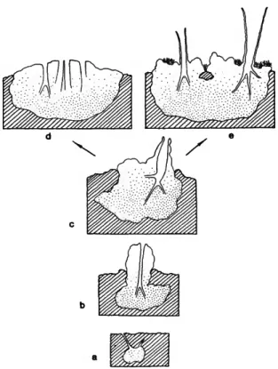
FIGURE 9.—Siphonodictyon coralliphagum, new species. Mor- phological variation of the forms in hypothetical sequence:
formae obruta (a), typica (b), typica (c), incrustans (d), tubu- losa (e).
NUMBER 77 11
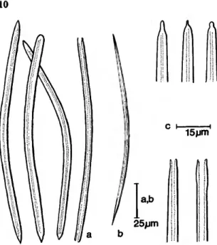
FIGURE 10.—Length versus width plots of spicule measurements for species and forms of Siphonodic- tyon. Means lie in the center of the diamonds; mean plus or minus one standard deviation is represented by bars; the values for mean plus or minus twice the standard error are connected to form the diamonds, a, Siphono- dictyon mucosum Bergquist;
b, Siphonodictyon cachacrouense, new species; c, Siphonodictyon coralliphagum, new species (hatchedzzforma obruta, blackzz forma typica, white=forma in- crustans, stippled •=. forma tubu- losa); d, Siphonodictyon sipho- num (Laubenfels).
200um
150 -
100
Siphonodictyon siphonum (Laubenfels), new combination
Siphonochalina siphona Laubenfels, 1949, pp. 11-13.
FIGURES 10d, 11; PLATES 7, 9d.
HOLOTYPE.—AMNH 468; Bimini, Bahamas; less than 1 m; 1 July 1948.
MATERIAL.-USNM 24106, USNM 24107; East of Turtle-Rocks, Bimini, Bahamas; 1 m; 7 August 1967.
DESCRIPTION.—Shape and size: Clusters of tubes and hollow processes protrude from the sand sub- stratum. They are irregular oval in cross section, 6-18 mm in diameter, and up to 70 mm high (from substrate level). The walls are 1-3 mm thick. Two to seven processes are fused together along their sides. The largest of the collected clusters extended over an area of 20 cm2, up to 20 were observed per m2 of sand bottom. Most of the basal mass of the
5 10 /jm choanosome is buried in the sand, only fragments were recovered.
Color: The exposed parts are chestnut brown, sometimes with a reddish tinge, the choanosome is greyish brown in life. They turn blackish brown in alcohol.
Consistency: The tubes are stiff but elastic, the choanosome is soft, compressible, and mucous.
Surface: Smooth to microhispid, particularly on top of the finger-shaped processes. Numerous wart- like swellings bearing a single pore (ostium?) can be noted.
Apertures: The oscula are more or less circular with irregular brush-like rims. They measure 5-12 mm in diameter. The ostia measure 150-300 /um.
Skeleton: A discontinuous surface reticulation is present. The tracts of spicules embedded in some spongin are 30-150 fim in diameter; they form meshes of 250-600 ju.ni. Below, the ectosomal reticu- lation consists of radiating primary fibers, brushing
out at the surface; these are connected by second- aries to form rectangular meshes. Fiber diameter, 75-125 /an; mesh diameter, 150-250 x 75-120 fim.
In the choanosome the spiculation is in confusion, with the exception of some irregular strands.
Spicules: The slender oxea are usually bent, with a tendency to being flexuous. A second size category is not distinct. In the ectosome they are frequently broken. The tips are rough to acuminate, some- times modified to styles or strongyles sometimes degenerated.
Measurements (of the present specimens and of the holotype): Given in Table 2.
REMARKS.—As Laubenfels (1949) has pointed out large accumulations of this sponge occur on (and in) shallow-water sand bottoms of Bimini. He also stated that the "basal mass" (actually the whole
a,b
25pm
A A
c d 15 pm
FIGURE U.—Siphonodictyon siphonum (Laubenfels): large (a,c) and small (b,d) oxea.
choanosome) is regularly buried beneath the sand and "its total size was never satisfactorily ascer- tained." Also in the present collection most of the mucous choanosome was left behind when the speci- mens were removed from the substratum. Recovered portions showed that fragments of dead coral em- bedded in the sand were clearly excavated where they interfered with the extending body of the sponge. This proves that the species not only has the morphological and anatomical features of Si- phonodictyon but also the excavating properties of the genus.
Histological Findings
On the level of the presently employed methods the histological picture of the various forms and species is quite similar. Exo- and endopinacoderm are formed in the usual manner by anucleolate cells.
The major canals, particularly in the ectosomal region are lined by a multiple layer of thin fibrous membranes (Spongin A? [Gross et al. 1956]) which are stained by aniline blue (Plate 8a).
These membrane sheets vary in thickness from 15-30 fim in the choanosome, to more than 500 fim in the ectosome. In the ectosome they contain spicule strands running crisscross in subsequent layers. Macerated preparations of primary spicule bundles show that they can be connected by such membranes. Thus it appears that the primary spi- cule fibers originate in membrane sheaths. They run parallel to the exhalant canals until somewhere along their course they radiate towards the sponge surface (see systematic section) .
In the extensive lacunar spaces between and attached to the membranes are numerous spindle- shaped nucleolate cells (15-35 fim long, 3-6 fim wide) filled with acidophilic and basophilic grains (Plate Sb). There is also another similar cell type which contains finely granular basophilic inclusions and sometimes a larger chromophobic grain. In peel preparations of membranes lining ocular tubes they can be seen in thick strands crowding bundles of spicules.
The ectosomal area of the spicule fiber network shows but little cellular material. Some of the same two cell types as in the membrane section are pres- ent as well as circular to oval nucleolate cells with acidophilic granules (6-17 x 5-8 fim. The most abundant cells, however, are anucleolate, 5-12 x 3-6
NUMBER 77
fim and amphophilic. They secrete a filamentous meshwork of basophilic ground substance. Both, the cells and the filamentous ground substance are par- ticularly dense in an area of regeneration of Si- phonodictyon coralliphagum forma typica (specimen YPM 6507). Part of the choanosome of this speci- men had obviously been exposed after perforation of the coral substratum, presumably by scraping fish
(Plate 3d). Some spicule tracts have also been re- generated in this area, the individual spicules being cemented by fairly large amounts of spongin B (Plate 8c). The presence of spongin in Siphono- dictyon had been overlooked in the original descrip- tion of the genus (Bergquist 1965), In fact, in normal specimens it occurs only in minute quanti- ties on the ectosomal spicules tracts. However, after careful search its presence could also be confirmed in the holotype of the type species. Apparently, spongin is used only during the first steps of skeleton formation to connect loose spicules and is later re- duced or at least not further developed.
Choanocyte chambers occur throughout the choanosome (Plate Sd). They are spherical and measure 16-20 fim in diameter. All three nucleolate cell types from the ectosome are also encountered in the choanosome in small numbers. Archeocytes containing 8-12 highly retractile spherules are common, particularly in specimens containing em- bryos (Plate 8e). The spherules occur also free in small aggregations. They measure 2-4 fim and are superficially eosinophilic. Their nature will have to be studied histochemically, but one can assume that they have nutritive function, since they are one of the main constituents of all embryos. Sperm cysts were only rarely found. They are oval, measure approximately 80 x 40 fim, and contain basophilic sperm.
The most conspicuous feature of the choanosome of all specimens are the spherical cell groups which are evenly distributed throughout the choanosome (Plates 8/, 9a). Each group is surrounded by an epithelium and a fine layer of fibrous basophilic material. These cysts measure 20-70 /urn in diameter and contain 5-20 large cells in each cross section.
The individual cells measure about 5-15 fim with a turgid nucleolate nucleus of 5-7 fim. Grains of chromatin form a loose net and sometimes fila- ments. These cell groups must be considered cysts of oogonia. Mature egg cells (Plate 9&) attain the size of 20-30 x 8-14 fim, with a nucleus of 7-8 fim
13 and a nucleolus of 2 jum. A single extremely large cell measured 40 x 27 fim; nucleus, 16 fim; nucleolus, 3 fim. They contain acidophilic granules of about 1 fim. Some were fixed in the state of leaving the cyst by means of developed amoeboid processes. Berg- quist (1965) suspected the cell groups mentioned as being responsible for mucus production. Unfortu- nately, attempts to conclusively support or disprove this view were unsuccessful during this study. The mucus (Plate 9c) is strongly metachromatic (tolui- dine blue). Some of the larger eggs cells show some metachromatic inclusions but so do some of the granular cells in the ectosomal membrane layers.
Large numbers of what look and stain like isolated collapsed nuclei can be found in mucus accumula- tions (Plate 9d). Individual mucus droplets are also abundant but no cellular material surrounding them was ever evident. It is striking to note that all embryos contain mucus in considerable quantity.
This could mean that the egg cells are capable of mucus secretion, since no other means of obtaining mucus were apparent. More work on the histology and histochemistry of this problem is definitely needed.
The embryos are oval and up to 1 mm in length.
They are surrounded by a sponginous capsule (Plates 8 e, 9 e) and an epithelium of roundish or somewhat polygonal cells measuring 14-20 fim, with a 3-4 jtim nucleus (Plate 9/). They are almost filled with the formerly mentioned highly refractive spherules, some of them in carrier cells and with mucus. Some amoeboidal basophilic cells are pres- ent throughout the embryo, nuclei are accumulated toward the periphery.
Ecological Observations and Distribution With respect to its ecology, available data indicate that Siphonodictyon is exclusively an inhabitant of tropical reef environments. Bimini, Bahamas is the northern-most locality where the sponges (S. coral- liphagum and S. siphomtm) were found during the present study. Careful search in numerous coral- reef patches off the Bermuda Islands did not reveal a single specimen. Thus, temperature could be con- sidered a limiting factor. In the surface layers of Bermuda it drops down to 15-17° C. in average winters (Laubenfels 1950).
S. coralliphagum is widely distributed throughout the Caribbean. However, it appears to be restricted
TABLE 2.—Spicule Dimensions in Micrometers (S.D., Standard Deviation; S.E., Standard Error).
Specimen Number
S iphonod ic tyon USNM 23697
(holotype) USNM 24105
S iphonodic tyon USNM 24094
(holotype) S iphonod ic tyon USNM 24095
(holotype) USNM 24096
USNM 24097
YFM 6487A
YPM 6507
S iphonod ic tyon USNM 24098
USNM 24099
USNM 24100
YPM 4398
S iphonod ic tyon USNM 24101
Oxea Size Category
mucosum I II I II
Length:
Range (Mean)
171.3-220.0(202.7) 142.5-207.5(187.0) 185.6-222.4(205.8) 153.6-198.4(175.1) cachacrouense
I II
185.0-217.5(203.9) 147.5-220.0(181.0) coralliphagum forma typica I
II I II I II I II I II
134.4-152.6(145.6) 109.0-145.6(129.0) 140.8-164.8(155.6) 124.8-153.6(142.9) 140.0-164.8(148.6) 121.6-150.4(136.5) 137.6-177.6(156.3) 102.4-160.0(137.1) 112.0-160.0(142.1) 83.2-147.2(129.0) coralliphagum forma obruta I
II I II I II I II
136.0-152.0(141.9) 113.6-144.0(129.3) 129.6-153.6(139.4) 105.6-139.2(127.4) 126.4-155.2(144.0) 112.0-144.0(129.4) 126.4-153.6(135.9) 107.2-134.4(118.9) coral liphastum forma tubulosa I
II
156.0-180.8(167.4) 121.6-174.4(152.2)
Length:
S.D. (S.E.)
12.0 16.0 10.4 13.8
8.0 15.5
6.7 10.7 6.7 9.4 7.0 7.2 8.6 17.4 11.7 14.7
5.6 8.2 6.6 8.0 7.7
7.8 5.5 6.9
6.6 11.2
(2.8) (3.5) (2.3) (3.3)
(1.2) (2.2)
(1.5) (2.4) (1.5) (2.1) (1.5) (1.6) (1.3) (4.3) (2.1) (2.9)
(1.2) (1.8) (1.8) (1.8) (1.8) (1.8) (0.8) (1.4)
(1.6) (2.6)
Width:
Range (Mean)
7.2-12.0(9.9) 2.4-8.0(4.2) 8.8-11.2(9.8) 1.6-6.4(4.7)
6.0-8.0(6.9) 1.3-4.8(3.0)
3.8-6.5(5.4) 1.0-4.0(2.2) 4.8-9.6(6.3) 1.3-3.2(2.3) 5.1-7.9(6.4) 1.6-3.5(2.6) 4.0-6.1(5.0) 1.3-3.2(2.2) 4.0-6.4(5.1) 1.6-3.5(2.7)
5.3-6.9(5.9) 1.6-2.9(2.1) 3.7-6.4(5.3) 1.6-3.5(2.7) 6.4-7.2(6.2) 1.3-4.0(2.7) 4.0-5.6(4.9) 1.3-3.5(2.3)
5.0-7.1(6.1) 1.3-4.0(2.6)
Width:
S.D. (S.E.)
1.0 1.4 0.6 1.5
0.6 1.1
0.7 0.6 0.7 0.7 0.8 0.6 0.4 0.6 0.5 0.7
0.8 0.5 0.4
0.6 0.6 0.8 0.3 0.6
0.7 0.8
(0.2) (0.3) (0.1) (0.4)
(0.1) (0.3)
(0.1) (0.2) (0.1) (0.2) (0.2) (0.1) (0.1) (0.2) (0.1) (0.1)
(0.2) (0.1) (0.1) (0.2) (0.2) (0.2) (0.1) (0.1)
(0.2) (0.2)
NUMBER 77
TABLE 2.—(Continued)
15
Specimen Number
Siphonodiotyon
USNM 24102
USNM 24103
USNM 24104
S iphonod ic tvon YPM 4757
YPM 4816
YPM 4831
YPM 4930 YPM 5346
YPM 5359
YPM 5388 YPM 5395 YPM 6496
YPM 6896
S iphonod ic tvon AMNH 468
(holotype) USNM 24106 USNM 24107
Oxea Size Category
Length:
Range (Mean)
coralliphaeum forma tubuloea --
I II I II I II
156.0-182.4(169.3) 121.6-161.6(148.2) 149.6-180.0(165.9) 116.8-168.0(151.2) 154.4-179.2(167.0) 120.0-161.6(144.5) coralliphagum forma incrustans I
II I II I II I I II I II I I I II I II
s iphonum I
I I
108.8-161.6(151.8) 118.4-156.8(140.5) 67.0-184.0(153.2) 123.2-160.0(143.8) 123.2-179.2(163.6) 123.2-166.4(149.7) 139.2-160.0(150.0) 136.0-176.0(163.4) 153.6-168.0(159.6) 137.6-177.6(162.3) 136.0-169.6(154.7) 148.8-174.4(163.0) 147.2-174.4(160.6) 148.8-177.6(164.5) 118.4-163.2(150.2) 147.2-185.6(164.9) 128.0-161.6(147.1)
121.6-206.4(170.9)
152.5-192.5(170.8) 152.0-138.8(172.8)
Length:
S.D. (S.E.)
Continued
7.8 10.4 8.0 11.5 7.5 12.3
8.5 8.8 19.1 10.5 9.4 10.8 6.6 8.7 3.7 8.6 10.2 5.8 7.8 6.6 12.4 8.9 12.1
17.2
11.1 16.0
(2.2) (2.4) (2.0) (2.7) (2.0) (2.7)
(1.2) (1.8) (3.2) (3.3) (1.3) (2.8) (1.0) (1.4) (1.0) (1.4) (2.6) (1.1) (1.7) (1.1) (3.2) (1.6) (2.8)
(4.3)
(2.7) (2.8)
Width:
Range (Mean)
5.7-7.6(6.9) 1.6-4.2(3.2) 5.1-7.6(6.4) 1.6-3.7(3.0) 5.2-8.1(6.8) 1.6-3.5(2.2)
3.2-6.4(4.7) 1.3-3.5(2.6) 4.0-6.4(5.0) 1.6-3.7(2.8) 3.5-6.7(4.8) 1.3-4.5(2.6) 4.8-6.4(5.6) 4.0-6.1(5.0) 2.1-3.2(2.8) 4.0-5.6(4.8) 1.3-3.5(2.3) 4.8-6.4(5.3) 3.7-6.1(5.2) 4.8-6.4(5.7) 1.3-6.1(2.8) 4.8-6.7(5.5) 1.3-3.7(2.3)
1.6-6.4(3.0)
2.4-4.8(3.6) 2.4-5.6(3.1)
Width:
S.D. (S.E.)
0.6 0.8 0.7 0.6 0.9 0.6
0.5 0.7 0.5 0.8 0.7 0.9 0.4 0.5 0.4 0.3 0.6 0.5 0.5 0.6 1.2 0.5 0.8
1.0
0.9 0.9
(0.2) (0.2) (0.2) (0.1) (0.2) (0.2)
(0.1) (0.1) (0.1) (0.2) (0.1) (0.2) (0.1) (0.1) (0.1) (0.1) (0.2) (0.1) (0.1) (0.1) (0.3) (0.1) (0.2)
(0.2)
(0.2) (0.2)
16
to the lower buttress zone, forereef, and forereef slope of fringing barrier-type reefs as they were described by Goreau (1959) and Goreau and Hart- man (1963) from the Jamaican north coast. These habitats are between 10 and 70 m, and are below the direct influence of wave action. Steady but moderate water currents together with the steep bottom gradient prevent accumulation of sediment masses, unless they are trapped in the current shadow of the reef framework. The shallowest rec- ord for S. coralliphagum (forma typica) is 1.5 m in Scotts Head Bay, Dominica. There, geomorphic features and reef zonation are very similar to the Jamaican north coast; but the semicircular bay at the southernmost tip of Dominica is unusually well protected, there is no Acropora palmata (Lamarck) zone, and the buttress zone starts at a depth of only 1 m. The nearby deepwater and the massive water exchange caused by currents generated in the Mar- tinique Channel on the other side of the Scotts Head peninsula prevent the bay from becoming a stagnant water body of lagoon character.
In the Bay, Porites astreoides (Lamarck) and Diploria strigosa (Dana) heads of an estimated 1- 3-liter volume, containing small specimens of the sponge, were lying loosely on the bottom. This situation must change considerably when a hur- ricane enters its usually leeward opening. As re- ported by natives of Cachacrou (Scotts Head Vil- lage) this occurred "a few years" before collecting time (1966). A perfect sandy beach surrounding the bay had then been washed out, leaving the pres- ent gravel shoreline. This may account for the absence of larger specimens of Siphonodictyon in shallow water, even in this protected habitat.
The depth range of the S. coralliphagum com- plex, based on the available data, is 1.5-57 m and the resulting mean depth is 29 m. The epilithic ectosomal sponge parts are fully exposed to the ambient light conditions, with exception of one specimen (YPM 5388) which was reported to be found under a shelving coral overhang. The oscular tubes and chimneys have a tendency to be oriented vertically, even if the substratum inclination is as much as 50-80 degrees. The light intensity on the substratum surfaces at noontime varied between 32% and 4% of the value just below the water surface (cadmium sulfide light-meter reading). The only specimen of S. cachacroiiense was found within the same depth range (35 m) as S. coralliphagum
and occurred under similar conditions and in the close neighborhood of formae typica and tubulosa.
Although, from the morphological appearance (Figure 9) it seems quite feasible that forma tubulosa derives from typica (in Dominica), no convincing transitional stages have been found. The same is true for formae incrustans and typica (in Jamaica). Still, it is likely that the encrusting as well as the tubular sponge are late stages, com- parable to the beta and gamma stages of some clionid sponges. The question remains why the shape of the grown specimens is so strikingly dif- ferent in the two locations. It is known that en- crusting sponges can develop oscular tubes in stag- nant water to prevent the expelled waste water from re-entering through the pores (Schafer 1956). Such extreme conditions however are certainly not ap- plicable here. Until more material from other locali- ties is available it seems safe to assume that the differences have a genetic rather than an environ- mental basis.
The Indo-Pacific 5. mucosum and the Caribbean S. siphonum have very similar ecological require- ments. Both occur in large groups on and in shallow (about 1 m) sand-bottoms in flat bays, in lagoons and in similarly wave-protected environments. They are unaffected by high sedimentation rates and live fully exposed to solar radiation. They are accord- ingly rather darkly pigmented. S. mucosum still ex- cavates burrows in dead coral, although these might be more or less buried in the sand. S. siphonum penetrates usually coarse sand but there is evidence on buried coral fragments that the potentiality of attacking limestone is present also in this species.
This leads us to the interesting burrowing habit of Siphonodictyon. The excavating technique of this genus as compared with that of clionid sponges will be discussed in a forthcoming communication (Riitzler, in preparation). Mention should be made, however, that in contrast to clionid sponges Siphono- dictyon coralliphagum as well as S. cacharouense attack living corals and harm the polyps Cliona infests, as a rule, the non-living basal parts of corals and only harms the living colony if the attachment is weakened and the coral breaks loose (Goreau and Hartman 1963). It could be a coincidence that the massive growing S. cachacroiiense and S. corallipha- gum forma tubulosa (no such information is avail- able on forma incrustans) are usually closely sur- rounded by living coral tissue. The sponges could
NUMBER 77 17 still have settled on a dead coral part and eventu-
ally, during growth, have reached and oppressed the polyp regions. Forma typica, however, has been observed alive in various growth stages and there is little doubt that in most cases settlement took place amongst the living coral polyps. Sponge chimneys protrude so regularly from otherwise perfectly healthy coral heads that it is unlikely that a dead spot (e.g., a barnacle burrow) had been found each time by the larva. The coral shows an early but obviously unsuccessful reaction to the intruder: a circular bulge is formed around the epilithic portion of the sponge (Plate 3e,f). The polyps located on this swelling have eventually died off. This could be due to small quantities of mucus which are ex- pelled through the osculum and run down to the sponge base; and since, as had been mentioned be- fore, even the embryos of Siphonodictyon are pro- vided with large amounts of mucus, it seems prob- able that the mucus protects the larva during settlement on the living coral surface and possibly promotes the killing of the polyps. Experimental evidence, however, will be needed to support these assumptions. It is evident in all coral burrowing species and forms that, once the sponge is estab- lished in the substratum, additional vents are formed by excavating from inside towards the sur- face (Plates 3b, 4g), where a new ectosomal struc- ture is formed.
Forma obrnta has been observed filling small cavities below living coral tissue but the openings of the vents were never localized. The preserved material on hand was exclusively collected from dead coral rubble.
The following living coral species were found infested: Stephanocoenin michelinii (Milne-Ed- wards and Haime) by Siphonodictyon corallipha- gum formae typica and tubulosa; Siderastrea siderea
(Ellis and Solander) by S.c. forma tubulosa; Porites astreoides (Lamarck) and Diploria strigosa (Dana) by S.c. forma typica; and Montastrea annularis
(Ellis and Solander) by S.c. forma typica and S.
cachacrouense.
No quantitative data are presently available on the extent and frequency of infestation but the breaking up of coral heads reveals many large
cavities, now filled with fine sediments, which can be attributed to former Siphonodictyon burrowing.
Indeed, samples of these sediments still contain spicules of the sponge. The formation of some
"microatolls" (i.e., coral heads with a dead center portion occupied by secondary settlers) can also be explained by the initial burrowing activity of Siphonodictyon.
Literature Cited
Bergquist, P. R.
1965. The Sponges of Micronesia, Part I: The Palao Archipelago. Pacific Science, 9:123-204.
Borojevic\ R., W. G. Fry, W. C. Jones, C. Lcvi, R.
Rasmont, M. Sara, and J. Vacelet
1968. Mise au point actuelle de la terminologie des eponges (A Reassessment of the Terminology for Sponges). Bulletin du Museum National d'Histoire Naturelle, 39:1224-1235.
Goreau, T. F.
1959. The Ecology of Jamaican Coral Reefs. I. Species Com- position and Zonation. Ecology, 40:67-90.
Goreau, T. F., and W. D. Hartman
1963. Boring Sponges as Controlling Factors in the Forma- tion and Maintenance of Coral Reefs. In Mecha- nisms of Hard Tissue Destruction, R. F. Sognnaes, editor. American Association for the Advancement of Science, Publication 75, pages 25—54.
Gross, J., Z. Sokal, and M. Rougvie
1965. Structural and Chemical Studies on the Connective Tissue of Marine Sponges. Journal of Histochemistry and Cytochemistry, 4:227-246.
Hubbs, C. L., and A. Perlmutter
1942. Biometric Comparison of Several Samples, with Par- ticular Reference to Racial Investigations. The American Naturalist, 75:582-592.
Laubenfels, M. W. de
1934. New Sponges from the Puerto Rican Deep. Smith- sonian Miscellaneous Collections, 91 (17): 1-28.
1949. Sponges of the Western Bahamas. American Museum Novitates, number 1431, 25 pages.
1950. An Ecological Discussion of the Sponges of Bermuda.
Transactions of the Zoological Society of London, 27:155-201.
Schafer, W.
1956. Dcr kritische Raum und die kritische Situation in der tierischen Sozietat. Aufsatze und Reden, Sen- ckenberg Naturforschende Gesellschaft, number 9, 38 pages.
PLATES
PLATE 1 .—Stphonodictyon mucosum Bergquist:
a,b Habitus of the Indian Ocean specimen (a:2.6 x> °'- 1-8 X) c Thick-section perpendicular to the surface of the tubes (36 d Surface net (36 x )
e Main fiber tracts (36 x )
NUMBER 77 21
NUMBER 77 23
PLATE 2.—Siphonodictyon cachacrouense, new species, holotype:
a Habitus of one ectosomal cone with two oscula (3 x ) -
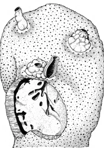
b Part of specimen cut through the center of one osculum. Note coral fragments to the right which indicate the level of the substratum (3 x ) -
c Underwater view of coral with sponge. Part of the sponge had been removed. Note mucus on the injured specimen and zone of dead coral exactly around it (0.8 x)-
d Surface fiber net (36 x)- e Primary fibers (36 x ) -
/ Spicule strands in canal lining membrane (36 x)-
PLATE S.—Siphonodictyon coralliphagum, new species, forma typica:
a Stephanocoenia michelinii infested by several specimens (Dominica) (0.5 x)- b Specimen USNM 24097 (paratype, Dominica), coral cut in half (0.5 x)- c Specimen YPM 6487 A (paratype, Jamaica) (0.8 x ) -
d Specimen YPM 6507 (paratype, Jamaica). Note exposed choanosomal portion (0.8 x)-
e,f Underwater view of holotype USNM 24095 (right) before and after it had been removed from the substratum (0.5 x ) -
NUMBER 77 25
NUMBER 77 27
PLATE 4.—Siphonodictyon coralliphagum, new species, forma typica:
a,b Surface fiber net as viewed from above (a) and below (b). The supporting main fibers are apparent (16 x ) -
c Spicule strands in canal lining membrane (36 x)- d Primary fibers (36 x)-
Forma obruta:
e-g Habitus of the sponges in their burrows (2 x ) -
PLATE 5.—Siphonodictyon coralliphagum, new species, forma tubulosa:
a Habitus, specimen USNM 24101 (Dominica) (0.8 x ) - b Habitus, specimen USNM 24102 (Dominica) (0.8 x ) -
Forma incrustans:
c Habitus, specimen YPM 6496 (Jamaica) (0.9 x)- d Habitus, specimen YPM 4831 (Jamaica) (0.8 x ) -
NUMBER 77 29