GATAE GENE
Thesis by Pei Yun Lee
In Partial Fulfillment of the Requirements for the Degree of
Doctor of Philosophy
California Institute of Technology Pasadena, California
2007
(Defended January 31, 2007)
2007 Pei Yun Lee All Rights Reserved
ACKNOWLEDGEMENTS
First and foremost, I would like to thank my advisor, Eric Davidson, for his vast knowledge on just about everything, for teaching me how to do biology quantitatively, and for his vision. My committee members Marianne Bronner-Fraser, Ellen Rothenberg, and David Anderson, for all their comments and questions regarding my project. Each and every one of you contributed greatly to my learning in graduate school.
Members of the Davidson lab, who provided a fun and intellectually stimulating environment. It has been a real pleasure working with such a group of talented individuals. A special thank you goes to Paola Oliveri for taking me under her wing when I first started in the lab and teaching me how to work with the sea urchin embryo.
Veronica Hinman for discussions on being a woman scientist. Jongmin Nam for his worldview and the hours discussing my project and the latest adventures of Jack Bauer.
Deanna Thomas and Jane Rigg for ensuring that the lab functions on a daily basis. Past and present residents of 017K, Jane Wyllie, Cesar Arenas-Mena for his “tell it like it is”
attitude, Takuya Minokawa for the daily Rathskeller lunches and for teaching me the
“new” in situ method, Qiang Tu for his graciousness in offering to let me use whatever equipment I needed as I was finishing up my degree, and especially to Roger Revilla, whom I have spend my entire graduate student life sharing an office and bay with, and who is not just a wonderful scientist, but also a great friend.
Bob Goldberg at UCLA, who first awakened my love for science with his unorthodox teaching methods, who first taught me how to do great science in his
laboratory, and for being a mentor on all things science and nonscience for the past ten years.
The men and women of the Angeles Chorale, who have kept my connection to the non-science world on Tuesday evenings, and especially to Don Neuen, for demanding precision, artistry and passion in every note and challenging me to be a better musician.
Debbie Goh and Jillian Baker, for being the best girlfriends anyone could ask for.
My parents, for their unconditional love and support in the last 30 years. My brother Min, for keeping me grounded in 27 of those years. And my fiancé Eric, for having traveled this entire journey with me, and with whom I look forward to even greater things.
ABSTRACT
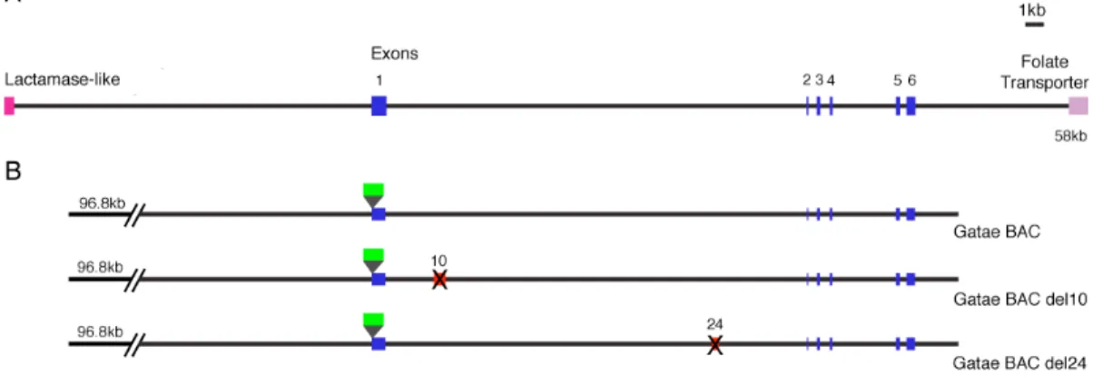
The Strongylocentrotus purpuratus gatae is orthologous to vertebrate gata4/5/6 genes. gatae is expressed throughout embryogenesis, beginning in the 15 h blastula in presumptive mesoderm cells, and at mesenchyme blastula, in endoderm and mesoderm cells of the veg2 lineage. During gastrulation, gatae is expressed in the midgut, hindgut and mesoderm, while in the pluteus expression it is limited to the midgut and coelomic pouches. Perturbation of gatae expression resulted in the lowered RNA levels for many endomesoderm transcription factors, including foxA, brachyury, and 1/2-otx, highlighting Gatae’s role as a regulator of transcription factors. gatae occupies an important node in the endomesoderm gene regulatory network, using its cross-regulatory interactions with otx to stabilize the endomesoderm gene expression program. Cis- regulatory analysis of gatae identified two modules responsible for its embryonic expression. Module 10 drives endomesoderm expression in the blastula, while module 24 activates gut expression in the gastrula and pluteus. Deletion of module 10 from a gatae GFP BAC resulted in a complete loss of blastula stage expression, demonstrating its necessity and sufficiency for early activity. Global cis-regulatory analysis of the gatae locus suggests that module usage is exclusionary; only one module can associate with the basal transcriptional apparatus and affect gene transcription at any given time. The endomesoderm gene regulatory network predicts that gatae is downstream of Otx and Notch signaling. Analysis of the sequence of module 10 identified Otx and Suppressor- of-Hairless (Su(H)) binding sites. Injection of Otx-engrailed RNA repressed the expression of module 10:GFP reporter; the effect is abolished when Otx binding sites
were mutated. Gel shifts demonstrated that the Otx protein binds to module 10. Module 10 expression was reduced under perturbation of Notch signaling. Mutations of either Otx or Su(H) binding sites resulted in lowered GFP RNA levels with no effect on spatial expression. Mutations of both Otx and Su(H) binding sites led to a further reduction but not elimination of reporter expression, suggesting that another input is involved. This unknown input was determined to be also downstream of Notch signaling and that gatae regulation functions via OR logic.
TABLE OF CONTENTS
Acknowledgements………...………iii Abstract………...v Table of Contents………...……..vii Introduction: Function of GATA Transcription Factors in Endoderm Specification…....1 Chapter 1: Expression of Spgatae, the Strongylocentrotus purpuratus Ortholog of Vertebrate GATA4/5/6 Factors………..33 Chapter 2:Exclusive Developmental Functions of gatae cis-Regulatory Modules in the Strongylocentrorus purpuratus Embryo………...……….45 Chapter 3: Use of OR Logic in the Regulation of a Sea Urchin GATA Factor………...77 Conclusions……….103 Appendix 1: A Genomic Regulatory Network for Development
Appendix 2: A Provisional Regulatory Gene Network for Specification of Endomeoderm in the Sea Urchin Embryo
Appendix 3: The Genome of the Sea Urchin Strongylocentrotus purpuratus
INTRODUCTION
Function of GATA Transcription Factors in Endoderm Specification
GATA factors are a class of zinc finger transcription factors named for binding to a GATA motif (Evans and Felsenfeld, 1989). Binding site studies have determined that GATA factors bind a WGATAR consensus sequence (Evans et al., 1988), although different family members have subtle differences in binding site preference (Ko and Engel, 1993; Merika and Orkin, 1993). The first GATA factor cloned was the chicken gata1 gene (Evans and Felsenfeld, 1989). Subsequently five more genes encoding GATA factors were identified in the vertebrates, named gata1-6, all of which play important roles during development. GATA factors are divided into two main classes:
members of the gata1/2/3 family function in hematopoiesis (Orkin and Zon, 1997), while the gata4/5/6 genes are widely expressed and utilized in the specification of endoderm and mesoderm specification as well as associated organ development.
Network based approach to GATA factor function
Traditionally, the study of a developmental process involves the generation of mutants through chemical or insertional mutagenesis followed by a screen for phenotypes pertaining to the process of interest. While this approach has identified many important genes and contributed greatly to our understanding of animal development, it is not without its limitations. Mutations manifested by dramatic phenotypes have turned out to
encode differentiation proteins, while mutations in genes encoding transcription factors are often lethal and not identified through mutant screens. Furthermore, phenotypic observations alone do not provide any information on the epistatic relationships between genes.
Transcription factors which are widely expressed during development, such as the GATA4/5/6 factors, are particularly difficult to study. Mouse knockouts in GATA genes result in early embryonic lethality, which precludes the analysis of their roles in development. A more informative approach is to study the function of GATA genes in the context of a gene regulatory network (GRN). Developmentally expressed genes do not function in parallel linear processes, rather they are integrated in complex networks containing activating and repressive interactions, signaling toggle switches, feed-back and feed-forward loops, to name a few. Presenting our current state of knowledge of GATA factors in the context of gene networks will further clarify their functions at the molecular level, and also provide broader views of their roles during endoderm specification. Comparison of similar gene networks involving GATA factors in different organisms may also lead to insights on regulatory circuitry conservation during evolution.
On a more general note, an understanding of developmental GRNs will also enable the definition of classical terms used by developmental biologists such as specification and commitment at the molecular level
In this review I will discuss the current state of knowledge pertaining to GATA transcription factor function and usage in endoderm specification and gut development. I have constructed a series of GRNs from six model organisms, the mouse, zebrafish, frog, worm, fly and sea urchin, using BioTapestry Editor (Longabaugh et al., 2005), focusing
on connections into and out of the GATA factors. All GRNs generated in this review are
“views from the genome,” meaning each includes interactions that take place over time and are not representative of events occurring in a single cell. In each GRN, connections originate from the upstream gene into a downstream one. Activating and repressive interactions are represented by arrows and bars respectively. Double arrows flanked by two circles denote intercellular signaling events. Any interaction that has been proven to be direct, either through transcription factor binding site mutation or otherwise, is indicated with a polygon of the same color as the upstream input. All of the GRNs are GATA-centric, and their main purposes are to highlight the role of GATA factors in each organism. Therefore while the connections into and out of the GATA factors are complete with respect to the current literature, none of the GRNs include all the gene interactions during endoderm specification.
While the extent and state of understanding of endoderm specification and gut development is different in each organism, a common theme emerges: the main function of GATA factors is to establish transcriptional domains through the regulation of endodermal transcription factors, in some cases by activating other GATA factors in a sequential fashion, and the direct activation of differentiation genes in later development.
Interestingly, engagement of GATA factors in cross- and autoregulatory loops suggest that one of their main roles is in the stabilization and lockdown of the transcriptional program for endoderm specification and organogenesis.
Biochemical properties of GATA factors
The GATA factors contain either one or two class IV zinc fingers, characterized by the CX2CX17CX2C motif (Evans et al., 1988). Biochemical studies have shown that the two zinc fingers have slightly different roles: the C terminal finger is required for binding site recognition (Morrisey et al., 1997b; Omichinski et al., 1993b; Visvader et al., 1995; Yang and Evans, 1992), while the N terminal finger contributes to binding specificity and stability (Yang and Evans, 1992) and increasing the spectrum of binding sites recognized (Merika and Orkin, 1993). In the GATA proteins that only possess a single zinc finger, it is the C terminal finger that is present (Lowry and Atchley, 2000), consistent with its necessity for DNA binding. NMR structural studies have shown that the C-terminal zinc finger interacts with the major groove of DNA, while the successive basic domain interacts with the minor groove of target sequences (Omichinski et al., 1993a). Studies of the GATA protein has implicated the C-terminal basic domain of the protein in transactivation (Nemer et al., 1999; Yang and Evans, 1992), while the N terminal domain plays an important role in protein-protein interactions with cofactors such as FOG (Svensson et al., 1999; Tevosian et al., 1999; Tsang et al., 1997).
Evolution of GATA transcription factors
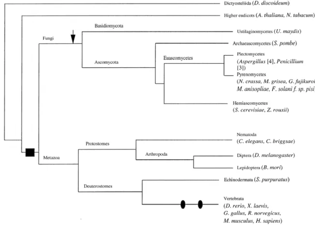
A comprehensive phylogenetic analysis of GATA factors was detailed in Lowry and Atchley (2000), which determined that all GATA factors evolved from an ancestral GATA factor contained a single zinc finger (Fig. 0.1). Zinc finger duplication occurred
before the split between metazoa and fungi, a conclusion supported by the fact that the N and C terminal zinc fingers in Drosophila are more closely related to its corresponding finger in the sea urchin and the vertebrates than to each other. Subsequent to zinc finger duplication, an insertion occurred between the two zinc fingers in the fungi, increasing the interfinger distance to 120 - 140 aa rather than the 30 aa observed in vertebrates. The ancestral deuterostome likely had two gata genes, supported by the fact that the echinoderms Asterina miniata (Hinman, pers. comm.) and Strongylocentrotus purpuratus (Pancer et al., 1999), and the urochordate Ciona intestinalis all have two GATA factors
Figure 0.1. Summary diagram for the evolution of GATA factors. The black box represents the duplication of zinc fingers before the divergence of fungi and metazoa. The arrow indicates the occurrence of a 100 aa insertion between the two zinc fingers; black ovals highlight genome duplication events in the vertebrates. From Lowry and Atchley, 2000.
(Yamada et al., 2003), while all jawed vertebrates studied thus far contain six gata genes.
C. elegans and Drosophila have eleven and five gata genes respectively, which could not be grouped with any certainty to gata1/2/3 or gata4/5/6.
Mouse
gata4/5/6 genes are widely expressed in the mouse during embryonic development and adulthood. In the adult mouse gata4/5/6 are expressed in the heart, lung, liver, gut, bladder, kidney and gonads, while in the embryo gata4/6 expression have been detected in extraembryonic visceral and parietal endoderm, definitive endoderm at the foregut and midgut junction, and lateral plate mesoderm. All three genes are also expressed in the intestine epithelium and developing endodermal and mesodermal organs (Arceci et al., 1993; Jacobsen et al., 2002; Koutsourakis et al., 1999; Morrisey et al., 1996; Morrisey et al., 1997a; Narita et al., 1996; Soudais et al., 1995) (Table 0.1).
Functional studies of the gata4/5/6 genes in the mouse have proven to be very difficult because of their importance in the development of extraembryonic endoderm.
Knockouts in gata4 resulted in embryos with no visceral endoderm and death at E8 with severe heart and gut defects (Kuo et al., 1997). gata6 knockouts die shortly after implantation, are much smaller in size, lack part of visceral endoderm and displayed embryonic ectoderm defects (Koutsourakis et al., 1999; Morrisey et al., 1998). Analyses using chimeric embryos demonstrated that the lethalities from gata4/6 knockouts were due to a lack of visceral endoderm differentiation (Koutsourakis et al., 1999; Morrisey et
al., 1998; Soudais et al., 1995), though gata4 mutants also possessed intrinsic defects in gut epithelium development (Jacobsen et al., 2002). Unlike gata4/6 mutants, gata5 knockouts were viable and displayed only defects in female genitouninary tract development (Molkentin et al., 2000) (Table 0.1).
gata4/6 are among the earliest genes expressed in the endoderm. The HMG transcription factor Sox7 activates gata4 transcription (Futaki et al., 2004) (Fig. 0.2).
Once activated, Gata4 feeds back and activates sox7 (Murakami et al., 2004), setting up the first cross-regulatory loop. Gata4, together with Sox7, have also been shown to directly activate fgf3 (Murakami et al., 2004). In addition, Gata4 also turns on gata6 (Fujikura et al., 2002) which is also regulated by retinoic acid (RA) signaling (Capo- Chichi et al., 2005). Once activated, Gata6 serves to reinforce gata4 expression in the second cross-regulatory loop in this network (Futaki et al., 2004; Morrisey et al., 1998).
Next, Gata4/6 activate a number of endoderm specific transcription factors, including hnf1, hnf3, hnf4, sox17 and gata4/5/6 (Fujikura et al., 2002; Futaki et al., 2004;
Morrisey et al., 1998; Murakami et al., 2004; Soudais et al., 1995). Cis-regulatory analysis on gut differentiation genes such as lactase-phlorizin hydrolase (LPH), and sucrase-isomaltase (SI) have shown that they are under the direct control of GATA factors and Hnf1 (Boudreau et al., 2002; van Wering et al., 2004).
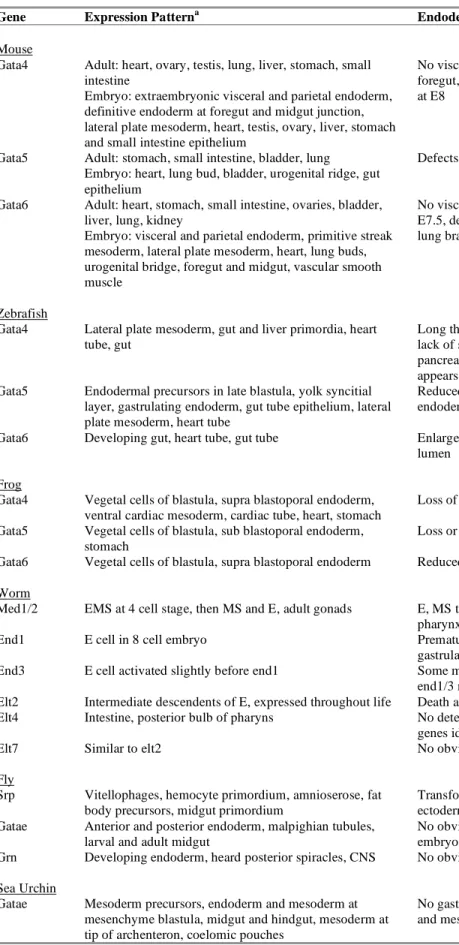
Table 0.1. Expression patterns and knockout phenotypes for GATA4/5/6 factors.
Gene Expression Patterna Endodermal Phenotype Mouse
Gata4 Adult: heart, ovary, testis, lung, liver, stomach, small intestine
Embryo: extraembryonic visceral and parietal endoderm, definitive endoderm at foregut and midgut junction, lateral plate mesoderm, heart, testis, ovary, liver, stomach and small intestine epithelium
No visceral endoderm, disorganized foregut, gastric epithelium defects, death at E8
Gata5 Adult: stomach, small intestine, bladder, lung Embryo: heart, lung bud, bladder, urogenital ridge, gut epithelium
Defects in genitourinary tract development
Gata6 Adult: heart, stomach, small intestine, ovaries, bladder, liver, lung, kidney
Embryo: visceral and parietal endoderm, primitive streak mesoderm, lateral plate mesoderm, heart, lung buds, urogenital bridge, foregut and midgut, vascular smooth muscle
No visceral endoderm, death at E6.5 - E7.5, defects in hepatic differentiation, lung branching morphogenesis
Zebrafish
Gata4 Lateral plate mesoderm, gut and liver primordia, heart tube, gut
Long thin gut tube with no epithelial folds, lack of severely reduced liver and pancreas, early endoderm specification appears normal
Gata5 Endodermal precursors in late blastula, yolk syncitial layer, gastrulating endoderm, gut tube epithelium, lateral plate mesoderm, heart tube
Reduced endoderm, lack of gut looping, endodermal organ defects
Gata6 Developing gut, heart tube, gut tube Enlarged gut, uncoiled intestine with no lumen
Frog
Gata4 Vegetal cells of blastula, supra blastoporal endoderm, ventral cardiac mesoderm, cardiac tube, heart, stomach
Loss of gut coiling and reduced gut tissue Gata5 Vegetal cells of blastula, sub blastoporal endoderm,
stomach
Loss or defect in gut coiling Gata6 Vegetal cells of blastula, supra blastoporal endoderm Reduced gut tissue with no coiling Worm
Med1/2 EMS at 4 cell stage, then MS and E, adult gonads E, MS to C transformation, no MS derived pharynx
End1 E cell in 8 cell embryo Premature division and defective
gastrulation
End3 E cell activated slightly before end1 Some mutants lack endoderm, lack of end1/3 most embryos no endoderm Elt2 Intermediate descendents of E, expressed throughout life Death at L1 with malformed gut Elt4 Intestine, posterior bulb of pharyns No detectable phenotype, no downstream
genes identified
Elt7 Similar to elt2 No obvious phenotype
Fly
Srp Vitellophages, hemocyte primordium, amnioserose, fat body precursors, midgut primordium
Transformation of endodermal midgut into ectodermal foregut and hindgut
Gatae Anterior and posterior endoderm, malpighian tubules, larval and adult midgut
No obvious morphological defects, most embryos do not hatch
Grn Developing endoderm, heard posterior spiracles, CNS No obvious endodermal phenotype Sea Urchin
Gatae Mesoderm precursors, endoderm and mesoderm at mesenchyme blastula, midgut and hindgut, mesoderm at tip of archenteron, coelomic pouches
No gastrulation, disorganized endoderm and mesoderm cells
a Gene expression patterns from the mouse, zebrafish, Xenopus, worm and fly and sea urchin are summarized together with any endodermal phenotypes from the perturbation of the genes. Phenotypes were obtained either from genetic knockouts, genetic mutations, RNAi and MASO injections.
Figure. 0.2. Network model outlining gene interactions underlying endoderm specification in the mouse, Mus musculus. Abbreviations of gene names are as follows: LPH, lactase-phlorizin hydrolase; SI, sucrase-isomaltase.
Zebrafish
As a model organism, the zebrafish Danio rerio is well suited for the study of animal development due to the ability to generate mutants, the ease of gene transfers and perturbation with mRNA and morpholino antisense oligonucleotides (MASO). As in the mouse, GATA factors in the zebrafish also display overlapping expression patterns.
Drgata4 is expressed in the lateral plate mesoderm, gut and liver primordia, heart tube
and gut (Table 0.1). gata5 is the earliest expressing GATA factor in the zebrafish, first observed in the endodermal precursors in the late blastula and yolk syncitial layer (Reiter et al., 2001). It is also expressed in the gastrulating endoderm, gut tube epithelium, lateral plate mesoderm and heart tube (Reiter et al., 1999). gata6 is also expressed in the developing gut, heart and gut tubes (Holtzinger and Evans, 2005). Perturbation of the expression of any of the gata genes by genetic mutation or MASO injections led to defects in gut looping and morphogenesis (Holtzinger and Evans, 2005; Peterkin et al., 2003). Genetic mutation of the gata5 (also known as faust) gene also led to reduced endoderm (Reiter et al., 1999). In addition to the gut, defects in organs such as heart, liver and pancreas were also observed (Holtzinger and Evans, 2005; Reiter et al., 2001).
Nodal signaling plays an important role in zebrafish endoderm formation.
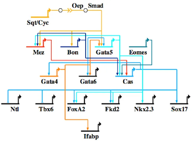
Mutations in the two zebrafish nodal genes, squint (sqt) and cyclops (cyc), or the Nodal co-receptor one-eyed pinhead (oep) lead to a complete loss of endoderm and mesoderm (Feldman et al., 1998; Gritsman et al., 1999). Downstream of Nodal signaling are three genes, mezzo (mez), bonnie and clyde (bon) and gata5 (Poulain and Lepage, 2002;
Rodaway et al., 1999) (Fig. 0.3). These three transcription factors, together with a maternal T box factor Eomesoderm (Eomes), activate the transcription of casanova (cas), which encodes a Sox-like protein whose mutation leads to a total loss of endoderm (Aoki et al., 2002; Bjornson et al., 2005; Kikuchi et al., 2001). Cas then activates gata5 in a cross-regulatory loop, and also activates the gata4 and gata6 genes (Alexander et al., 1999). Like Cas, Gata5 also feeds back and cross-regulates with gata4, and plays a role in the activation of gata6. In addition, Cas also activates endoderm transcription factors
Figure 0.3. Network model outlining gene interactions underlying endoderm specification in the zebrafish Danio rerio. Gene abbreviations are as follows: Bon, bonnie and clyde; Cas, casanova; Cyc, cyclops; Eomes, eomesodermin; Mez, mezzo; Ntl, no tail; Oep, one-eyed pinhead; Ifabp, intestinal fatty acid binding protein; Sqt, squint.
axial/foxA2, fkd2, nkx2.3 and sox17 (Alexander et al., 1999; Kikuchi et al., 2001; Reiter et al., 2001) and functions in the repression of mesoderm genes ntl and tbx6 (Aoki et al., 2002). It is unclear whether the gata genes play direct roles in activating any of these transcription factors, though Gata4 has been implicated in the activation of the intestinal fatty acid binding protein gene, ifabp (Holtzinger and Evans, 2005).
Frog
In the Xenopus laevis embryo, gata4/5/6 are all expressed in the vegetal pole during blastula stages. gata4/6 are expressed in the involuting supra blastoporal endoderm until gastrulation, while gata5 is expressed in the non-involuting sub blastoporal endoderm in midgastrula (Afouda et al., 2005; Weber et al., 2000) (Table 0.1). Gata4 RNA has also been detected in the developing ventral cardiac mesoderm, cardiac tube, heart, stomach and other endodermally derived organs (Kelley et al., 1993).
Translational inhibition of all three gata genes with MASO injections led to the same phenotype: a reduction in the amount of endodermal tissue and a loss or defect in gut coiling (Afouda et al., 2005).
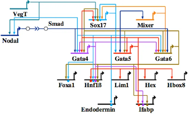
The Xenopus endoderm specification program is initiated by the maternal T-box transcription factor VegT, which, when depleted, resulted in embryos that did not express many endodermal markers (Zhang et al., 1998). VegT is upstream of Nodal signaling, which plays a role in the activation of all three GATA factors (Hilton et al., 2003;
Xanthos et al., 2001). VegT activates the sox17 gene cell autonomously (Clements and Woodland, 2003) (Fig. 0.4). Once the gata genes are activated by Nodal signaling, they take over the regulation of sox17 in the Nodal dependent phase of sox17 expression (Afouda et al., 2005; Weber et al., 2000). In return, Sox17 engages the gata5/6 genes in cross-regulatory loops (Sinner et al., 2006). A homeodomain transcription factor, Mixer, also regulates gata6 and sox17. In turn, the three gata genes auto- and cross-regulate (Afouda et al., 2005). Once the Gata proteins are available, they activate endoderm
Figure 0.4. Network model outlining gene interactions underlying endoderm specification in the frog Xenopus laevis. Gene abbreviations are as follows: Ifabp, intestinal fatty acid binding protein.
transcription factors like foxa1, hnf1, lim1, hex and hbox8 (Afouda et al., 2005; Weber et al., 2000). At least one of these interactions, into hnf1, is direct. In the gut differentiation gene battery, GATA factors have also been shown to be a direct activator for the intestinal fatty acid binding protein (ifabp) (Gao et al., 1998).
Worm
GATA factors have been studied extensively in the nematode C. elegans. The C.
elegans genome encodes eleven GATA factors, seven of which are expressed in the endoderm. Usage of GATA factors appears to be at least partially redundant. The earliest expressing GATA factors are the med-1/2 genes, which are activated in the EMS
cell at the four-cell stage. They are transiently expressed in the MS and E cells, which develop into the endodermal gut and body wall muscle (Table 0.1). In addition, they are also expressed in the adult gonads (Maduro et al., 2001). In med-1/2 mutants, some of the embryos display E,MS to C (hypodermis) transformation. Recently, Maduro et al.
(2006) demonstrated that the low penetrance of the zygotic med-1/2 mutation was due to a maternal contribution to the Med-1/2 function.
The end-1/3 genes are the earliest expressing genes in the E lineage, expressed in the E cell from the 8-cell embryo, with end-3 activated slightly before end-1 (Maduro et al., 2006; Maduro et al., 2005a; Maduro and Rothman, 2002; Zhu et al., 1997).
Mutations in both end-1 and end-3 genes led to an E to C transformation, in which most embryos did not have any endoderm, whereas mutation of either one of the genes resulted in much weaker phenotypes (Maduro et al., 2005a; Zhu et al., 1997).
The final group of GATA factors transcribed are the elt genes. Three elt genes are expressed in the endoderm, elt-2, which is expressed from the 2E cell stages and whose expression lasts through the life of the animal (Fukushige et al., 1998). This includes elt-4, which is expressed in the intestine of the embryo and posterior bulb of the pharynx in late embryogenesis; and elt-7, which is expressed in a similar pattern as elt-2 (Fukushige et al., 2003; Maduro and Rothman, 2002). Mutation of the elt-2 gene led to death at the larval L1 stage with malformed guts, whereas mutations in elt-4/7 did not result in a detectable phenotypes. To date, elt-4 has not been demonstrated to be upstream of any gene and appears to be non-functional (Fukushige et al., 2003).
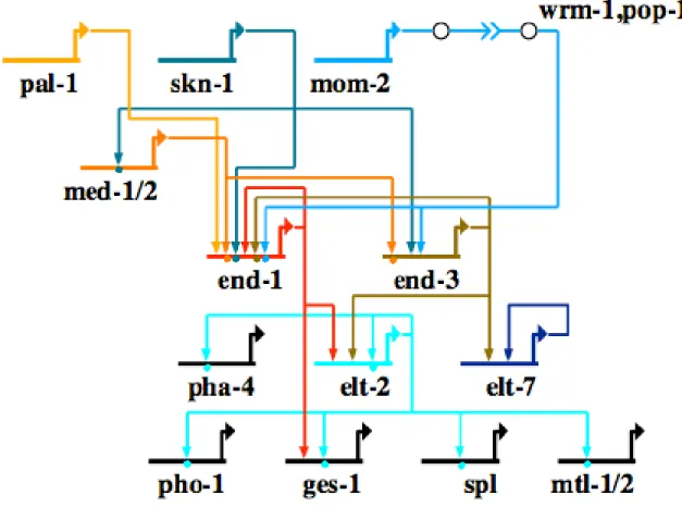
Figure 0.5. Network model outlining gene interactions underlying endoderm specification in the nematode Caenorhabditis elegans. Gene abbreviations are as follows: ges-1, gut esterase; mtl, metallothionein; pho-1, essential acid phosphatase; spl, Sphingosine-1-phosphate Lyase.
Notably, endoderm specification in C. elegans occurs through the sequential activation of the GATA factors. A bZIP maternal transcription factor, Skn-1, is responsible for turning on med-1/2 in the EMS cell at the 4-cell stage (Maduro et al., 2001) (Fig. 0.5). Med1/2 have been demonstrated to directly bind the cis-regulatory region of end-1/3 (Broitman-Maduro et al., 2005). In addition to activation by Med-1/2, end-1/3 expression are also dependent on Wnt signaling. In the presence of Wnt signaling, Pop-1, a Lef homolog is switched from a repressor to an activator that activates end-1/3 (Calvo et al., 2001; Maduro et al., 2005b). End-3, the earlier expressing of the
two end genes, also activates end-1 (Maduro et al., 2006; Maduro et al., 2005b). A direct input from Skn-1 has also been identified in end-1 (Maduro et al., 2005b). End-1/3 then activate the final pair of GATA factors involved in endoderm specification, elt-2 and elt- 7, which autoregulate and whose main roles are to activate differentiation gene batteries in the gut (Fukushige et al., 2005; Fukushige et al., 1998; Fukushige et al., 1999; Kalb et al., 1998; Maduro and Rothman, 2002; Moilanen et al., 1999; Oskouian et al., 2005;
Peterkin et al., 2003; Zhu et al., 1998).
Fly
The Drosophila genome has five gata genes, pannier, serpent (srp), grain (grn), gatad and gatae. pnr, grn and srp are part of a cluster of genes on chromosome three (Okumura et al., 2005). Among that Drosophila GATA factors, srp, grn and gatae are expressed in the endoderm. In Drosophila only the midgut is derived from endoderm, whereas the fore- and hindguts are ectodermal derivatives. Of the three genes, srp is the earliest expressing GATA factor, first detected in the prospective endoderm and ceases to be expressed before obvious midgut differentiation. In addition to the endoderm, srp is also expressed in the vitellophages, hemocyte primordium, amnioserosa and fat body precursors, reflecting its numerous functions in Drosophila development (Rehorn et al., 1996; Sam et al., 1996) (Table 0.1). Two isoforms of Srp exist, one containing two zinc fingers and the other with one zinc finger. Both isoforms have the same expression pattern, though they appear to regulate genes differently (Waltzer et al., 2002). srp mutants exhibited a midgut to fore- and hindgut transformation (Reuter, 1994).
Drosophila gatae expression begins in the endoderm at stage 8 and continues in the midgut throughout the life of the animal. In addition, it is also expressed in the malpighian tubules. gatae mutants displayed no obvious morphological defects, but lacked expression of many midgut specific genes (Okumura et al., 2005). grn is expressed in the developing endoderm, head, posterior spiracles and central nervous system, though grn mutants had no discernable endodermal phenotype (Brown and Castelli-Gair Hombria, 2000; Lin et al., 1995).
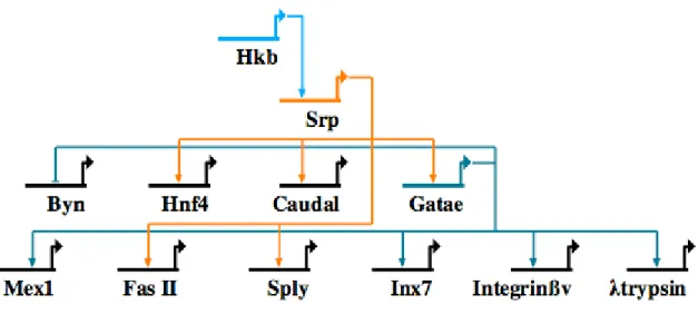
Drosophila endodermal transcriptional program begins with the gap gene hkb, which itself is downstream of Torso RTK signaling. It activates srp, which in turn activates gatae in a sequential manner (Okumura et al., 2005) (Fig. 0.6). In addition, Srp activates zygotic caudal and hnf4 expression in the endoderm (Reuter, 1994). Both srp and gatae have been demonstrated to be upstream of intestinal differentiation genes,
Figure 0.6. Network model outlining gene interactions underlying endoderm specification in the fruit fly Drosophila melanogaster. Gene abbreviations are as follows: Byn, brachyenteron; Cau, caudal; Fas II, fasciclin; Hkb, huckebein; Inx7, innexin; Sply, Sphingosine-1-phosphate Lyase; Srp, serpent.
though the precise epistatic relationships remain unclear. In case in the sply gene, srp regulation is not mediated by Gatae, suggesting that both srp and gatae directly activate differentiation genes (Okumura et al., 2005; Okumura et al., 2007). In addition to activation of endodermal differentiation genes, Gatae also plays a role in repressing the expression of brachyenteron, normally expressed in the hindgut, from the midgut (Okumura et al., 2005). Furthermore, Gatae has also been shown to directly activate immune genes in the intestine of the Drosophila larva (Senger et al., 2006).
Sea Urchin
The purple sea urchin Strongylocentrotus purpuratus has two GATA factors, gatae, orthologous to gata4/5/6, and gatac, orthologous to gata1/2/3 (Pancer et al., 1999).
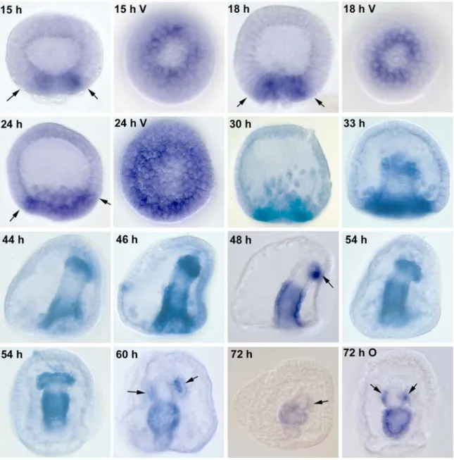
gatac is expressed in a subset of mesoderm cells known as blastocoelar cells in the blastula and in the adult coelomyctes (Pancer et al., 1999 and J.P. Rast, unpublished data). gatae is expressed throughout embryogeneis, first detected in the prospective secondary mesenchyme cells in the 15 h blastula. By the mesenchyme blastula it is expressed in both endoderm and mesoderm, and in the gastrulating gut and mesoderm at the tip of the archenteron until the end of embryogenesis (Lee and Davidson, 2004).
Perturbation of gatae translation by MASO injection resulted in embryos that failed to gastrulate and which exhibited severely compromised organization of the endomesoderm (Table 0.1). Even so, pigment expressing cells, presumably of mesodermal origin were still visible. In situ hybridizations on MASO injected embryos showed that the disorganized cells within the blastocoel expressed endoderm and mesoderm markers.
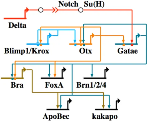
To determine the position of gatae in the endomesoderm gene network, fertilized eggs were injected with gatae MASO and the expression of genes were quantified in 18 h and 24 h embryos. Figure 0.7 is a graphical representation of the genes whose expression in response of gatae perturbation had been analyzed. A large number of endomesoderm expressing genes are downstream of gatae, including many transcription factors. In addition to Gatae’s role as an activator of other endomesoderm transcription factors, it is also engaged in a cross-regulatory loop with otx, functioning to lock down the endomesoderm specification program (Davidson et al., 2002a; Davidson et al., 2002b) (Fig. 0.8). This interaction has been verified at the DNA level through binding site mutations (Lee and Davidson, 2007; Yuh et al., 2004). Differentiation genes, such as apobec and kakapo, have also been demonstrated to be downstream of gatae, though it is not clear if Gatae regulates them directly or via brachyury.
GATA factors as regulators of endoderm transcription factors
In all organisms studied, GATA factors are widely expressed in both the mesoderm and endoderm. Attempts to determine function based on phenotype alone were challenging in many organisms due to the multitude and severity of defects in the mutants or knockdowns. Even though the states of understanding of endoderm specification in the different organisms surveyed are at different levels of completion, all the GRNs have one thing in common: GATA factors are activators of other endoderm specific transcription factors. Examples of genes encoding transcription factors downstream of the GATA factors include the hnf3/forkhead genes, Sox class of HMG
Figure 0.7. Graphs depicting the expression pattern of genes in (A) 18 h, and (B) 24 h embryos after injection with an Spgatae MASO. RNA was extracted and QPCR performed on embryos injected with either gatae or control MASO. Ct was calculated from gatae vs. control MASO injected embryos after normalization to ubiquitin to account for differing embryo numbers in each reaction. Ct is defined as the cycle number at a threshold when the PCR product is accumulating exponentially. The two dashed lines (positive and negative) indicate a Ct of 1.7 between gatae and control MASO injected embryos. Due to variances between embryo batches, only perturbation effects at or above this level are considered to be significant. The genes are grouped into the following categories based on the color of the bars: blue, endomesoderm transcription factors; lavender, endomesoderm differentiation genes; purple, oral ectoderm genes, pink, ectoderm (other) genes; yellow, skeletogenic mesenchyme gene.
Figure 0.8. Network model outlining gene interactions underlying endoderm specification in the sea urchin Strongylocentrotus purpuratus. Abbreviations for genes are as follows: Bra, brachyury; Brn, brain.
transcription factors, homeodomain proteins such as Nkx, Hex, Otx, Lim and Caudal. In addition, GATA factors also play a role in the repression of non-endodermal states, such as the repression of byn in Drosophila.
During development transcriptional domains are initiated and progressively refined, and in every organism the GATA factors function near the top of this hierarchy, often the immediate downstream target of a maternal transcription factor or in direct response to signaling toggle switches. A few examples of signaling cascades that they are under the control of include Nodal in vertebrates (Feldman et al., 1998), Wnt in C.
elegans (Maduro et al., 2005b), and Notch in the sea urchin (Lee and Davidson, 2007).
GATA factors are not exclusive regulators of other transcription factors however, they have also been shown to directly activate the expression of differentiation genes, particularly in later development and the adult animal (Bossard and Zaret, 1998;
Boudreau et al., 2002).
GATA factors as stabilizers of the endoderm transcriptional state
An interesting observation is that, at least in the deuterostome lineage, GATA factors are involved in cross-regulatory loops with other early acting transcription factors like to facilitate the “lockdown” of the endodermal transcriptional states. These feedback loops involve gata4 and sox7 in the mouse (Futaki et al., 2004; Murakami et al., 2004), gata5 and cas in the zebrafish (Alexander et al., 1999; Kikuchi et al., 2001), gata5/6 and sox17 in Xenopus (Sinner et al., 2006) and gatae and otx in the sea urchin (Davidson et al., 2002a; Davidson et al., 2002b). While the partners for GATA factors in these cross-
regulatory loops are different for each organism, it is notable that in the vertebrates, they all involved members of the Sox family of transcription factors. Such feedback loops are not observed in the two ecdysozoans Drosophila and C. elegans, and may reflect a newly acquired function of the GATA factors in the deuterostome lineage, although it is also possible that those connections have simply not been identified in the ecdysozoa.
However, Drosophila and C. elegans both have multiple GATA factors that are activated sequentially, and which could not be grouped into either the 1/2/3 or 4/5/6 GATA sub- families by phylogenetic analysis. This observation is also reflected in the function of these factors. For example, Drosophila srp is involved in both hematopoiesis and endoderm development, a feature not observed in deuterstome GATAs.
Summary
GATA factors play functionally conserved roles in setting up the endodermal transcriptional state through its activation of other transcription factors. Furthermore, in the deuterostomes they are also participants in cross-regulatory feedback loops that serve to “lockdown” the developmental program. These networks are by no means complete even in the best studied case of the sea urchin. However, even with the limited information available it is still possible to draw conclusions regarding the function of GATA factors in endoderm development.
Why do GATA factors function near the top of the developmental gene regulatory hierarchy in every organism studied? One possible explanation is that the protostome/deuterostome ancestor utilized GATA factors in such a fashion, and this
function has been conserved and fixed in the GRNs over time. Another possibility is that properties of the GATA protein, for example, its ability to interact with multiple co- factors to affect gene expression in subtle ways, are important for and lead to their active selection as pan endodermesodermal activators. As more details regarding endoderm specification become available, we will be able to better understand not just the developmental process, but also how the regulatory circuitries have evolved in the Bilateria.
List of transcription factors represented in GRNs
Transcription Factor Family
Bon Mix homeodomain
Bra/Byn/Ntl T box
Blimp1/Krox Zinc finger
Brn-1/2/4 POU homeodomain
Cas Sox HMG
Caudal/Pal-1 Homeodomain
End-1/3 GATA zinc finger
Elt-2/7 GATA zinc finger
Eomes T box
Fkd2 Forkhead GATA4/5/6/Gatae (Sp) GATA zinc finger
Gatae (Dm) GATA zinc finger
Hbox8 Homeodomain Hex Homeodomain
Hkb Zinc finger
Hnf1/ POU-homeodomain
Hnf3/FoxA/Pha-4 Forkhead
Hnf4 Orphan nuclear receptor
Lim Lim homeodomain
Med-1/2 GATA zinc finger
Mezzo Mix homeodomain
Nkx2.3 Nk-2 homeodomain
Pop-1 HMG Skn-1 bZIP
Sox 7 Sox HMG
Sox17 Sox HMG
Srp GATA zinc finger
Tbx6 T box
VegT T box
References
Afouda, B.A., Ciau-Uitz, A. and Patient, R., 2005. GATA4, 5 and 6 mediate TGFbeta maintenance of endodermal gene expression in Xenopus embryos. Development 132, 763-74.
Alexander, J., Rothenberg, M., Henry, G.L. and Stainier, D.Y., 1999. casanova plays an early and essential role in endoderm formation in zebrafish. Dev Biol 215, 343- 57.
Aoki, T.O., David, N.B., Minchiotti, G., Saint-Etienne, L., Dickmeis, T., Persico, G.M., Strahle, U., Mourrain, P. and Rosa, F.M., 2002. Molecular integration of casanova in the Nodal signalling pathway controlling endoderm formation. Development 129, 275-86.
Arceci, R.J., King, A.A., Simon, M.C., Orkin, S.H. and Wilson, D.B., 1993. Mouse GATA-4: a retinoic acid-inducible GATA-binding transcription factor expressed in endodermally derived tissues and heart. Mol Cell Biol 13, 2235-46.
Bjornson, C.R., Griffin, K.J., Farr, G.H., 3rd, Terashima, A., Himeda, C., Kikuchi, Y. and Kimelman, D., 2005. Eomesodermin is a localized maternal determinant required for endoderm induction in zebrafish. Dev Cell 9, 523-33.
Bossard, P. and Zaret, K.S., 1998. GATA transcription factors as potentiators of gut endoderm differentiation. Development 125, 4909-17.
Boudreau, F., Rings, E.H., van Wering, H.M., Kim, R.K., Swain, G.P., Krasinski, S.D., Moffett, J., Grand, R.J., Suh, E.R. and Traber, P.G., 2002. Hepatocyte nuclear factor-1 alpha, GATA-4, and caudal related homeodomain protein Cdx2 interact functionally to modulate intestinal gene transcription. Implication for the developmental regulation of the sucrase-isomaltase gene. J Biol Chem 277, 31909-17.
Broitman-Maduro, G., Maduro, M.F. and Rothman, J.H., 2005. The noncanonical binding site of the MED-1 GATA factor defines differentially regulated target genes in the C. elegans mesendoderm. Dev Cell 8, 427-33.
Brown, S. and Castelli-Gair Hombria, J., 2000. Drosophila grain encodes a GATA transcription factor required for cell rearrangement during morphogenesis.
Development 127, 4867-76.
Calvo, D., Victor, M., Gay, F., Sui, G., Luke, M.P., Dufourcq, P., Wen, G., Maduro, M., Rothman, J. and Shi, Y., 2001. A POP-1 repressor complex restricts inappropriate cell type-specific gene transcription during Caenorhabditis elegans embryogenesis. Embo J 20, 7197-208.
Capo-Chichi, C.D., Rula, M.E., Smedberg, J.L., Vanderveer, L., Parmacek, M.S., Morrisey, E.E., Godwin, A.K. and Xu, X.X., 2005. Perception of differentiation cues by GATA factors in primitive endoderm lineage determination of mouse embryonic stem cells. Dev Biol 286, 574-86.
Clements, D. and Woodland, H.R., 2003. VegT induces endoderm by a self-limiting mechanism and by changing the competence of cells to respond to TGF-beta signals. Dev Biol 258, 454-63.
Davidson, E.H., Rast, J.P., Oliveri, P., Ransick, A., Calestani, C., Yuh, C.H., Minokawa, T., Amore, G., Hinman, V., Arenas-Mena, C., Otim, O., Brown, C.T., Livi, C.B., Lee, P.Y., Revilla, R., Rust, A.G., Pan, Z., Schilstra, M.J., Clarke, P.J., Arnone, M.I., Rowen, L., Cameron, R.A., McClay, D.R., Hood, L. and Bolouri, H., 2002a.
A genomic regulatory network for development. Science 295, 1669-78.
Davidson, E.H., Rast, J.P., Oliveri, P., Ransick, A., Calestani, C., Yuh, C.H., Minokawa, T., Amore, G., Hinman, V., Arenas-Mena, C., Otim, O., Brown, C.T., Livi, C.B., Lee, P.Y., Revilla, R., Schilstra, M.J., Clarke, P.J., Rust, A.G., Pan, Z., Arnone, M.I., Rowen, L., Cameron, R.A., McClay, D.R., Hood, L. and Bolouri, H., 2002b.
A provisional regulatory gene network for specification of endomesoderm in the sea urchin embryo. Dev Biol 246, 162-90.
Evans, T. and Felsenfeld, G., 1989. The erythroid-specific transcription factor Eryf1: a new finger protein. Cell 58, 877-85.
Evans, T., Reitman, M. and Felsenfeld, G., 1988. An erythrocyte-specific DNA-binding factor recognizes a regulatory sequence common to all chicken globin genes. Proc Natl Acad Sci U S A 85, 5976-80.
Feldman, B., Gates, M.A., Egan, E.S., Dougan, S.T., Rennebeck, G., Sirotkin, H.I., Schier, A.F. and Talbot, W.S., 1998. Zebrafish organizer development and germ- layer formation require nodal-related signals. Nature 395, 181-5.
Fujikura, J., Yamato, E., Yonemura, S., Hosoda, K., Masui, S., Nakao, K., Miyazaki Ji, J.
and Niwa, H., 2002. Differentiation of embryonic stem cells is induced by GATA factors. Genes Dev 16, 784-9.
Fukushige, T., Goszczynski, B., Tian, H. and McGhee, J.D., 2003. The evolutionary duplication and probable demise of an endodermal GATA factor in Caenorhabditis elegans. Genetics 165, 575-88.
Fukushige, T., Goszczynski, B., Yan, J. and McGhee, J.D., 2005. Transcriptional control and patterning of the pho-1 gene, an essential acid phosphatase expressed in the C. elegans intestine. Dev Biol 279, 446-61.
Fukushige, T., Hawkins, M.G. and McGhee, J.D., 1998. The GATA-factor elt-2 is essential for formation of the Caenorhabditis elegans intestine. Dev Biol 198, 286-302.
Fukushige, T., Hendzel, M.J., Bazett-Jones, D.P. and McGhee, J.D., 1999. Direct visualization of the elt-2 gut-specific GATA factor binding to a target promoter inside the living Caenorhabditis elegans embryo. Proc Natl Acad Sci U S A 96, 11883-8.
Futaki, S., Hayashi, Y., Emoto, T., Weber, C.N. and Sekiguchi, K., 2004. Sox7 plays crucial roles in parietal endoderm differentiation in F9 embryonal carcinoma cells through regulating Gata-4 and Gata-6 expression. Mol Cell Biol 24, 10492-503.
Gao, X., Sedgwick, T., Shi, Y.B. and Evans, T., 1998. Distinct functions are implicated for the GATA-4, -5, and -6 transcription factors in the regulation of intestine epithelial cell differentiation. Mol Cell Biol 18, 2901-11.
Gritsman, K., Zhang, J., Cheng, S., Heckscher, E., Talbot, W.S. and Schier, A.F., 1999.
The EGF-CFC protein one-eyed pinhead is essential for nodal signaling. Cell 97, 121-32.
Hilton, E., Rex, M. and Old, R., 2003. VegT activation of the early zygotic gene Xnr5 requires lifting of Tcf-mediated repression in the Xenopus blastula. Mech Dev 120, 1127-38.
Hinman, V., pers. comm. personal communication.
Holtzinger, A. and Evans, T., 2005. Gata4 regulates the formation of multiple organs.
Development 132, 4005-14.
Jacobsen, C.M., Narita, N., Bielinska, M., Syder, A.J., Gordon, J.I. and Wilson, D.B., 2002. Genetic mosaic analysis reveals that GATA-4 is required for proper differentiation of mouse gastric epithelium. Dev Biol 241, 34-46.
Kalb, J.M., Lau, K.K., Goszczynski, B., Fukushige, T., Moons, D., Okkema, P.G. and McGhee, J.D., 1998. pha-4 is Ce-fkh-1, a fork head/HNF-3alpha,beta,gamma homolog that functions in organogenesis of the C. elegans pharynx. Development 125, 2171-80.
Kelley, C., Blumberg, H., Zon, L.I. and Evans, T., 1993. GATA-4 is a novel transcription factor expressed in endocardium of the developing heart. Development 118, 817- 27.
Kikuchi, Y., Agathon, A., Alexander, J., Thisse, C., Waldron, S., Yelon, D., Thisse, B.
and Stainier, D.Y., 2001. casanova encodes a novel Sox-related protein necessary and sufficient for early endoderm formation in zebrafish. Genes Dev 15, 1493- 505.
Ko, L.J. and Engel, J.D., 1993. DNA-binding specificities of the GATA transcription factor family. Mol Cell Biol 13, 4011-22.
Koutsourakis, M., Langeveld, A., Patient, R., Beddington, R. and Grosveld, F., 1999. The transcription factor GATA6 is essential for early extraembryonic development.
Development 126, 723-32.
Kuo, C.T., Morrisey, E.E., Anandappa, R., Sigrist, K., Lu, M.M., Parmacek, M.S., Soudais, C. and Leiden, J.M., 1997. GATA4 transcription factor is required for ventral morphogenesis and heart tube formation. Genes Dev 11, 1048-60.
Lee, P.Y. and Davidson, E.H., 2004. Expression of Spgatae, the Strongylocentrotus purpuratus ortholog of vertebrate GATA4/5/6 factors. Gene Expr Patterns 5, 161- 5.
Lee, P.Y. and Davidson, E.H., 2007. Use of OR logic in the regulation of a sea urchin GATA factor (in preparation).
Lin, W.H., Huang, L.H., Yeh, J.Y., Hoheisel, J., Lehrach, H., Sun, Y.H. and Tsai, S.F., 1995. Expression of a Drosophila GATA transcription factor in multiple tissues in the developing embryos. Identification of homozygous lethal mutants with P- element insertion at the promoter region. J Biol Chem 270, 25150-8.
Longabaugh, W.J., Davidson, E.H. and Bolouri, H., 2005. Computational representation of developmental genetic regulatory networks. Dev Biol 283, 1-16.
Lowry, J.A. and Atchley, W.R., 2000. Molecular evolution of the GATA family of transcription factors: conservation within the DNA-binding domain. J Mol Evol 50, 103-15.
Maduro, M.F., Broitman-Maduro, G., Mengarelli, I. and Rothman, J.H., 2006. Maternal deployment of the embryonic SKN-1-->MED-1,2 cell specification pathway in C.
elegans. Dev Biol.
Maduro, M.F., Hill, R.J., Heid, P.J., Newman-Smith, E.D., Zhu, J., Priess, J.R. and Rothman, J.H., 2005a. Genetic redundancy in endoderm specification within the genus Caenorhabditis. Dev Biol 284, 509-22.
Maduro, M.F., Kasmir, J.J., Zhu, J. and Rothman, J.H., 2005b. The Wnt effector POP-1 and the PAL-1/Caudal homeoprotein collaborate with SKN-1 to activate C.
elegans endoderm development. Dev Biol 285, 510-23.
Maduro, M.F., Meneghini, M.D., Bowerman, B., Broitman-Maduro, G. and Rothman, J.H., 2001. Restriction of mesendoderm to a single blastomere by the combined action of SKN-1 and a GSK-3beta homolog is mediated by MED-1 and -2 in C.
elegans. Mol Cell 7, 475-85.
Maduro, M.F. and Rothman, J.H., 2002. Making worm guts: the gene regulatory network of the Caenorhabditis elegans endoderm. Dev Biol 246, 68-85.
Merika, M. and Orkin, S.H., 1993. DNA-binding specificity of GATA family transcription factors. Mol Cell Biol 13, 3999-4010.
Moilanen, L.H., Fukushige, T. and Freedman, J.H., 1999. Regulation of metallothionein gene transcription. Identification of upstream regulatory elements and transcription factors responsible for cell-specific expression of the metallothionein genes from Caenorhabditis elegans. J Biol Chem 274, 29655-65.
Molkentin, J.D., Tymitz, K.M., Richardson, J.A. and Olson, E.N., 2000. Abnormalities of the genitourinary tract in female mice lacking GATA5. Mol Cell Biol 20, 5256- 60.
Morrisey, E.E., Ip, H.S., Lu, M.M. and Parmacek, M.S., 1996. GATA-6: a zinc finger transcription factor that is expressed in multiple cell lineages derived from lateral mesoderm. Dev Biol 177, 309-22.
Morrisey, E.E., Ip, H.S., Tang, Z., Lu, M.M. and Parmacek, M.S., 1997a. GATA-5: a transcriptional activator expressed in a novel temporally and spatially-restricted pattern during embryonic development. Dev Biol 183, 21-36.
Morrisey, E.E., Ip, H.S., Tang, Z. and Parmacek, M.S., 1997b. GATA-4 activates transcription via two novel domains that are conserved within the GATA-4/5/6 subfamily. J Biol Chem 272, 8515-24.
Morrisey, E.E., Tang, Z., Sigrist, K., Lu, M.M., Jiang, F., Ip, H.S. and Parmacek, M.S., 1998. GATA6 regulates HNF4 and is required for differentiation of visceral endoderm in the mouse embryo. Genes Dev 12, 3579-90.
Murakami, A., Shen, H., Ishida, S. and Dickson, C., 2004. SOX7 and GATA-4 are competitive activators of Fgf-3 transcription. J Biol Chem 279, 28564-73.
Narita, N., Heikinheimo, M., Bielinska, M., White, R.A. and Wilson, D.B., 1996. The gene for transcription factor GATA-6 resides on mouse chromosome 18 and is expressed in myocardium and vascular smooth muscle. Genomics 36, 345-8.
Nemer, G., Qureshi, S.T., Malo, D. and Nemer, M., 1999. Functional analysis and chromosomal mapping of Gata5, a gene encoding a zinc finger DNA-binding protein. Mamm Genome 10, 993-9.
Okumura, T., Matsumoto, A., Tanimura, T. and Murakami, R., 2005. An endoderm- specific GATA factor gene, dGATAe, is required for the terminal differentiation of the Drosophila endoderm. Dev Biol 278, 576-86.
Okumura, T., Tajiri, R., Kojima, T., Saigo, K. and Murakami, R., 2007. GATAe- dependent and -independent expressions of genes in the differentiated endodermal midgut of Drosophila. Gene Expr Patterns 7, 178-86.
Omichinski, J.G., Clore, G.M., Schaad, O., Felsenfeld, G., Trainor, C., Appella, E., Stahl, S.J. and Gronenborn, A.M., 1993a. NMR structure of a specific DNA complex of Zn-containing DNA binding domain of GATA-1. Science 261, 438-46.
Omichinski, J.G., Trainor, C., Evans, T., Gronenborn, A.M., Clore, G.M. and Felsenfeld, G., 1993b. A small single-"finger" peptide from the erythroid transcription factor GATA-1 binds specifically to DNA as a zinc or iron complex. Proc Natl Acad Sci U S A 90, 1676-80.
Orkin, S.H. and Zon, L.I., 1997. Genetics of erythropoiesis: induced mutations in mice and zebrafish. Annu Rev Genet 31, 33-60.
Oskouian, B., Mendel, J., Shocron, E., Lee, M.A., Jr., Fyrst, H. and Saba, J.D., 2005.
Regulation of sphingosine-1-phosphate lyase gene expression by members of the GATA family of transcription factors. J Biol Chem 280, 18403-10.
Pancer, Z., Rast, J.P. and Davidson, E.H., 1999. Origins of immunity: transcription factors and homologues of effector genes of the vertebrate immune system expressed in sea urchin coelomocytes. Immunogenetics 49, 773-86.
Peterkin, T., Gibson, A. and Patient, R., 2003. GATA-6 maintains BMP-4 and Nkx2 expression during cardiomyocyte precursor maturation. Embo J 22, 4260-73.
Poulain, M. and Lepage, T., 2002. Mezzo, a paired-like homeobox protein is an immediate target of Nodal signalling and regulates endoderm specification in zebrafish. Development 129, 4901-14.
Rehorn, K.P., Thelen, H., Michelson, A.M. and Reuter, R., 1996. A molecular aspect of hematopoiesis and endoderm development common to vertebrates and Drosophila. Development 122, 4023-31.
Reiter, J.F., Alexander, J., Rodaway, A., Yelon, D., Patient, R., Holder, N. and Stainier, D.Y., 1999. Gata5 is required for the development of the heart and endoderm in zebrafish. Genes Dev 13, 2983-95.
Reiter, J.F., Kikuchi, Y. and Stainier, D.Y., 2001. Multiple roles for Gata5 in zebrafish endoderm formation. Development 128, 125-35.
Reuter, R., 1994. The gene serpent has homeotic properties and specifies endoderm versus ectoderm within the Drosophila gut. Development 120, 1123-35.
Rodaway, A., Takeda, H., Koshida, S., Broadbent, J., Price, B., Smith, J.C., Patient, R.
and Holder, N., 1999. Induction of the mesendoderm in the zebrafish germ ring by yolk cell-derived TGF-beta family signals and discrimination of mesoderm and endoderm by FGF. Development 126, 3067-78.
Sam, S., Leise, W. and Hoshizaki, D.K., 1996. The serpent gene is necessary for progression through the early stages of fat-body development. Mech Dev 60, 197- 205.
Senger, K., Harris, K. and Levine, M., 2006. GATA factors participate in tissue-specific immune responses in Drosophila larvae. Proc Natl Acad Sci U S A 103, 15957- 62.
Sinner, D., Kirilenko, P., Rankin, S., Wei, E., Howard, L., Kofron, M., Heasman, J., Woodland, H.R. and Zorn, A.M., 2006. Global analysis of the transcriptional network controlling Xenopus endoderm formation. Development 133, 1955-66.
Soudais, C., Bielinska, M., Heikinheimo, M., MacArthur, C.A., Narita, N., Saffitz, J.E., Simon, M.C., Leiden, J.M. and Wilson, D.B., 1995. Targeted mutagenesis of the transcription factor GATA-4 gene in mouse embryonic stem cells disrupts visceral endoderm differentiation in vitro. Development 121, 3877-88.
Svensson, E.C., Tufts, R.L., Polk, C.E. and Leiden, J.M., 1999. Molecular cloning of FOG-2: a modulator of transcription factor GATA-4 in cardiomyocytes. Proc Natl Acad Sci U S A 96, 956-61.
Tevosian, S.G., Deconinck, A.E., Cantor, A.B., Rieff, H.I., Fujiwara, Y., Corfas, G. and Orkin, S.H., 1999. FOG-2: A novel GATA-family cofactor related to multitype zinc-finger proteins Friend of GATA-1 and U-shaped. Proc Natl Acad Sci U S A 96, 950-5.
Tsang, A.P., Visvader, J.E., Turner, C.A., Fujiwara, Y., Yu, C., Weiss, M.J., Crossley, M.
and Orkin, S.H., 1997. FOG, a multitype zinc finger protein, acts as a cofactor for transcription factor GATA-1 in erythroid and megakaryocytic differentiation. Cell 90, 109-19.
van Wering, H.M., Bosse, T., Musters, A., de Jong, E., de Jong, N., Hogen Esch, C.E., Boudreau, F., Swain, G.P., Dowling, L.N., Montgomery, R.K., Grand, R.J. and Krasinski, S.D., 2004. Complex regulation of the lactase-phlorizin hydrolase promoter by GATA-4. Am J Physiol Gastrointest Liver Physiol 287, G899-909.
Visvader, J.E., Crossley, M., Hill, J., Orkin, S.H. and Adams, J.M., 1995. The C-terminal zinc finger of GATA-1 or GATA-2 is sufficient to induce megakaryocytic differentiation of an early myeloid cell line. Mol Cell Biol 15, 634-41.
Waltzer, L., Bataille, L., Peyrefitte, S. and Haenlin, M., 2002. Two isoforms of Serpent containing either one or two GATA zinc fingers have different roles in Drosophila haematopoiesis. Embo J 21, 5477-86.
Weber, H., Symes, C.E., Walmsley, M.E., Rodaway, A.R. and Patient, R.K., 2000. A role for GATA5 in Xenopus endoderm specification. Development 127, 4345-60.
Xanthos, J.B., Kofron, M., Wylie, C. and Heasman, J., 2001. Maternal VegT is the initiator of a molecular network specifying endoderm in Xenopus laevis.
Development 128, 167-80.
Yamada, L., Kobayashi, K., Degnan, B., Satoh, N. and Satou, Y., 2003. A genomewide survey of developmentally relevant genes in Ciona intestinalis. IV. Genes for HMG transcriptional regulators, bZip and GATA/Gli/Zic/Snail. Dev Genes Evol 213, 245-53.
Yang, H.Y. and Evans, T., 1992. Distinct roles for the two cGATA-1 finger domains.
Mol Cell Biol 12, 4562-70.
Yuh, C.H., Dorman, E.R., Howard, M.L. and Davidson, E.H., 2004. An otx cis- regulatory module: a key node in the sea urchin endomesoderm gene regulatory network. Dev Biol 269, 536-51.
Zhang, J., Houston, D.W., King, M.L., Payne, C., Wylie, C. and Heasman, J., 1998. The role of maternal VegT in establishing the primary germ layers in Xenopus embryos. Cell 94, 515-24.
Zhu, J., Fukushige, T., McGhee, J.D. and Rothman, J.H., 1998. Reprogramming of early embryonic blastomeres into endodermal progenitors by a Caenorhabditis elegans GATA factor. Genes Dev 12, 3809-14.
Zhu, J., Hill, R.J., Heid, P.J., Fukuyama, M., Sugimoto, A., Priess, J.R. and Rothman, J.H., 1997. end-1 encodes an apparent GATA factor that specifies the endoderm precursor in Caenorhabditis elegans embryos. Genes Dev 11, 2883-96.
CHAPTER 1
Expression of Spgatae, the Strongylocentrotus purpuratus Ortholog of Vertebrate GATA4/5/6 Factors
Pei Yun Lee and Eric H. Davidson
Published in Gene Expression Patterns 5, 161-5 (2004).
Abstract
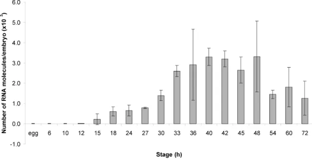
Spgatae is the sea urchin ortholog of the vertebrate gata4/5/6 genes, as confirmed by phylogenetic analysis. The accumulation of Spgatae transcripts during embryonic development and the spatial pattern of expression are reported here. Expression was first detected in the 15 h blastula. The number of Spgatae RNA molecules increases steadily during blastula stages, with expression peaking during gastrulation. After gastrulation is complete, the level of expression decreases until the end of embryogenesis. Whole mount in situ hybridization showed that Spgatae transcripts were first detected in a ring of prospective mesoderm cells in the vegetal plate. Spgatae expression then expands to include the entire vegetal plate at the mesenchyme blastula stage. During gastrulation Spgatae is expressed at the blastopore, and at prism stage strongly in the hindgut and midgut but not foregut, and also in mesoderm cells at the tip of the archenteron. Toward
the end of embryogenesis, expression in the hindgut decreases. The terminal pattern of expression is in midgut plus coelomic pouches.
Keywords: Sea urchin, Strongylocentrotus purpuratus, Gata factors, Endomesoderm specification, Endomesoderm gene network, Spgatae
RESULTS AND DISCUSSION
GATA factors are a class of DNA binding zinc finger transcriptional regulators which are named for the WGATAR sequence its members recognize. The GATA factors contain class IV zinc fingers which are characterized by the sequence CX2CX17-18CX2C (Lowry and Atchley, 2000). There are two vertebrate families of GATA factors.
Members of the gata1/2/3 family play important roles in hematopoiesis (Bockamp et al., 1994), while members of the gata4/5/6 family are indispensable for endoderm and mesoderm development. In the zebrafish, gata5 is required for heart and endoderm development (Reiter et al., 1999). In Caenorhabditis elegans, pairs of GATA transcription factors, encoded by the med, end, and elt genes, operate sequentially in the specification of the EMS and then the endoderm lineages (reviewed by Maduro and Rothman, 2002). In Drosophila the gata gene serpent is involved in gut specification (Reuter, 1994). Genes of the gata family and their orthologs thus operate in the process of endoderm specification and development across the Bilateria.
Two GATA factors were identified in the sea urchin (Pancer et al., 1999).
Spgatac is expressed in coelomycytes (Pancer et al., 1999), and this gene is an ortholog of the vertebrate gata1/2/3 genes, while Spgatae is the ortholog of the vertebrate gata4/5/6 genes. Functional analysis of Spgatae had shown that it plays an important role in endomesoderm specification during sea urchin embryogenesis. Perturbation of Spgatae expression using a morpholino antisense oligonucleotide revealed many genes which are positioned downstream of Spgatae in the endomesoderm gene regulatory network. The SpGatae transcription factor proves to be an important regulator of the
expression of many other endoderm and mesoderm regulatory genes; prominent examples include the Spbrachyury and Spfoxa genes. Most importantly, gene network analysis has shown that Spgatae participates in a cross regulatory loop that also involves Spkrox and SpOtx. The function of this gene regulatory loop is to lock down the expression of these three genes and drive the process of development forward (Davidson et al., 2002a; Davidson et al., 2002b).
Spgatae was initially isolated from a 48 h S. purpuratus cDNA library (Pancer et al., 1999). Identification and sequencing of a full length cDNA clone (Genbank accession No. AY623814) and mRNA blot hybridization showed that the length of the transcript is 4.3 kb (data not shown). Both the appearance of a single band on the mRNA blot (data not shown) and the search of an S. purpuratus EST catalog (Poustka et al., 2003) suggest that there are no alternative splice variants of Spgatae. A search of the traces from the S. purpuratus genome project did not identify any other paralogs. This, combined with the information that screens of S. purpuratus BAC and cDNA libraries identified no gata4/5/6 sequences other than Spgatae clones, suggest that it is the only sea urchin ortholog of the vertebrate gata4/5/6 genes.
A conceptual translation of the coding sequence revealed a protein that contains 567 amino acids (Fig. 1.1A) and two class IV zinc fingers. The zinc finger regions of Spgatae were aligned with those of various other GATA proteins, including Asterina miniata gatae, Mus musculus gata4/5/6, Drosophila melanogaster pannier and Spgatac, and a phylogenetic tree was constructed using neighbor joining analysis. The phylogenetic analysis confirmed that Spgatae is the S. purpuratus ortholog to vertebrate gata4/5/6 genes, Amgatae, Dmpannier and is paralogous to the vertebrate gata1/2/3