Introduction
Development of mouse model that replicates the metastasis process
Investigation of targeted, single-agent therapeutic nanoparticles in
Overall summary and conclusions
Recent developments in cancer treatment: opportunities and
- Brain metastases as emerging threats to long-term
- Human epidermal growth factor 2 (HER2)-positive
Incidence of brain metastases in patients with HER2-positive breast cancer as documented in retrospective studies. Since then, considerable clinical data have been collected on the incidence and outcomes of brain metastases in HER2-positive breast cancer patients.

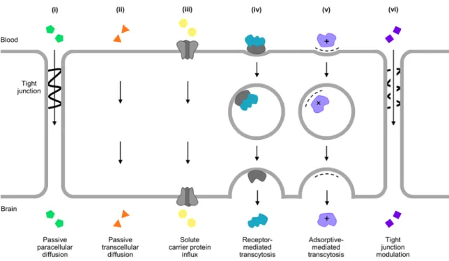
The blood-brain and blood tumor barriers (BBB and BTB)
- Structure and function of the BBB
- Solute transport at the BBB: regulation, not isolation
- Barrier integrity in HER2-positive breast cancer brain
However, the BBB endothelium contains a number of specific transport proteins to supply these substances to the brain (42). There is considerable debate in the field of brain metastasis research regarding the extent to which the BBB remains intact with brain metastases in the form of the blood tumor barrier (BTB).

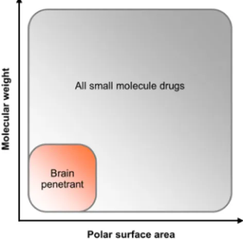
Current approaches for drug delivery to the brain
- Physically bypassing the BBB
- Transiently disrupting the BBB
- Exploiting endogenous solute transport systems at the
Another aspect that affects the delivery of small molecules to the brain - either systemically or by invasive bypassing or disruption of the BBB - is the presence of efflux pumps on the basal side of the endothelium. This strategy of “masking” the therapeutic agent has been dubbed the “Trojan horse” approach to smuggling drugs into the brain (40).
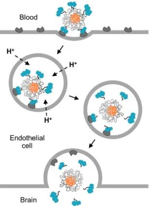
Transferrin receptor (TfR)-targeted drug delivery to the brain
- Receptor-mediated transcytosis (RMT) of transferrin
- Drug delivery across the BBB using anti-TfR
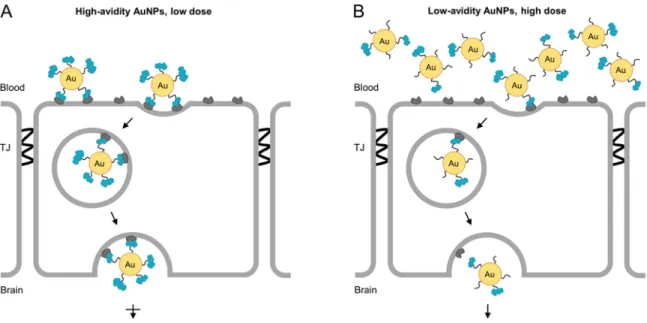
- Transport of TfR-targeted gold nanoparticles at the
The first of these studies showed that reducing the affinity of anti-TfR Abs to TfR maximizes their uptake into the brain parenchyma (94). High-affinity anti-TfR Abs are trafficked to the lysosome, while lower-affinity variants are more capable of transcytosis (95) (Fig. 1.9).

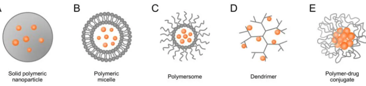
Nanoparticle drug delivery systems
- Polymeric nanoparticle formulations
- Passive and active targeting of nanoparticles
- Stimuli-responsive systems
Two approaches are primarily used to facilitate nanoparticle homing to the desired site: passive and active targeting. A number of types of affinity ligands have been investigated for active targeting of polymeric nanoparticles, including peptides and antibodies (112,113).
Thesis objectives and organization
Then, the newly developed model and two commonly used models from the literature were used to evaluate the efficacy and brain uptake of TfR-targeted single-agent. TfR-targeted therapeutic nanoparticles showed significant accumulation in brain metastases and led to enhanced antitumor activity compared to free drug and non-targeted nanoparticles in all models examined.
Yonemori K, Tsuta K, Ono M, Shimizu C, Hirakawa A, et al. 2010) Disruption of the blood-brain barrier by brain metastases from triple-negative and basal breast cancer, but not from HER2/neu-positive breast cancer. Kreuter J, Shamenkov D, Petrov V, Ramge P, Cychutek K, et al. 2002) Apolipoprotein-mediated transport of nanoparticle-bound drugs across the blood-brain barrier. Lajoie JM, Shusta EV (2015) Targeting receptor-mediated transport for delivery of biologics across the blood-brain barrier.
Limited transport of anti-transferrin receptor antibody (OX26) across the blood-brain barrier in the rat.
Introduction
- HER2-positive breast cancer brain metastasis
- The BBB/BTB debate
- Animal models of breast cancer brain metastasis
Extracts from this chapter are reprinted from Wyatt EA, Davis ME (2018) A method to identify breast cancer brain metastases affects brain uptake of targeted therapeutic nanoparticles. Furthermore, an investigation of breast cancer subtypes showed that brain metastases removed from patients with HER2-positive breast cancer do not disrupt the important barrier (8). One of the most commonly used methods for studying brain metastases of breast cancer involves the direct injection of human breast cancer cells into the parenchyma of the mouse brain (Figure 2.1A).
Here, we present a novel murine model of HER2-positive breast cancer brain metastasis involving intravenous (IV) injection of breast cancer cells into Rag2-/-;Il2rg-/- mice (Fig. 2.1C).

Results and discussion
- Rag2 -/- ; Il2rg -/- mice are permissive to brain metastasis
- IV model of HER2-positive breast cancer brain
We compared BT474-Gluc metastatic brain tumors diagnosed with the IV method versus the standard IC method in Rag2-/-;Il2rg-/- mice. Metastatic Brain Tumor Growth of BT474-Gluc When Established by IC and IV Injection of Breast Cancer Cells as Monitored by MRI. Identification of metastasis in Rag2-/-;Il2rg-/- mice after IV injection of BT474-Gluc breast cancer cells.
Metastatic ability of human BT474-Gluc breast cancer cells in Rag2-/-; Il2rg-/- mice after IV injection.

Conclusions
Materials and methods
The tissues were then incubated in melted paraffin (3 x 1 hour) at 60 °C, then placed in a paraffin mold and stored at 4 °C until sectioned. Tissues were mounted using Permount (Fisher) and images were acquired on an Olympus IX50 microscope using a 10x CPlan objective and QCapture Pro 6 imaging software (QImaging). For HER2 identification, tissues were incubated with a 1:100 dilution of an anti-human HER2 rabbit primary Ab (Dako A0485) in PBST for 1 h at room temperature, washed with PBST (2 x 5 min), followed by incubation with A 1:100 of an HRP-conjugated anti-rabbit goat secondary Ab (Abcam ab97051) in PBST for 1 h at room temperature, and finally washed with PBST (2 x 5 min).
Tissues were incubated in A4P0 hydrogel monomer solution (4% acrylamide in PBS, pH 7.4) overnight with shaking (acrylamide solution, BioRad; thermal initiator, Wako).
Lacroix M, Leclercq G (2004) The importance of breast cancer cell lines as models for breast tumors: an update. Bos PD, Nguyen DX, Massagué J (2010) Modeling metastasis in mice. 2009) Genes mediating breast cancer brain metastasis. Kim LS, Huang S, Lu W, Lev DC, Price JE (2004) Expression of vascular endothelial growth factor promotes the growth of breast cancer brain metastases in nude mice.
Multiorgan metastasis of human HER-2+ breast cancer in Rag2−/−;Il2rg−/− mice and treatment with PI3K inhibitor.
Introduction
- Intracellular trafficking at the BBB
- Investigation of TfR-targeted, therapeutic nanoparticles
However, despite a more detailed understanding of the properties that promote transcytosis, several challenges exist in translating anti-TfR Abs to the clinic, including the need to: (i) dose very high amounts (5), (ii ) reduce effector function driven safety concerns (10) and (iii) develop species-specific Abs (11). After endocytosis, rapid acidification of the endosome causes the separation of Tf ligands from the nanoparticle core, allowing free diffusion of the nanoparticle into the brain parenchyma after transcytosis. We focused on HER2-positive brain metastases in breast cancer due to the inadequate drug concentrations achieved in these tumors in the clinical setting.
We found that this targeted nanoparticle delivery system could be used to deliver CPT to HER2-positive breast cancer brain metastases.
Results
- Synthesis and characterization of TfR-targeted and non-
- Specific binding of TfR allows targeted nanoparticles to
- Brain tumors show significant delay in growth with
- Brain uptake of therapeutics differs in tumor, but not
To prepare the TfR-targeted and non-targeted MAP-CPT nanoparticles, either Tf-PEG-nitroPBA or OMe-PEG-nitroPBA was added to the nanoparticles at 20 molar excess (Fig. 3.2). Apical to basal transport of non-targeted and TfR-targeted MAP-CPT nanoparticles in model BBB. Interestingly, we observed a modest response with CPT treatment but not with non-targeted MAP-CPT nanoparticles (although this difference was not significant).
Importantly, TfR-targeted MAP-CPT nanoparticles showed the highest accumulation in brain tumor tissue identified by IC-, ICD-, and IV.
Discussion
Furthermore, we show that TfR-targeted nanoparticles are capable of delivering a small chemotherapeutic molecule, CPT, to HER2-positive breast cancer brain metastases. We observed that TfR-targeted MAP-CPT nanoparticles significantly slowed tumor growth in the brain and showed increased accumulation of brain metastases compared to free drug and non-targeted nanoparticles. Thus, it is encouraging to observe tumor growth delay upon delivering CPT via targeted nanoparticles to the BT474-Gluc brain metastases.
Furthermore, it is important to note that TfR-targeted nanoparticles accumulated in significant amounts in healthy brain tissue compared to free drug and non-targeted nanoparticles in all three models.
Conclusions
This observed whole-brain penetration has implications for the selection of therapies to include in this delivery system and for target diseases. In the case of brain cancer, the ability to penetrate not only tumor tissue but also healthy tissue could be advantageous in gaining access to micrometastases or fingers of glioma tumors that are often the reason for treatment failure. For other brain diseases where therapeutic exposure of the whole brain is highly desirable, such as neurodegenerative diseases, this targeted nanoparticle system may provide a compelling approach to delivering therapies across an intact BBB.
Furthermore, TfR-targeted nanoparticles showed an enhanced ability to cross an intact BBB, resulting in whole-brain therapeutic accumulation.
Materials and methods
To this was added anhydrous N,N-diisopropylethylamine (205 µL, 4 equiv) dried over molecular sieves and the solution was stirred under argon at room temperature for 42 h. 3-Carboxy-5-nitrophenyl boronic acid (nitroPBA, 100 mg, 1 equiv, Alfa Aesar) was added to an oven-dried 10 ml round-bottom flask. Boronic acid 3-acyl chloride-5-nitrophenyl (46 mg, 2 equiv) was added to an oven-dried 25 mL round-bottom flask.
Acetic acid-PEG-amine (5 kDa, 500 mg, 1 equiv, JenKem) was added to a separate 10 ml oven-dried round-bottom flask.
Initially focusing on drug delivery to HER2-positive breast cancer brain metastases, we addressed two important aspects of the development and translation of new therapies. We have developed a novel mouse model of HER2-positive breast cancer brain metastasis involving IV injection of human breast cancer cells in an attempt to create a clinically representative, impermeable BBB/BTB to standard therapeutics. Second, we demonstrated a methodology for delivering a small molecule drug across the BBB/BTB to breast cancer brain metastases in mice.
In summary, this work describes the development of a new mouse model of HER2-positive breast cancer brain metastases with high clinical relevance, enabling more meaningful translational studies of therapeutic brain penetration.
-Ly N, Yu YJ, Bumbaca D, Elstrott J, Boswell CA, et al. 2014) Transferrin receptor (TfR) trafficking determines brain uptake of TfR antibody affinity variants. Wiley DT, Webster P, Gale A, Davis ME (2013) Transcytosis and brain uptake of transferrin-containing nanoparticles by tuning transferrin receptor avidity. Clark AJ, Davis ME (2015) Enhanced brain uptake of targeted nanoparticles by adding an acid-cleavable linker between transferrin and the nanoparticle core.
Wyatt EA, Davis ME (2018) Method of detecting breast cancer brain metastases influences brain uptake and efficacy of targeted, therapeutic nanoparticles.
Preamble
Introduction
Furthermore, brain metastases in this PET study showed modest but highly variable uptake of [11C]lapatinib. Furthermore, in their earliest stages, where one would expect the greatest change in effective treatment, brain metastases are hidden behind an intact BBB. The aim of this work was to prepare TfR-targeted lapatinib-loaded nanoparticles for future use as a single-agent therapeutic or in combination with assembled trastuzumab.
Here, we synthesize two MAP-modified polymer scaffolds used to prepare urea- and carbamate-based lapatinib polymer drug conjugates that allow the assembly of TfR-targeted lapatinib-loaded nanoparticles.
Results and discussion
- Lapatinib displays increased in vitro cytotoxicity in
- Synthesis of MAP-amidoethanamine and MAP-
- Addition of lapatinib to MAP-amidoethanamine and
- Preparation and characterization of lapatinib-loaded,
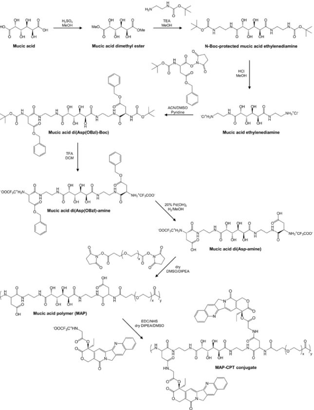
Lapatinib contains only secondary amine moieties (magenta) that can be used for conjugation to the polymer. The MAP, N-Boc-ethylenediamine, 2-(benzyloxy)ethan-1-amine and intermediate reaction products leading to the preparation of MAP-amidoethanamine and MAP-amidoethanol were fully characterized by NMR (Fig. A.7 to A.13) ). Reactivity of the lapatinib secondary amines was first investigated before adding the molecule to the MAP derivatives.
Magnified regions of 1H NMR of lapatinib (grey) and crude product (black) for peaks s and e are provided (inset).

Conclusions
Materials and methods
Hydrogen gas was added through a double-layer balloon and the reaction was stirred at room temperature for 24 hours. Deuterated dimethyl sulfoxide (0.5 ml) was added under argon to dissolve lapatinib (5 mg, 1 equiv, Sigma) in a 4 ml glass vial closed with a septum. Anhydrous dimethyl sulfoxide (1 mL) was added under argon to dissolve lapatinib (7.6 mg, 1.2 equiv, Sigma) in a 10 mL round-bottom flask.
To this was added anhydrous N,N-diisopropylethylamine (35 μL, 2 equiv) dried over molecular sieves and anhydrous DCM (5 mL) to dissolve the PEG.
Konecny GE, Pegram MD, Venkatesan N, Finn R, Yang G, et al. 2006) Activity of the dual kinase inhibitor lapatinib (GW572016) against HER-2-overexpressing and trastuzumab-treated breast cancer cells. Saleem A, Searle GE, Kenny LM, Huiban M, Kozlowski K, et al. 2015) Lapatinib access to normal brain and brain metastases in patients with Her-2-overexpressing breast cancer. Morikawa A, Peereboom DM, Thorsheim HR, Samala R, Balyan R, et al. 2015) Uptake of capecitabine and lapatinib in surgically resected brain metastases from patients with metastatic breast cancer: a prospective study.
తస్కర్ KS, రుదరాజు V, మిట్టపల్లి RK, సామల R, Thorsheim HR, మరియు ఇతరులు. 2012) HER2 వాట్ ఎక్స్పెరిమెంటల్ బ్రీన్మెటాస్టేసెస్ వాన్ బోర్స్కంకర్ ఊరుయిట్డ్రుక్లో లాపటినిబ్-వర్సెస్ప్రెడింగ్.
Preamble
Image selected for 2017 NCI Cancer Close Up
Image selected for NCI Visuals Online
Materials and methods
For vasculature identification, brain samples were incubated with a 1:200 dilution of an anti-CD31 rabbit primary Ab (Abcam ab28364) and a 1:200 dilution of an AlexaFlor 594-conjugated anti-rabbit donkey secondary Ab (Jackson ImmunoResearch with 0 .02% NaN3 in PBST for 7 days, each with shaking to visualize the vasculature. Immunostains were replaced with a fresh cocktail every two days, and tissues were washed for two days with a minimum of four exchanges in PBST with 0.02% NaN3 Samples were incubated in RIMS (prepared with Histodenz, Sigma-Aldrich, RI = 1.46) with gentle shaking for one day.
Samples were placed inside the spacer, followed by light overflow of fresh RIMS and a coverslip.