Targeting DNA repeat sequences with Py-Im polyamides
Thesis by JeenJoo Sophia Kang
In Partial Fulfillment of the Requirements for the degree of
Doctor of Philosophy
California Institute of Technology Pasadena, California
2015
(Defended March 27, 2015)
© 2015 JeenJoo Sophia Kang
All Rights Reserved
Dedicated to my family
Acknowledgements
I learned a great deal in graduate school, and a good measure of it is from the people I have interacted with over the years. Many people on- and off- campus that have positively influenced my years at Caltech. I would like to acknowledge at least some of them here.
I would like to thank my advisor Prof. Peter Dervan for his scientific guidance during my graduate studies. The intellectual freedom that he promotes in the lab, balanced by his advice on risk, has taught me to explore different areas of research in a considered manner. He has been supportive of, and appreciated the joy of, pursuing self- generated ideas even as I kept going “off the ranch”. I expect the scientific judgment and understanding of new technologies that I have gained during my graduate education will prove essential to my future career. I would also like to thank my graduating committee chaired by Prof. Bob Grubbs and joined by Prof. Jim Heath and Prof. Linda Hsieh- Wilson. Their advice and support has been indispensible to my development in graduate school. Beyond science, the genuine interest of professors in the welfare of students makes Caltech an exceptional place.
My education was made much richer by my interactions with my labmates in the Dervan lab. In my earlier years, I learned a great deal from Katy Muzikar, Jim Puckett, and Jordan Meier who took the time to transfer their knowledge of some very time- intensive techniques. With all my colleagues over the years, I have appreciated the discussions of science, academia, culture, and our futures held over drinks, meals, and
late nights in the lab. My fellow graduate students: Alexis Kurmis, Ben Li, Thomas Martinez, Dave Montgomery, Jerzy Szabowski, Fei Yang; and post-doctoral colleagues:
Alissa Hare, Amanda Hargrove, Jevgenij Raskatov, Dan Gubler, Amanda Silberstein, Jamie Wang, Sam Weisbrod, and Tim Welch were wonderful labmates. We are all so different, yet the work of this lab brought us all together for a short while— there is something special in that, and I look forward to when we might cross paths again.
My work would not have progressed without the help of Igor Antoshechkin in the Sequencing facility; and Shelley Diamond and Diana Perez in the Flow cytometry facility. They provided not only scientific help, but also moral support in the face of difficulties. Similarly, Vicky Brennan, Joe Drew, and Agnes Tong fix everything.
Thank you to all my friends, but in particular those in chemistry and the Korean (sometimes-) Tennis club. You have injected fun and relief into these years; my time spent with you defines a great deal of my Caltech experience. Some of our memories—
grillin’n’chillin, “stop it, Myles”, “Leonard”, poker nights, “yeah, we’re three people”, nyan cat, tennis, “definitely a halibut”— I hope to build on over a lifetime of friendship.
Lastly, my family—Mom, Dad, and Alex, and my extended family… without you guys, I probably never would have started and I certainly couldn’t have finished. It is with your love and support that I have the confidence to try more things than I fail at.
There is nothing I can say to fully express my gratitude, so, I’ll just say I love you.
Abstract
Hairpin pyrrole-imdazole polyamides are cell-permeable, sequence-programmable oligomers that bind in the minor groove of DNA. This thesis describes studies of Py-Im polyamides targeted to biologically important DNA repeat sequences for the purpose of modulating disease states. Design of a hairpin polyamide that binds the CG dyad, a site of DNA methylation that can become dysregulated in cancer, is described. We report the synthesis of a DNA methylation antagonist, its sequence specificity and affinity informed by Bind-n-Seq and iteratively designed, which improves inhibitory activity in a cell-free assay by 1000-fold to low nanomolar IC50. Additionally, a hairpin polyamide targeted to the telomeric sequence is found to trigger a slow necrotic-type cell death with the release of inflammatory molecules in a model of B cell lymphoma. The effects of the polyamide are unique in this class of oligomers; its effects are characterized and a functional assay of phagocytosis by macrophages is described. Additionally, hairpin polyamides targeted to pathologically expanded CTG•CAG triplet repeat DNA sequences, the molecular cause of myotonic dystrophy type 1, are synthesized and assessed for toxicity. Lastly, ChIP-seq of Hypoxia-Inducible Factor is performed under hypoxia-induced conditions.
The study results show that ChIP-seq can be employed to study the genome-wide perturbation of Hypoxia-Inducible Factor occupancy by a Py-Im polyamide.
Table of Contents
Acknowledgements ... iv
Abstract ... vi
Table of Contents ... vii
List of Figures and Tables ... ix
Chapter 1 ... 1
1.1 Complexity coded in DNA ... 2
1.2 Epigenetic Organization of DNA ... 2
1.3 Transcriptional Regulation ... 5
1.4 Molecular recognition of DNA ... 7
1.5 Biological modulation with Py-Im polyamides ... 12
1.6 Scope of Work ... 15
1.7 References ... 17
Chapter 2 ... 21
Abstract ... 22
2.1 Introduction ... 23
2.2 Results ... 26
2.3 Discussion ... 39
2.4 Materials and Methods ... 41
2.5 Acknowledgment ... 45
2.6 References ... 47
Chapter 3 ... 50
Abstract ... 51
3.1 Introduction ... 52
3.2 Results ... 54
3.3 Discussion ... 71
3.4 Materials and Methods ... 73
3.5 Acknowledgments ... 82
3.6 References ... 94
Chapter 4 ... 98
Abstract ... 99
4.1 Introduction ... 100
4.2 Results ... 103
4.3 Discussion ... 107
4.4 Materials and Methods ... 109
4.5 References ... 112
Chapter 5 ... 114
Abstract ... 115
5.1 Introduction ... 116
5.2 Results ... 121
5.3 Discussion ... 127
5.4 Materials and Methods ... 127
5.5 References ... 130
List of Figures and Tables
Chapter 1 ... 1
Figure 1.1 ... 3
Figure 1.2 ... 6
Figure 1.3 ... 7
Figure 1.4 ... 8
Figure 1.5 ... 9
Figure 1.6 ... 10
Figure 1.7 ... 11
Figure 1.8 ... 12
Figure 1.9 ... 13
Figure 1.10 ... 14
Chapter 2 ... 21
Figure 2.1 ... 25
Figure 2.2 ... 27
Figure 2.3 ... 28
Table 2.1 ... 29
Figure 2.4 ... 30
Figure 2.5 ... 32
Figure 2.6 ... 33
Table 2.2 ... 34
Figure 2.7 ... 35
Figure 2.8 ... 36
Figure 2.9 ... 38
Table 2.3 ... 42
Chapter 3 ... 50
Figure 3.1 ... 53
Figure 3.2 ... 55
Figure 3.3 ... 56
Figure 3.4 ... 58
Figure 3.5 ... 59
Figure 3.6 ... 61
Figure 3.7 ... 63
Figure 3.8 ... 65
Table 3.1 ... 68
Figure 3.9 ... 69
Figure 3.10 ... 70
Table 3.6 ... 74
Figure 3.11 ... 75
Table 3.7 ... 76
Table 3.8 ... 76
Table 3.9 ... 77
Table 3.2 ... 83
Table 3.3 ... 89
Table 3.4 ... 91
Table 3.5 ... 92
Chapter 4 ... 98
Figure 4.1 ... 100
Figure 4.2 ... 102
Table 4.1 ... 103
Figure 4.3 ... 104
Figure 4.4 ... 106
Table 4.2 ... 107
Figure 4.5 ... 108
Table 4.3 ... 110
Chapter 5 ... 114
Figure 4.1 ... 117
Figure 4.2 ... 119
Figure 4.3 ... 121
Table 4.1 ... 122
Figure 4.4 ... 123
Figure 4.5 ... 125
Figure 4.6 ... 126
Chapter 1
Introduction
1.1 Complexity coded in DNA
The blueprint for living organisms encoded in deoxyribose nucleic acid (DNA) determines every possible transcriptomic and proteomic state for all the cells in an organism. According to the central dogma of molecular biology put forth in 1958, DNA is the foundational molecule from which ribonucleic acid (RNA) is transcribed, and protein is translated from the RNA transcript (Figure 1.1A).1 The coordination and control of these molecules are critical to the spatiotemporal organization of cell activity and tissue maintenance in organismal health. In humans, the 3 billion letter genome is organized in 23 chromosomes and encodes over 20,000 protein-coding genes.2 In addition, over 15,000 long non-coding RNA transcripts have been identified and are thought to play a regulatory role.3 The structural organization of DNA and its gene expression profile is specific to each cell-type and is accomplished by layers of exquisitely controlled processes. The etiology of many diseases can be found in misregulated DNA. Modulation by chemical means of DNA regulation may yield strategies for therapeutic benefit.
1.2 Epigenetic Organization of DNA
Eukaryotic DNA is condensed and regulated as chromatin to expose appropriate genes for transcription and maintain genome integrity in the face of a genotoxic environment (Figure 1.1A). The fundamental unit of organization is the nucleosome core particle, a histone octamer protein assembly wrapped by 147 base pairs of DNA (Figure 1.1B).4 The wrapped DNA can be compacted densely into heterochromatin, which is less
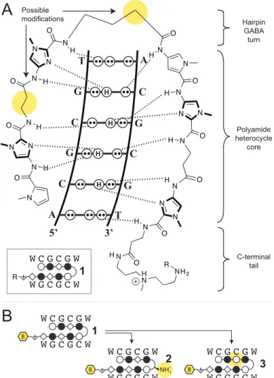
Figure 1.1 Structural organization of DNA in the cell. A) DNA (in black) is wrapped around the nucleosome core particle and organized as compact heterochromatin or transcriptionally active euchromatin. Transcription factors (TF) bind DNA and recruit cofactors (CF) and RNA polymerase II (RNAP) to transcribe mRNA from DNA. mRNA is translated into protein by ribosomes. B) The nucleosome core particle is comprised 147 base pairs of DNA wrapped around a histone octamer. Epigenetic modifications, including methylation (Me), acetylation (Ac), ubiquitiniylation, and SUMO-lation on the tails of histones govern chromatin structure.
Methylated DNA is recognized by methyl-binding proteins (MBP) which recruit histone deacetylases (HDAC) to modify chromatin into a transcriptionally inactive state. C) Chromosomes end in the telomeric 5’-TTAGGG-3’ hexamer repeat sequence, which can trigger a DNA damage response due to its similarity to a DNA double-stranded break. Telomeres form a
RNAP
RNA
Protein
Translation
Nucleus
DNA Euchromatin
Heterochromatin Cytoplasm
A
B C
Transcription TF RNAP
TF TF
TFCF
TTAGGG-repeat
DNA break signal from exposed end 3’5’
3’5’
TRF1 TRF2 POT1
TRF1TRF2
TRF1 TRF2 TRF1 TRF2
TRF1TRF2
MBP
Me Me
Me
Me HDAC Me
Ac Ac
loop structure with invasion by the 5’ strand to conceal the DNA end. TRF1 and TRF2 bind the telomere sequence and POT1 stabilizes the single-stranded DNA.
accessible for transcription and the requisite transcription machinery. Alternatively it can be packed more sparsely into euchromatin, which is a more transcriptionally active structure. The regulation of chromatin structure occurs by chemical annotation of the histones and DNA at the associated loci.5 Histones can be methylated, acetylated, ubiquitinylated, phosphorylated, and SUMO-lated to modulate transcription or to respond to genotoxic stress. Proteins bound to enhancers modify, or recruit other enzymes that modify, the proximal histones that determine the transcriptional landscape of the cells.
These epigenetic modifications are heritable and strongly correlate with the transcriptional programs that determine cell fate.6 The key element of the epigenetic code on mammalian DNA is the methylation of the 5’ carbon of cytosines in the palindromic CG dyad. This two base pair motif is concentrated in sequences called CpG islands, often associated with promoter sites.7 The methylation of these CpG islands is transcriptionally repressive; methylated cytosines are bound by methyl binding proteins which recruit histone modifying enzymes to annotate the proximal histones into transcriptionally repressive heterochromatin (Figure 1.1B).8 In this manner, DNA is purposefully structured and regulated for function.
In this organization of DNA, chromosome ends present a danger of being detected as DNA double-stranded breaks. Histones are sensitive to DNA damage and become phosphorylated upon detection of double stranded breaks, signaling towards repair or cell death.9 To resolve the “end-protection problem”, nature provides a structural solution.10 The telomere is comprised of 6- base pair repeats of 5’-TTAGGG-3’ spanning 5-30
kilobases with a 5’ single strand overhang.10 The repeating overhang is looped back to invade the double-stranded repeat region (Figure 1.1C).11 The sequence repetition ensures complementarity and the end is hidden in the loop. Proteins including TRF1 and TRF2 of the shelterin complex, as well as POT1, which binds the single stranded portion, clamp this structure together.11 The maintenance of this structure is important to maintain genome integrity, as its disruption is known to cause genotoxic stress signals and deleterious recombination.12
1.3 Transcriptional Regulation
In the context of this regulated DNA structure, transcription is enabled when transcription factor proteins bind enhancer regions coded in the DNA sequence.
Transcription factors are proteins that bind DNA in a sequence-specific manner and function to either activate or repress transcription.13 The enhancers of genes under a transcription factor’s control contain the conserved cognate binding sequence for the protein. Transcription factors achieve specificity through their DNA binding domains (DBD). There are several conserved structural motifs for DNA binding and they include basic-helix-loop-helix, zinc finger, and high mobility group box DBDs (Figure 1.2).14-16 These DBDs are linked to protein domains that transactivate transcription.
In mammals, these transcription factors do not bind in isolation but bind cooperatively to recruit cofactors and the RNA Polymerase II machinery to the transcription start site. The assemblage of multiple factors integrates various signaling
Figure 1.2 Crystal structures of transcription factors bound to DNA. DNA binding domains of transcription factors fall into several families, including basic-helix-loop-helix (Myc/Max, PDB 1NKP), zinc-finger (glucocorticoid receptor, PDB 1R40), and high mobility group (Lef-1, PDB 2LEF).55
cascades for precise detection of environmental cues.13 Further, the expanded footprint of the bound proteins provides gene specificity. The interferon β (IFNB) gene enhancer is an example of the coordinate binding of multiple transcription factors for transcriptional activation. The DNA serves as a sequence-encoded scaffold for the binding of ATF-2, c- Jun, IRF-3A, IRF-7B, IRF-3C, IRF-7D, p50, and RelA at the enhancer of IFNB (Figure 1.3).13 These factors in turn recruit the coactivators CBP/p300 in a multivalent fashion.
The composite enhanceosome modifies chromatin and recruits RNA polymerase II to transcribe the IFNB gene into messenger RNA. Combining all these elements encoded in both the sequence and structure of DNA achieves exquisite control of the transcriptomic program.
Myc/Max Glucocorticoid
Receptor Lef-1
Figure 1.3 Model of the interferon β enhanceosome. 17
1.4 Molecular recognition of DNA
The sequence-specific molecular recognition of DNA is key to this regulatory system. DNA is comprised of four nucleotides linked by anti-parallel phosphodiester backbones in a double helix (Figure 1.4). In the B-form DNA typically found in nature, the four nucleotides hydrogen-bond by the Watson-Crick pairings: adenosine (A) pairs with thymine (T) through two hydrogen bond interactions; cytosine (C) pairs with
5 ’ - T A A A T G A C A T A G G A A A A C T G A A A G G G A G A A G T G A A A G T G G G A A A T T C C T C T G - 3 ’ 3 ’ - T T T A C T G T A T C C T T T T G A C T T T C C C T C T T C A C T T T C A C C C T T T A A G G A G A C A - 5 ’
ATF-2 IRF-3A IRF-3C
IRF-7D
p50
RelA IRF-7B
c-Jun
-102 -70 -51
Figure 1.4 Structure of deoxyribose nucleic acid. DNA comprised of four nucleic acid bases linked by a phophodiester backbone in a double helix (PDB 1BNA) is shown.59
guanosine (G) through 3 hydrogen bonding interactions.18 This specific pairing pattern provides for heritability of the genetic code as identical daughter genomes are synthesized during replication following the base complementarity. The exposed edges of the base pair form a wide major groove and a narrow minor groove on opposite sides of the double helix (Figure 1.4). Each of the four base pairs exposes a unique stereo- electronic edge in the grooves (Figure 1.5A). These sequence-specific stereo-electronics are recognized by DNA binding proteins. In addition, they are recognized by small molecule ligands which may be used to modulate the binding activity of the proteins.
minor groovemajor groove
N N N N
H NH
N N N
O H H
N N O
O
N N N N O
HN H H H
A
T G C
Figure 1.5 Each base pair has a unique stereo-electronic edge in both the major and minor groove.55 Hydrogen- bonding patterns are shown for both the major and minor groove for each of the base pairs. Circles with two dots represent lone pairs and circles with an H represent a hydrogen on the exocyclic amine of guanine.
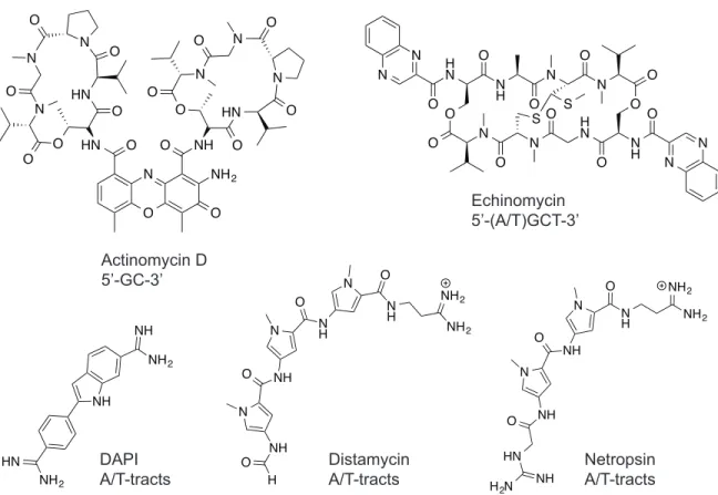
There are DNA-binding natural products with known sequence specificity that have been successfully employed as therapeutic drugs and scientific tools. Actinomycin D is a DNA-intercalator with a sequence preference for 5’-GC-3’ that was one of the earliest chemotherapeutics for cancer (Figure 1.6).19 Echinomycin is a bis-intercalating minor groove binding ligand that preferentially binds 5’-(A/T)CGT-3’ and was found to downregulate hypoxia inducible factor transcriptional activity (Figure 1.6).20-22 Fluorescent DNA-binding ligands such as DAPI have seen wide use as nuclear stains in microscopy (Figure 1.6).23,24 Among DNA minor groove-binding ligands, distamycin and netropsin lend themselves to rational design and chemical modification due to their modular structure (Figure 1.6).25
major groove
minor groove
G G C
N NN
O
N N
N
O N
N H
H H
H H
major groove
minor groove
A
NT
N N
N N
N N O
O H H
H
H
H major groove
minor groove
G
N NC
N
O
N N N
O N
N
H H H
H H
H H H
major groove
minor groove
A
T
N NO NN NNNO H H
H
H
Figure 1.6 Structures of DNA binding small molecule ligands and their preferred binding sequences.
The minor groove surface of DNA presents a binding target for effecting modulation of dysfunctional biology in disease states. Distamycin preferentially binds AT-tracts, as there is steric hindrance from the exocyclic amine of guanosines in C•G/G•C on the minor groove floor (Figure 1.7A).26 The introduction of a N-methyl imidazole ring (Im) to replace a N-methyl pyrrole ring (Py) provides a “hole” for the
“bump” of the guanine residue.27,28 In particular, the 2:1 binding of distamycin in the minor groove of DNA29 (Figure 1.7B) suggested that linked Py-Im polyamides can target sequences through cofacial arrangement of the aromatic ring pairs to distinguish the edges of the four Watson-Crick base pairs.30 Pairing rules for programmable specificity have been determined: Im/Py specifies a G•C base pair, Hp/Py codes for T•A base pairs
O N O
HN O NH
O O
O N
N HN
O O
O N O
O
N N
O O
HN O NH2
N O O
N N H N
O N
H O
N
O N
O
O O O
O N
O N O H
N O N
H O
N N S S
Actinomycin D 5’-GC-3’
DAPIA/T-tracts Distamycin
A/T-tracts Netropsin
A/T-tracts Echinomycin
5’-(A/T)GCT-3’
NH2
N NH2
O NH
O NH
O
N
NH
HN H2N NH NH2
N
NH N
NH2
NH O
O H O NH
O N
NH
NH
NH2
NH
NH2 HN
Figure 1.7 Crystal structures of distamycin bound in the minor groove of DNA. A) Distamycin bound 1:1 (PDB 2DND) and B) 2:1 in an antiparallel orientation (PDB 378D).59
and Py/Py binds both T•A/A•T (Figure 1.8).27,28,31-34 This pairing register of the antiparallel polyamide strands can be covalently enforced with a γ-amino butyric acid linker to form a hairpin architecture.35 However, Py-Im polyamide strands, particularly those containing many imidazoles, were found to be over-curved compared to natural DNA.36 β-alanine residues were introduced to relieve the curvature in such cases, which allowed targeting longer sequences and sequences with more C•G/G•C residues.37-41 The development of this molecular recognition technology has enabled sequence-specific targeting of DNA with affinities similar to that of transcription factors.42
Distamycin 1:1
Distamycin 2:1
A
B
Figure 1.8 Pairing rules for Py-Im polyamide DNA sequence-specificity. A) Hydrogen- bonding pattern of the minor groove is depicted as described above. Model for the binding of a ImHpPyPy-γ-DABA-ImHpPyPy-tri-IPA polyamide bound to 5’-AGTACT-3’.
Hydrogen bonds shown with dashed lines. B) Ball and stick notation for polyamides, with legend in C).
1.5 Biological modulation with Py-Im polyamides
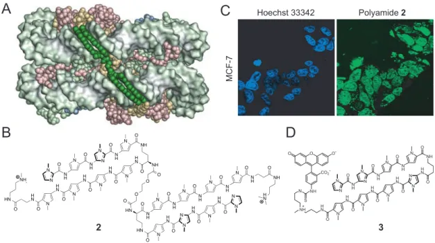
Studies support Py-Im polyamides effecting biological modulation through its DNA binding capacity. The accessibility of DNA in the context of chromatin for Py-Im polyamide binding was demonstrated in a crystal structure of a polyamide bound to the nucleosome core particle (Figure 1.9A). In live cell culture experiments, the cell- permeability and nuclear localization of dye-conjugated polyamides were observed
N
N
N O
N
N O H
H
N O H N
N N N
O N N
O H
H O
N H N H
N O
N
N N N
O H
O
H
OH
HO
A
. .
. .
.
3’
. .
. .
.
.
. .
.
5’
. .
G
. .
. . ..
Py/Im
..
targets C•G Py/Hp targets A•T
.
..
.
Im/Py targets
G•C
.
Hp/Py targets T•A
. ..
T C A T
H
H γ-turn
targets A•T/T•A
targetstail A•T/T•A
A
G T
C A
T
N
HN O O
OH
Hairpin γ-turn
NH3
C-Terminal Tail Polyamide Heterocyclic Core
NH N
H N β
H O
Im
+ Dp
NH N
H N
H tri
+ γ- DABA
NH
O NH3
Hp
N O
NH OH N Py
O
NH N
O
NH
H
+
+
+ IPA
H H
5 ' - A G T A C T - 3 ’
3 ' - T C A T G A - 5 ’ OH
O O
A
1
1
B
C
IPA
N
Figure 1.9 Py-Im polyamides access chromatin. A) Crystal structure of a nucleosome core particle with B) polyamide dimer 1 bound in the adjacent minor grooves.60 C) Confocal microscopy of MCF-7 breast cancer cell line treated with nuclear-stain Hoechst or D) dye- conjugate polyamide 2.61 Images show co-localization to the nucleus.
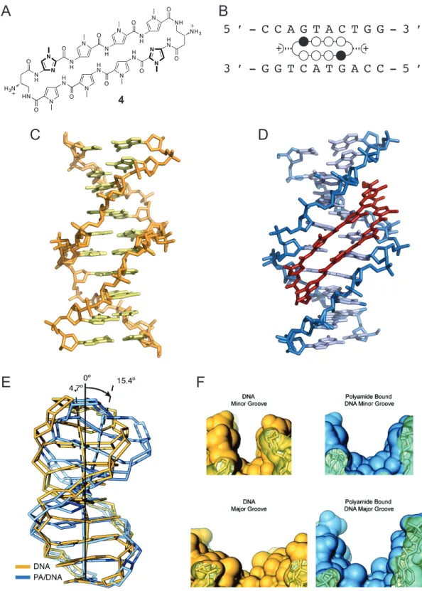
through confocal microscopy in a wide range of cell lines (Figure 1.9C).43-44 This demonstrated that access to chromatin with no external transfection was possible with these oligomers. Transcriptional modulation was demonstrated in cell culture by Py-Im polyamides targeted to the respective consensus binding sequences of the transcription factors hypoxia-inducible factor, androgen receptor, glucocorticoid receptor, and NF- κB.46-50 An X-ray crystal structure of a cyclic polyamide bound to DNA provided a rationale for the exclusion of major-groove binding proteins upon minor groove binding by the polyamide.51 The crystal structure revealed that polyamide binding caused an expansion of the minor groove and compression of the major groove (Figure 1.10). Py-Im polyamides were also found to disrupt processive DNA enzymes. For example, a high
Hoechst 33342
MCF-7
Polyamide 2
3 2
A C
D B
HN O
HN O NH O N
N NH
O N
N
NH O N
NH O N
N HN
O HN
O N
N HN
O
N
NH
O HN
NH O
NH O
HN O N
N
HN O N
N HN
O N HN
O N
N NH O
NH N O
N NH O N
HN
O O NH
O O
O NH
HN
Figure 1.10 Allosteric disruption of major groove by minor groove binding cyclic polyamide.51 A) Structure of cyclic polyamide 4. B) Ball-and-stick notation for
polyamide 4 shown with the sequence of the DNA oligonucleotide. C) Crystal structure of native B-form DNA (PDB 1D8G). D) Crystal structure of cyclic polyamide 4 bound in the minor groove of DNA (PDB 3OMJ). E) Binding of polyamide 4 causes a distortion to the native DNA structure. F) The binding of polyamide 4 causes a widening of the minor
N O
HN
O N HN
O N
N HN
O N NH
O
NH O N
NH O N
NH O
N N
HN
N O HN
NH3
NH O
H3N
+ +
5 ’ - C C A G T A C T G G - 3 ’
3 ’ - G G T C A T G A C C - 5 ’
N O
O N HN
N O HN
NH H3N
A
4
B
C D
E F
groove (top), and a compression of the major groove (bottom). Native structure shown in yellow, polyamide-bound structure shown in blue.
affinity hairpin targeted to the androgen response element also disrupted RNA polymerase II elongation, causing cytotoxicity and p53 activation without DNA damage.52 Another study showed that high doses of this polyamide can disrupt helicase activity and can cause replicative stress.53 These results indicate DNA minor groove binding Py-Im polyamides can cause a variety of alterations to biological activity.
Studies in mouse models have shown that many of these effects observed in cell culture translate to the mouse with systemic treatment of the polyamide.
Pharmacokinetics studies have shown that Py-Im polyamides are bioavailable and have reasonable exposure.54 Cyclic architecture and acetylation of the turn of the same core aromatic oligomer sequence have each been explored and shown to have different pharmacokinetic and toxicity profiles.55,56 Gene expression has been measured in the tumor of mouse xenograft models and shown to be modulated by polyamide treatment similarly to the experiments done in cell culture.57 In a xenograft model of prostate cancer, the high-affinity Py-Im polyamide targeted to the androgen receptor response element reduced tumor size.52 Indeed, a C-14 labeled analog of the molecule showed preferential localization to the tumor xenograft.58 In sum, Py-Im polyamides are a class of molecules well-suited for the study of biological perturbation through DNA binding.
1.6 Scope of Work
This thesis describes studies of Py-Im polyamides targeted to biologically important DNA repeat sequences for the purpose of modulating disease states. In chapter
2, a Py-Im polyamide is designed to bind a sequence that contains two CG dyads and is tested as a DNA methylation antagonist. Chapter 3 explores the immunogenic signaling triggered by a polyamide that is targeted to the telomere repeat sequence. Chapter 4 describes work in assessing the animal toxicity of hairpin polyamides targeted to the CAG/CTG triplet repeat expansion to disrupt the transcription of the associated RNA, which is the molecular cause of myotonic dystrophy type 1. Chapter 5 describes a ChIP- seq experiment that explores the genome-wide perturbation of hypoxia-inducible factor occupancy induced under hypoxia by a Py-Im polyamide targeted to the consensus sequence of the hypoxia response element.
1.7 References
(1) Crick, F. H. Symposia. of the. Society for Experimental Biology. 1958, 12, 138.
(2) Harrow, J.; Frankish, A.; Gonzalez, J. M.; Tapanari, E.; Diekhans, M.;
Kokocinski, F.; Aken, B. L.; Barrell, D.; Zadissa, A.; Searle, S.; Barnes, I.;
Bignell, A.; Boychenko, V.; Hunt, T.; Kay, M.; Mukherjee, G.; Rajan, J.;
Despacio-Reyes, G.; Saunders, G.; Steward, C.; Harte, R.; Lin, M.; Howald, C.;
Tanzer, A.; Derrien, T.; Chrast, J.; Walters, N.; Balasubramanian, S.; Pei, B.;
Tress, M.; Rodriguez, J. M.; Ezkurdia, I.; van Baren, J.; Brent, M.; Haussler, D.;
Kellis, M.; Valencia, A.; Reymond, A.; Gerstein, M.; Guigó, R.; Hubbard, T. J.
Genome Res. 2012, 22, 1760-1774.
(3) Derrien, T.; Johnson, R.; Bussotti, G.; Tanzer, A.; Djebali, S.; Tilgner, H.;
Guernec, G.; Martin, D.; Merkel, A.; Knowles, D. G.; Lagarde, J.; Veeravalli, L.;
Ruan, X.; Ruan, Y.; Lassmann, T.; Carninci, P.; Brown, J. B.; Lipovich, L.;
Gonzalez, J. M.; Thomas, M.; Davis, C. A.; Shiekhattar, R.; Gingeras, T. R.;
Hubbard, T. J.; Notredame, C.; Harrow, J.; Guigó, R. Genome Res. 2012, 22, 1775-1789.
(4) Kornberg, R. D. Science. 1974, 184, 868-871.
(5) Berger, S. L. Nature. 2007, 447, 407-412.
(6) Heintzman, N. D.; Hon, G. C.; Hawkins, R. D.; Kheradpour, P.; Stark, A.; Harp, L. F.; Ye, Z.; Lee, L. K.; Stuart, R. K.; Ching, C. W.; Ching, K. A.; Antosiewicz- Bourget, J. E.; Liu, H.; Zhang, X.; Green, R. D.; Lobanenkov, V. V.; Stewart, R.;
Thomson, J. A.; Crawford, G. E.; Kellis, M.; Ren, B. Nature. 2009, 459, 108-112.
(7) Bird, A. P. Nature. 1986, 321, 209-213.
(8) Nan, X.; Ng, H. H.; Johnson, C. A.; Laherty, C. D.; Turner, B. M.; Eisenman, R.
N.; Bird, A. Nature. 1998, 393, 386-389.
(9) Mah, L. J.; El-Osta, A.; Karagiannis, T. C. Leukemia. 2010, 24, 679-686.
(10) de Lange, T. Cold Sprin. Harb. Symp. Quant. Biol. 2010, 75, 167-177.
(11) de Lange, T. Science. 2009, 326, 948-952.
(12) Sfeir, A.; de Lange, T. Science. 2012, 336, 593-597.
(13) Tjian, R.; Maniatis, T. Cell. 1994, 77, 5-8.
(14) Williams, D. C.; Cai, M.; and Clore, G. M. J. Biol. Chem. 2004, 279, 1449.
(15) Juo, Z. S.; Chiu, T. K.; Leiberman, P. M.; Baikalov, I.; Berk, A. J.; Dickerson, R.
E. J. Mol. Biol. 1996, 261, 239.
(16) Love, J. J.; Li, X.; Case, D. A.; Giese, K.; Grosschedl, R.; Wright, P. E. Nature 1995, 376, 791.
(17) Panne, D. Curr. Opin. Struct. Biol. 2008, 18, 236-242.
(18) Watson, J. D.; Crick, F. H. Nature. 1953, 171, 737-738.
(19) Kamitori, S.; Takusagawa, F. J. Mol. Biol. 1992, 225, 445-456.
(20) Waring; M., J.; Wakelin; G., L. P.; Waring, M. J.; Wakelin, L. P. G. Nature.
1974, 252, 653-657.
(21) VanDyke, M. M.; Dervan, P. B. Science. 1984, 225, 1122-1127.
(22) Kong, D.; Park, E. J.; Stephen, A. G.; Calvani, M.; Cardellina, J. H.; Monks, A.;
Fisher, R. J.; Shoemaker, R. H.; Melillo, G. Cancer Res. 2005, 65, 9047-9055.
(23) Larsen, T. A.; Goodsell, D. S.; Cascio, D.; Grzeskowiak, K.; Dickerson, R. E. J Biomol. Struct. Dyn. 1989, 7, 477-491.
(24) Otto, F. Methods Cell. Bio. 1990, 33, 105-110.
(25) Kopka, M. L.; Yoon, C.; Goodsell, D.; Pjura, P.; Dickerson, R. E. Proc. Natl.
Acad. Sci. U. S. A. 1985, 82, 1376-1380.
(26) Pelton, J. G.; Wemmer, D. E. Biochemistry. 1988, 27, 8088-8096.
(27) Wade, W. S.; Mrksich, M.; Dervan, P. B. J. Am. Chem. Soc. 1992, 114, 8783- 8794.
(28) Kielkopf, C. L.; Baird, E. E.; Dervan, P. B.; Rees, D. C. Nat. Struct. Biol. 1998, 5, 104-109.
(29) Mitra, S. N.; Wahl, M. C.; Sundaralingam, M. Acta Crystallogr. D. Biol.
Crystallogr. 1999, 55, 602-609.
(30) Dervan, P. B.; Edelson, B. S. Curr. Opin. Struct. Biol. 2003, 13, 284-299.
(31) Trauger, J. W.; Baird, E. E.; Dervan, P. B. Nature. 1996, 382, 559-561.
(32) White, S.; Baird, E. E.; Dervan, P. B. Chem. Biol. 1997, 4, 569-578.
(33) White, S.; Szewczyk, J. W.; Turner, J. M.; Baird, E. E.; Dervan, P. B. Nature.
1998, 391, 468-471.
(34) Kielkopf, C. L.; White, S.; Szewczyk, J. W.; Turner, J. M.; Baird, E. E.; Dervan, P. B.; Rees, D. C. Science. 1998, 282, 111-115.
(35) Mrksich, M.; Parks, M. E.; Dervan, P. B. J. Am. Chem. Soc. 1994, 116, 7983- 7988.
(36) Kelly, J. J.; Baird, E. E.; Dervan, P. B. Proc. Natl. Acad. Sci. U. S. A. 1996, 93, 6981-6985.
(37) Trauger, J. W.; Baird, E. E.; Mrksich, M.; Dervan, P. B. J. Am. Chem. Soc. 1996, 118, 6160-6166.
(38) Swalley, S. E.; Baird, E. E.; Dervan, P. B. Chem- Eur. J. 1997, 3, 1600-1607.
(39) Trauger, J. W.; Baird, E. E.; Dervan, P. B. Ange. Chem. Int. Edit. 1998, 37, 1421- 1423.
(40) Turner, J. M.; Swalley, S. E.; Baird, E. E.; Dervan, P. B. J. Am. Chem. Soc. 1998, 120, 6219-6226.
(41) Wang, C. C.; Dervan, P. B. J. Am. Chem. Soc. 2001, 123, 8657-8661.
(42) Hsu, C. F.; Phillips, J. W.; Trauger, J. W.; Farkas, M. E.; Belitsky, J. M.; Heckel, A.; Olenyuk, B. Z.; Puckett, J. W.; Wang, C. C.; Dervan, P. B. Tetrahedron.
2007, 63, 6146-6151.
(43) Dudouet; Burnett; Dickinson; Wood; Melander; Belitsky; Edelson; Wurtz;
Briehn; Dervan; Gottesfeld Chem. Biol. 2003, 10, 859-867.
(44) Edelson, B. S.; Best, T. P.; Olenyuk, B.; Nickols, N. G.; Doss, R. M.; Foister, S.;
Heckel, A.; Dervan, P. B. Nucleic Acids Res.. 2004, 32, 2802-2818.
(45) Nickols, N. G.; Jacobs, C. S.; Farkas, M. E.; Dervan, P. B. Nucleic Acids Res.
2007, 35, 363-370.
(46) Olenyuk, B. Z.; Zhang, G. J.; Klco, J. M.; Nickols, N. G.; Kaelin, W. G.; Dervan, P. B. Proc. Natl. Acad. Sci. U. S. A. 2004, 101, 16768-16773.
(47) Nickols, N. G.; Jacobs, C. S.; Farkas, M. E.; Dervan, P. B. ACS Chem. Biol. 2007, 2, 561-571.
(48) Nickols, N. G.; Dervan, P. B. Proc. Natl. Acad. Sci. U. S. A. 2007, 104, 10418- 10423.
(49) Muzikar, K. A.; Nickols, N. G.; Dervan, P. B. Proc. Natl. Acad. Sci. U. S. A.
2009, 106, 16598-16603.
(50) Raskatov, J. A.; Meier, J. L.; Puckett, J. W.; Yang, F.; Ramakrishnan, P.; Dervan, P. B. Proc. Natl. Acad. Sci. U. S. A. 2012, 109, 1023-1028.
(51) Chenoweth, D. M.; Dervan, P. B. J. Am. Chem. Soc. 2010, 132, 14521-14529.
(52) Yang, F.; Nickols, N. G.; Li, B. C.; Marinov, G. K.; Said, J. W.; Dervan, P. B.
Proc. Natl. Acad. Sci. U. S. A. 2013, 110, 1863-1868.
(53) Martínez, T. F.; Phillips, J. W.; Karanja, K. K.; Polaczek, P.; Wang, C. -M.; Li, B.
C.; Campbell, J. L.; Dervan, P. B. Nucleic Acids Res. 2015, 42, 11546-11559.
(54) Synold, T. W.; Xi, B.; Wu, J.; Yen, Y.; Li, B. C.; Yang, F.; Phillips, J. W.;
Nickols, N. G.; Dervan, P. B. Cancer Chemother. Pharmacol. 2012, 70, 617-625.
(55) Raskatov, J. A.; Hargrove, A. E.; So, A. Y.; Dervan, P. B. J. Am. Chem. Soc.
2012, 134, 7995-7999.
(56) Yang, F.; Nickols, N. G.; Li, B. C.; Szablowski, J. O.; Hamilton, S. R.; Meier, J.
L.; Wang, C. -M.; Dervan, P. B. J. Med. Chem. 2013, 56, 7449-7457.
(57) Nickols, N. G.; Szablowski, J. O.; Hargrove, A. E.; Li, B. C.; Raskatov, J. A.;
Dervan, P. B. Mol. Cancer. Ther. 2013, 12, 675-684.
(58) Raskatov, J. A.; Puckett, J. W.; Dervan, P. B. Bioorg. Med. Chem. 2014, 22, 4371-4375.
(59) Muzikar, K. A. Repression of DNA-binding-dependent glucocorticoid receptor- mediated gene expression. Ph. D. Thesis, 2011. Figures used with permission.
(60) Edayathumangalam, R. S.; Weyermann, P.; Gottesfeld, J. M.; Dervan, P. B.;
Luger, K. Proc. Natl. Acad. Sci. U. S. A. 2004, 101, 6864-6869.
(61) Best, T. P.; Edelson, B. S.; Nickols, N. G.; Dervan, P. B. Proc. Natl. Acad. Sci. U.
S. A. 2003, 100, 12063-12068.
Chapter 2
Design of Sequence-Specific DNA Binding Molecules for DNA Methyltransferase Inhibition
The text of this chapter is taken in part from a manuscript co-authored with Jordan L.
Meier and Peter B. Dervan (California Institute of Technology).
(Kang, J. S.; Meier, J. L.; Dervan, P. B. J. Am. Chem. Soc. 2014, 136, 3687-3694.)
Abstract
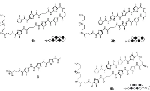
The CpG dyad, an important genomic feature in DNA methylation and transcriptional regulation, is an attractive target for small molecules. To assess the utility of minor groove binding oligomers for CpG recognition, we screened a small library of hairpin pyrrole-imidazole polyamides targeting the sequence 5′-CGCG-3′ and assessed their sequence specificity using an unbiased next-generation sequencing assay. Our findings indicate that hairpin polyamide of sequence PyImβIm-γ-PyImβIm (1), previously identified as a high affinity 5′-CGCG-3′ binder, favors 5′-GCGC-3′ in an unanticipated reverse binding orientation. Replacement of one β alanine with Py to afford PyImPyIm- γ-PyImβIm (3) restores the preference for 5′-CGCG-3′ binding in a forward orientation.
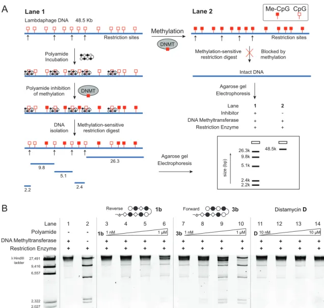
The minor groove binding hairpin 3 inhibits DNA methyltransferase activity in the major groove at its target site more effectively than 1, providing a molecular basis for design of sequence-specific antagonists of CpG methylation.
2.1 Introduction
The role of epigenetic dysregulation in cancer has motivated interest in DNA methylation and methods for its modulation.1,2 In mammals, DNA methylation occurs in the major groove of DNA at the 5′ position of both cytosine residues in the palindromic CG dyad (CpG). CpG’s are rare in the genome and 70% methylated, with nearly all unmethylated CpG’s clustered in G,C-rich regions called “CpG islands”.3 Approximately 60% of RNA Polymerase II transcribed human genes contain CpG islands,4 and their methylation causes transcriptional repression.5 In cancer, for example, otherwise functional tumor suppressor genes can be silenced by hypermethylation in their associated CpG island.6 Importantly, inhibition of DNA methylation at tumor suppressor genes has been shown to reactivate apoptotic pathways and sensitize cancer cells to previously ineffective chemotherapy.7,8
The most effective demethylation agents are cytidine analogs such as 5-aza- deoxycytidine, which find limited use due to significant side effects.1 These cytidine analogs are suicide inhibitors incorporated into DNA to form covalent methyltransferase- DNA adducts.9 The methyltransferase is sequestered and unavailable to methylate CpG’s, resulting in genome-wide demethylation. DNA binding molecules, such as the bis-intercalating natural product echinomycin,10 can disrupt CpG methylation in vitro but have dose-limiting toxicities that have abrogated further clinical advancement.11 While other CpG methylation inhibitors are under investigation,12-14 none of these agents have demonstrated the ability to inhibit DNA methylation in a sequence-specific fashion.
Hairpin pyrrole-imidazole (Py-Im) polyamides are a class of sequence-specific oligomers that bind in the minor groove of DNA.15-20 Programmable sequence preference is accomplished by side-by-side pairings of aromatic amino acids that distinguish the edges of the four Watson-Crick base pairs.15-20 Referred as the pairing rules, Im/Py codes for G•C base pairs, Hp/Py codes for T•A base pairs, and Py/Py binds both T•A/A•T in preference to G•C/C•G. Eight-ring hairpin oligomers linked by a central aliphatic γ- aminobutyric acid unit have affinities for match sites with Ka ~108 to 1010 M-1.16,21 These binding energetics are comparable to natural transcription factors, and like natural DNA binding proteins, are sensitive to differences in the sequence-dependent microstructure of DNA. To relax the curvature of all ring hairpins, β alanine (β) can be substituted for Py- rings in some cases such that β/β pairs replace Py/Py for T•A/A•T specificity, and Im/β replaces Im/Py pairs in strategic locations while retaining specificity for G•C base pair.22-
26 Hairpin Py-Im polyamides usually bind with the N-to-C terminus aligned in the 5′-to- 3′ direction of DNA, referred to as “forward orientation”.27 This modest forward binding preference can be enforced by substitution of the prochiral α position in the γ-turn, i.e., replacement of γ-aminobutyric acid by (R)-2,4-diaminobutyric acid.28 Hairpin architectures containing β/β pairs and β/ring pairs have been found in some cases to prefer the N to C terminus aligned in a 3′-to-5′ direction of DNA.29 While adhering to the pairing rules, this reverse hairpin orientation would bind a different DNA sequence.
Recently we used massively parallel sequencing methods in conjunction with biotin- tagged hairpins, termed Bind-n-Seq, to scan genome-size DNA sequence space for
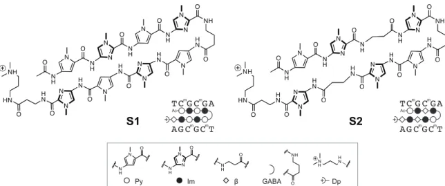
Figure 2.1 Structure of Py-Im polyamides S1 and S2 previously reported to bind methylated 5’- CGCG-3’ oligonucleotide duplex.32 Legend for ball-and-stick notation.
hairpin high affinity sites.30 Although the canonical pairing rules are remarkably predictive of polyamide DNA binding specificity, we identified high affinity DNA binding sites in the reverse orientation for several polyamides containing β/Im pairs.30
Eight-ring hairpin Py-Im polyamides have been shown to discriminate 5′-GGGG- 3′, 5′-GCGC-3′ and 5′-GGCC-3′ with appropriate arrangement of four Im/Py pairs.31 From experience, sequences with CpG steps such as 5′-CGCG-3′ are not as readily accessed for reasons not well understood. In an effort to improve the affinity of an eight- ring hairpin polyamide for the sequence 5′-CGCG-3′, Sugiyama and coworkers replaced two Im/Py pairs with Im/β pairs (Figure 2.1). A change from PyImPyIm-γ-PyImPyIm (S1) to PyImβIm-γ-PyImβIm (S2) afforded a 65-fold increase in affinity for 5′-CGCG- 3′.32 Both hairpins conform to the pairing rules and would bind 5′-CGCG-3′ in the forward orientation. In this study, we employ a high-throughput sequencing assay of polyamide-DNA association to revisit targeting the 5′-CGCG-3′ sequence. Our findings indicate that hairpin polyamides of sequence PyImβIm-γ-PyImβIm S2 favor 5′-GCGC-3′,
S2
HN
O
HN
O NH
O
NH
N O N
H N O
N
NH N O
N
HN
O HN
O N N
N HN
O
N N
O HN
NH N
H O
TC G C GAm m
AG C GC Tm m
β
NH O
GABA Dp
NH
Im O N N
O
NH
Py
N O NH
Ac
+
TC G C GAm m
AG C GC Tm m Ac
+
+
S1
HN
O
HN
O NH
N O
NH
N O N
H N O
N
NH N O
N
N HN
O HN
O N N
N HN
O
N N
O HN
NH
HN NH NH
O
a reverse binding mode. The issue of designing a hairpin polyamide sequence that prefers 5′-CGCG-3′ to 5′-GCGC-3′ remains to be solved. Using Bind-n-Seq methods30 as our screen for a library of polyamide-biotin conjugates, we find that replacement of one β alanine with Py to afford PyImPyIm-γ-PyImβIm restores the preference for forward binding 5′-CGCG-3′. Recent structural work has shown that a cyclic Py-Im polyamide binding in the minor groove causes significant widening of the minor groove width of DNA,33,34 and provides a mechanistic rationale for disruption of DNA-binding proteins in the major groove. We demonstrate the ability of our 5′-CGCG-3′ specific minor groove binding hairpin polyamides to inhibit enzymatic CpG methylation in the major groove of a 5′-CGCG-3′ sequence.
2.2 Results
Sequence Based Analysis of PyImβIm-γ-PyImβIm Specificity. The 5′-CGCG- 3′ sequence is a compelling DNA target for an 8-ring hairpin Py-Im polyamide because it is one of the least represented 6-bp sequence patterns in the human genome, potentially promoting greater genomic specificity.30 Minoshima and coworkers have previously targeted this sequence and shown that polyamide S2 (Figure 2.1) can bind the fully methylated sequence.32 In their study, the substitution of two β’s for Py moieties resulted in improved affinity for 5′-CGCG-3′ over the eight-aromatic ring architecture S1 (Figure 2.1). In light of recent Bind-n-Seq studies, however, we wondered whether these changes may have also had the unintended effect of reducing the preference of the polyamide for binding in the forward orientation.30 Bind-n-Seq is a high-throughput sequencing method
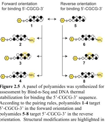
Figure 2.2 A) Scheme of Bind-n-Seq method.30 Polyamide- biotin conjugate is incubated in a genome-sized library of all possible 21mers, enriched, sequenced, and the resulting dataset analyzed with motif-finding software.30 B) Polyamide 1 could potentially bind in the forward orientation or the reverse orientation. The highest affinity binding sequence of 1 is the reverse orientation binding 5’-GCGC-3’.
that allows facile identification of high affinity binding sites of biotin-labeled Py-Im polyamides by affinity purification followed by sequencing (Figure 2.2A).30 As a first step, we synthesized an analog of S2 and examined polyamide-biotin conjugate 1 of sequence PyImβIm-γ-PyImβIm (Figure 2.2B), which has a biotin affinity tag appended at the C-terminus of the heterocyclic oligomer (Figure 2.3). Polyamide-biotin conjugate 1 was incubated at 50 nM in a library of all possible 21 base pair DNA sequences,
B +
B +
1
0 1 2
bits 1AT
2
G
3C
4G
5C
6TA
7AT0 1 2
bits 1AT
2
G
3C
4G
5C
6TA 7AT5’-W C G C G W-3’
3’-W G C G C W-5’
5’-W G C G C W-3’
3’-W C G C G W-5’+ B 1
Binding orientation Forward
(not observed)
Reverse (observed)
Bind-n-Seq
Highest affinity motif
A
B
B+
dsDNA randomer
enriched dsDNA
sequencing by synthesis
motif finding specificity analysis barcode
(”AAA”) illumina primer
degenerate (N x 21)
illumina primer
Figure 2.3 Structures of Py-Im polyamides 1-3.
enriched, and sequenced to identify polyamide-bound sequences. This dataset was then analyzed by the DREME algorithm to construct a motif logo summarizing the highest affinity sequences. A binding preference for 5′-GCGC-3′ was revealed, suggestive of a reverse binding mode (Table 1).
Redesign Hairpin for CGCG versus GCGC Preference. In order to restore the preference for binding 5′-CGCG-3′ in the forward orientation, we considered two possible points of modification (Figure 2.4A). First, we made a single modification to 1 at the turn unit, replacing the GABA turn to a chiral γ-amino GABA, affording 2 (Figure 2.3 and 2.4B). The γ-amino GABA turn has previously been shown to restore forward orientation and increase affinity, including in β-containing polyamides.28-30 This effect is thought to arise from a steric interaction with the floor of the minor groove when the chiral α-amino GABA turn unit is bound in the reverse orientation.23,28 Assessment of polyamide 2 by Bind-n-Seq found that this modification improved the reverse/forward ratio but was insufficient to restore a forward orientation binding preference (Table 2.1).
To confirm the high-throughput sequencing findings, we performed a thermal DNA
HN
O
HN
O NH O
NH O
N N
H O N
N
NH O N
N
HN
O HN
O N N
N HN
O N
N
O HN O
O O
HN
O
NH HN
S O
HN NH
O O
O
HN
O NH N O
NH O
N N
H N O
N
NH O N
N
HN O
HN
O N N
N HN
O N
N HN
O HN O
O O
HN
O
NH HN
S
HN NH
O O
HN
O
HN
O NH O
NH O
N N
H O N
N
NH O N
N
HN
O HN
O N N
N HN
O N
N
O HN O
O O
HN
O
NH HN
S O
HN NH
O O
NH3
2
1 B B NH3+ 3 B
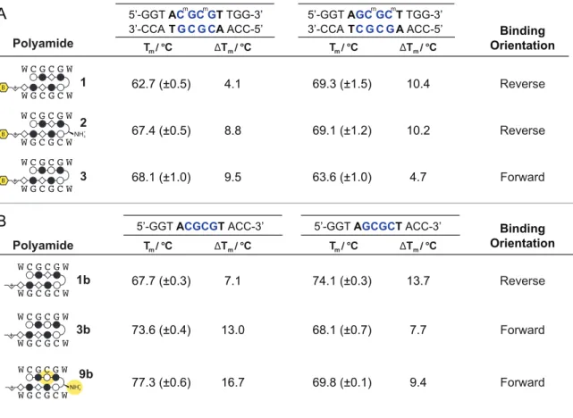
Table 2.1 The preferred binding orientations of polyamides 1-8 were queried with Bind-n-Seq to generate the highest affinity sequence motif. Polyamide-mediated thermal stabilization (ΔTm) of 12 base pair oligonucleotides of the forward (5′-CGCG-3′) and reverse 5′-GCGC-3′ sequences were use to validate the revealed motifs. Melting temperatures reflect the mean and standard deviation of quadruplicate measurements.
denaturation study, as previous studies have shown the thermal stabilization (ΔTm) of duplex DNA by Py-Im polyamides correlates well with binding affinity.35 Assays were performed with DNA oligonucleotides differing only in the central binding sequence (5’-
B
3 1
B
2
NH3+
B
7
B
6
B NH3+
5
B
4
NH3+
B
8
B NH3+
Bind-n-Seq<