TARGETING VASCULAR ENDOTHELIAL GLUTAMINASE IN TRIPLE NEGATIVE BREAST CANCER
By
Verra Manka’a Ngwa
Dissertation
Submitted to the Faculty of the Graduate School of Vanderbilt University
in partial fulfillment of the requirements for the degree of
DOCTOR OF PHILOSOPHY in
Cancer Biology August 12, 2022 Nashville, Tennessee
Approved:
Barbara Fingleton, Ph.D. (Chair) Julie Rhoades, Ph.D.
Linda Sealy, Ph.D.
Jin Chen, M.D., Ph.D. (Advisor)
ii
Copyright © 2022 by Verra Manka’a Ngwa All Rights Reserved
iii DEDICATION
This dissertation is dedicated to all breast cancer patients fighting for their lives and cancer survivors who are winning their battles daily and giving hope to scientists.
In Memoriam:
To my parents, Rose and Mathew Ngwa
iv
ACKNOWLEDGMENT
I would like to express my sincere gratitude and appreciation to the following people, without whom this work would not have been possible.
First and foremost, I am extremely grateful to my mentor, Dr. Jin Chen, for giving me the opportunity to train in her lab as well as her valuable advice, continuous support and guidance throughout my PhD study. Thank you for believing in me and the constant reminders of how good I am especially the high-fives. Being under Jin’s mentorship has been an incredible experience and has provided memories and lessons which will stay with me for a lifetime. I would like to thank my committee members both past and present: Drs. Barbara Fingleton, Julie Rhoades, Linda Sealy, and Rebecca Cook for their expertise, scientific guidance, and counsel throughout my training.
I want to thank the present members of the Chen lab: Dr. Deanna Edwards, Yoonha Hwang, Dr.
Xiagong Wang, and Breelyn Karno, as well as the past members: Drs. Wenqiang Song, Dana Brantley-Sieders, Shan Wang, Laura Kim, and Eileen Shiuan. I am thankful for the scientific knowledge, critiques, encouragement and most of all their friendship during my graduate training. A special thanks to Dr Deanna Edwards for being an instrumental part of my training.
From when I rotated in the lab until my last experiments, Deanna has always listened to my questions and provided me with guidance on how to proceed. I would also like to highlight Dr.
Shan Wang for being patient with me as a new-graduate student and for teaching me most of the techniques that were used in this thesis project.
I would also like to thank the Initiative for Maximizing Student Diversity (IMSD) program, specifically the leadership under Drs. Linda Sealy, Roger Chalkley, and Christina Keeton. To Roger and Linda, thank you for sitting and listening to me practice my presentations before my
v
qualifying exams and providing me with great feedback. It was through the IMSD program that I met my very good friends in graduate school; for that I say thank you!
I would like to thank my previous mentors Drs. Carole Chrestensen and Jonathan McMurry at Kennesaw State University. They both instill in me the love for research. Carole took me into her lab as an undergraduate and taught me the first steps of what research would look like as a career. Jonathan believed in me and allow me to explore my dreams in the lab on a brand-new project. He has been instrumental in my graduate career writing letters of recommendation to every application I did in graduate school and he will always include one of the protein-protein interaction figures from my master’s thesis to the letters.
I would like to thank those who supported me financially during my training, specifically Dr. Jin Chen through the T32 Microenvironmental Influences in Cancer Training Program (MICTP) and the National Cancer Institute for the Predoctoral Ruth L. Kirchstein National Research Service Award (F31). Additionally, I want to that the leaders and members of the Program in Cancer Biology for support especially the Cancer Biology Student Association (CBSA).
Last but not the least, I want to thank friends for their love and support throughout my training. I want to thank my friends (Vera Endah, Grace Nebane, Irene Nwatum, Jocelyne Woopong, and Doris Mbua) back home in Cameroon for their constant love and support. For the text messages telling me how strong I am and that they know I could do it. For the Cameroonian community in Nashville, thank you for making life in Nashville not too far away from home.
Finally, I want to thank my family. First to my parents (deceased) for inspiring me especially my mother who passed with breast cancer. To my family back in Cameroon for their constantly encouraging but kept asking me when I will be done with school. To my uncle and his wife Drs.
vi
Mercy (Aunty Mercy) and Emmanuel Chebe thank you for always supporting me throughout my graduate career. Thank you for always encouraging and telling me how far I could go. A special thanks to Aunties Mercy and Perpetual for always praying for me to succeed in graduate school and life as a whole. To my extended family and in-laws, thank you for your constant love and support. I am grateful especially to Dr Gerard Shu Tangyie and his wife Mrs Comfort Anih, for showing me such an amazing love, making sure I was okay after every failed experiment and encouraging me to keep working hard. Truly, it takes a village! Lastly, to my wonderful husband, Adrian, I cannot thank you enough for your unwavering support especially with the children. I could not have done this without you. I love you!
vii
TABLE OF CONTENTS
DEDICATION... iii
ACKNOWLEDGEMENTS ... iv
LIST OF TABLES ... x
LIST OF FIGURES ... xi
LIST OF ABBREVIATIONS ... xiv
CHAPTERS I. INTRODUCTION ...1
Overview ...1
Breast Cancer and Therapeutic approach ...3
Metabolic Reprograming in Cancer ...6
Glutamine Metabolism and Transporters in Cancer …...6
Regulation of Glutaminase in Cancer ………...11
Targeting Glutaminolysis in Cancer ….……….12
Blood Vessel Formation ………...16
Angiogenesis ……….…....17
Tumor Angiogenesis ………. 20
Tumor Vessel Normalization ………... 21
Metabolism in Normal Endothelial and Tumor Endothelial Cells ………... 23
Glucose Metabolism ……….23
Fatty Acid Metabolism ………. 24
Glutamine Metabolism ………. 25
Anti-angiogenic Drugs for Cancer Therapy ………...… 26
Limitations of Anti-angiogenic Therapy ………. 30
Summary and Thesis Projects ………. 31
II. MATERIALS AND METHODS ... 33
Animals ...………... 33
Genotyping ………. 33
Cell lines and cell culture ………...…... 34
Endothelial cells isolation and culture from tumor bearing mice ………. 35
viii
Tumor model and Metastasis ………... 36
Immunofluorescence ……… 36
Immunohistochemistry ………...….. 38
Tumor hypoxia and blood vessel perfusion assays ………. 39
Treatment of E0771 tumors with chemotherapeutic agents and GLS inhibitor ……… ………. 40
Leptin Treatment of E0771 tumor mice ………. 40
Cytokine array ……… 41
Western blot ………... 42
ELISA ……….. 42
Glutamate assay ……… 43
qRT-PCR assay ……… 43
RNA Sequencing ………... 44
Flow cytometry ……….…. 44
Statistical analysis ………. 45
III. LOSS OF VASCULAR ENDOTHELIAL GLUTAMINASE INHIBITS TUMOR GROWTH AND METASTASIS, AND INCREASES SENSITIVITY TO CHEMOTHERAPY ………. 50
Summary………. 50
Significance ………...……… 51
Introduction ……… 51
Results ………..………. 53
Loss of vascular endothelial glutaminase reduces breast cancer growth and metastasis ………...………... 53
Endothelial GLS deletion reduces tumor vascular density and normalizes tumor vessels………...………. 59
Decreased Leptin in GLSECKO tumors and Leptin treatment rescued tumor growth defects in GLSECKO mice …………...………..….. 63
Loss of endothelial GLS promotes delivery of chemotherapeutical agents ………. 67
Pharmacological inhibition of GLS enhance efficacy of chemotherapeutic agents……….... 70
ix
Discussion ………... 71
IV. CONCLUSIONS AND FUTURE DIRECTIONS ……….... 74
Conclusions ………74
Future Directions ………...77
1. Open Questions ……….77
How does loss of GLS in the endothelium affect leptin secretion in the tumor cells? ………77
What role does other metabolic pathways contribute to the observed phenotype in this study? ………...81
How does loss of GLS in the endothelium contribute to the immune profiles of the tumor microenvironment? ………. 84
Is the microbiome playing a role on the immune phenotype? .... 90
2. Translation Potential of my Thesis Work ……….. 92
3. Study limitations ……… 96
Concluding Remarks ……… 97
REFERENCES ... 99
x
LIST OF TABLES
Table Page
1.1 Molecular subtypes of breast cancer and biomarker expression ………... 5
1.2 Anti-angiogenic Drugs Use in Cancer Treatment ……… 29
2.1 Mouse Cytokine Antibody Array G-Series 3 Map ……… 42
2.2 Gating strategies use in flow cytometry analysis.……… 46
2.3 Antibodies used in flow cytometry………... 47
xi
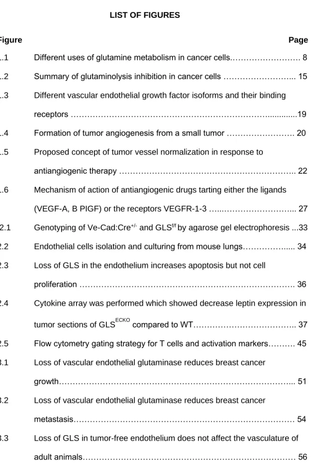
LIST OF FIGURES
Figure Page
1.1 Different uses of glutamine metabolism in cancer cells.………. 8 1.2 Summary of glutaminolysis inhibition in cancer cells ………... 15 1.3 Different vascular endothelial growth factor isoforms and their binding
receptors ………...19 1.4 Formation of tumor angiogenesis from a small tumor ………. 20 1.5 Proposed concept of tumor vessel normalization in response to
antiangiogenic therapy ……….. 22 1.6 Mechanism of action of antiangiogenic drugs tarting either the ligands
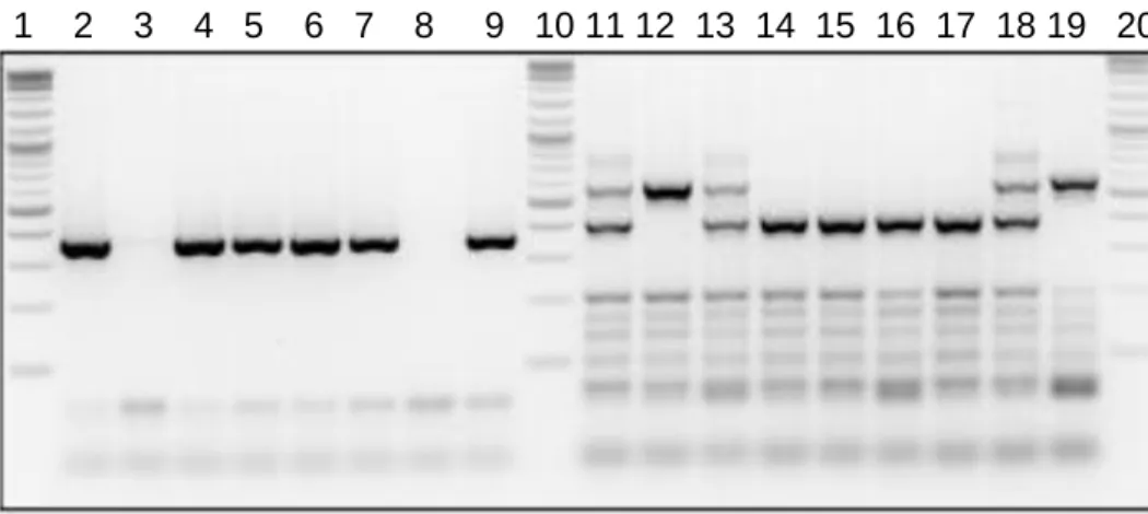
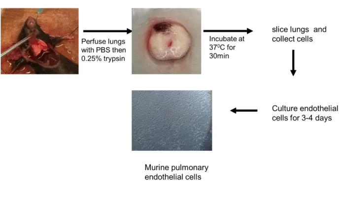
(VEGF-A, B PIGF) or the receptors VEGFR-1-3 …...………... 27 2.1 Genotyping of Ve-Cad:Cre+/- and GLSf/f by agarose gel electrophoresis ...33 2.2 Endothelial cells isolation and culturing from mouse lungs………... 34 2.3 Loss of GLS in the endothelium increases apoptosis but not cell
proliferation ………. 36 2.4 Cytokine array was performed which showed decrease leptin expression in tumor sections of GLSECKO compared to WT……….. 37 2.5 Flow cytometry gating strategy for T cells and activation markers………. 45 3.1 Loss of vascular endothelial glutaminase reduces breast cancer
growth………... 51 3.2 Loss of vascular endothelial glutaminase reduces breast cancer
metastasis……… 54 3.3 Loss of GLS in tumor-free endothelium does not affect the vasculature of
adult animals……… 56
xii
3.4 Endothelial GLS deletion reduces tumor vascular density and normalizes tumor vessels …..………... 57 3.5 Percentages of different cell types within the E0771 tumors are presented
in the pie chart ….………..………. 60 3.6 Leptin treatment rescues tumor growth defects in GLSECKO mice .………. 61 3.7 Loss of endothelial GLS enhances the efficacy of chemotherapeutic
agents………... 64 3.8 CB-839 treatment on WT mice improves drug delivery ………... 69 4.1 Proposed model of endothelial GLS deletion in breast tumor
vasculature………..…. 74 4.2 CoCL2 and hypoxia increase HIF-1α which in turn induce leptin mRNA and
protein expression……….. 76 4.3 Loss of GLS in the endothelium does not affect subcutaneous tumor
growth………... 78 4.4 RNA-seq showing PEDF secretion from tumor cells isolated from WT vs
GLSECKO……… 78
4.5 RNA seq analysis of WT and GLS KO endothelial cells ….……… 81 4.6 Endothelial GLS deletion does not appear to affect tumor infiltrating
immune cells ... 85 4.7 Cytokine array showing differentially expressed proteins in WT versus
GLSECKO tumor lysates ………..……… 86 4.8 Endothelial GLS deletion does not appear to affect eosinophil tumor
infiltrating but activation is increased in GLSECKO ………..………… 87 4.9 T cells activation markers in WT vs GLSECKO………. 90
xiii
4.10 Proposed overall contribution of endothelial GLS deletion in breast tumor vasculature ………..………..………. 94
xiv
LIST OF ABBREVIATIONS
2HG 2-hydrocyglutarate
ACK Ammonium-chloride-potassium acyl-CoA Cholesterol acyltransferase Akt Protein kinase B
α-KG Alpha-ketoglutarate Ang Angiopoietin
ANOVA Analysis of variance
ASCT2 Alanine-serine-cysteine transporter 2 ATP Adenosine triphosphate
AXL Anexelekto
BCH System-L inhibitor 2-amino-2-norbornanecarboxylic acid BPTES Bis-2-(5-phenylacetamido-1,3,4-thiadiazol-2-yl)ethyl sulfide CAAs Cancer-associated adipocytes
CAD Carbamoyl-phosphate synthetase 2, Aspartate transcarbamoylase, and Dihydroorotase CAFs Cancer-associated Fibroblasts
CB-839 Telaglenastat
CCL C-C chemokine ligand CD Cluster of differentiation
CDH5 Cadherin 5
CDK Cyclin-dependent kinase
CHIP-seq Chromatin immunoprecipitation sequencing CoCL2 Cobalt chloride
xv
Cpt Cisplatin
CPT1a Carnitine palmitoyltransferase 1a CRC Colorectal cancer
Cre Carbapenem-resistant enterobacteriaceae CSFR1 Colony stimulating factor 1 receptor
CTLA4 Cytotoxic T-lymphocyte-associated protein 4 CXCL C-X-C motif chemokine ligand
DAB 3,3′-diaminobenzidine
DAPI 4’,6-diamidino-2-phenylindole DC101 Anti-VEGF receptor 2 antibody DCA Deoxycholic acid
DEG Differentially expressed genes DLL4 Delta like canonical notch ligand 4 DON 6-diazo-5-oxo-L-norleucine
DOXO Doxorubicin HCL EC Endothelial cell
DMEM Dulbecco’s modified eagle’s medium DNase Deoxyribonuclease
EGF Epidermal growth factor
EGFR Epidermal growth factor receptor ELISA Enzyme-linked immunosorbent assay EPHA2 Ephrin type-a receptor 2
ER Estrogen receptor
ERK Extracellular signal-regulated kinase FACS Fluorescence-activated cell sorting
xvi FAO Fatty acid oxidation
FASN Fatty acid synthase FBS Fetal bovine serum FGF Fibroblast growth factor
FGFR Fibroblast growth factor receptor FITC Fluorescein isothiocyanate FLT3 Fms-like tyrosine kinase 3 FMO Fluorescence minus one
GAC Glutaminase C
GBM Glioblastoma multiforme GDH Glutamate dehydrogenase GIST Gastrointestinal tumor
GLS Glutaminase
GLSECKO Endothelial glutaminase deletion GLUD Glutamate dehydrogenase GLUL Glutamate-ammonia ligase
GOT Glutamate–oxaloacetate transaminase GPNA Gamma-l-glutamyl-p-nitroanilide GSEA Gene set enrichment analysis GSH Reduced glutathione
GSK3α/β Glycogen Synthase Kinase 3 alpha/Beta
GZMB Granzyme B
HCC Hepatocellular carcinoma
HER2 Human epidermal growth factor receptor 2 HIF-1α Hypoxia-inducible factor-1 alpha
xvii HRE Hypoxia response element
IACUC Institutional Animal Care and Utilization Committee ICAM-1 Intercellular adhesion molecule 1
ICI Immune checkpoint inhibitor ICIs Immune checkpoint inhibitors IDH Isocitrate dehydrogenase
IF Immunofluorescence
IFN Interferon
IHC Immunohistochemistry IL Interleukin
JAK c-Jun N-terminal kinase
JHU-083 Ethyl 2-(2-Amino-4-methylpentanamido)-DON KEGG Kyoto Encyclopedia of Genes and Genomes KGA Kidney glutaminase
LAT Large Amino acid Transporter LDHA Lactate dehydrogenase A
Lep Leptin
LepR Leptin receptor
LL202 (E)-15,22-Dioxa-4,11-diaza-5(2,5)-thiadiazola-10(3,6)-pyridazina-1,14(1,3)- dibenzenacyclodocosaphan-18-ene-3,12-dione
LLC Lewis lung carcinoma LPS Lipopolysaccharide
Ly6G Lymphocyte antigen 6 complex, locus G Ly6C Lymphocyte antigen 6 complex, locus C M.O.M Mouse-on-mouse
xviii M1-like Classically activated macrophages M2-like Alternatively activated macrophages MHC Major histocompatibility complex MAPK Mitogen-activated protein kinase mCRC Metastatic colorectal cancer MDSC Myeloid-derived suppressor cell
MEK Mitogen-activated protein kinase kinase M-MDSC Mononuclear myeloid-derived suppressor cell MMTV-Neu Mouse mammary tumor virus- rat ERBB2
MMTV-PyMT Mouse mammary tumor virus-polyoma middle tumor-antigen MPMEC Mouse pulmonary microvascular endothelial cells
mRNA Messenger Ribonucleic acid
mTORC1 Mammalian target of rapamycin complex 1 NADPH Nicotinamide adenine dinucleotide phosphate NEC Normal endothelial cells
NES Normalized enrichment score NG2 Neural/glial antigen 2
NIH National Institutes of Health NK Natural killer
NOTCH1 Notch receptor 1 NRP-1 Neuropilin receptor-1 NSCLC Non-small cell lung cancer OCT Optimal cutting temperature OS Overall survival
OXPHOS Oxidative phosphorylation
xix PBS Phosphate buffered saline PCR Polymerase chain reaction
PD1 Programmed death 1
PDGF Platelet-derived growth factor PD-L1 Programmed death ligand PDX Patient-derived Xenograft
PE Phycoerythrin
PEDF Pigment epithelium-derived factor PerCP Peridinin chlorophyll protein PFA Paraformaldehyde
PFKFB3 6-phosphofructo-2-kinase/fructose-2,6-biphosphatase 3 PI3K Phosphatidylinositol 3-kinase
PIGF Placental growth factor
PMN-MDSC Polymorphonuclear myeloid-derived suppressor cell PR Progesterone receptor
qRT-PCR Quantitative real-time polymerase chain reaction R5P Ribose-5-phosphate
RAC Ras-related C3 botulinum toxin substrate RAF Rapidly accelerated fibrosarcoma
RCC Renal cell cancer
RET Rearranged during Transfection Rgs5 Regulator of g protein signaling 5 RIPA Radioimmunoprecipitation assay RNAse Ribonuclease
RNAseq RNA-sequencing
xx RPMI Roswell Park Memorial Institute RTK Receptor tyrosine kinase
S6K1 Ribosomal protein S6 kinase beta-1
SDS-PAGE Sodium dodecyl sulfate polyacrylamide gel electrophoresis SEM Standard Error of Mean
shRNA Short hairpin RNA
SLC1A5 Solute carrier family 1 member 5 SLC7A11 Solute carrier family 7 member 11 SLC7A5 Solute carrier family 7 member 5 SMA Smooth Muscle Actin
SNAT2 Sodium-dependent neutral amino acid transporter-2 STAT Signal transducer and activator of transcription TAp63 Tumor protein p63
TAp73 Tumor protein p73
TAZ WW domain containing transcription regulator 1 TCA Tricarboxylic acid
TCR T cell receptor
TEAD4 TEA domain transcription factor 4 TECs Tumor endothelial cells
Tek TEK Receptor tyrosine kinase, or Tie2 TGF-β Transforming growth factor-beta Th T helper cells
TIE Angiopoietin receptor TKI Tyrosine kinase inhibitor TME Tumor microenvironment
xxi TNBC Triple-negative breast cancer TNF Tumor necrosis factor
TP53 Tumor protein 53 Treg Regulatory T cell
TVN Tumor vessel normalization VCAM-1 Vascular cell adhesion molecule 1 VE-Cad Vascular endothelial cadherin VEGF-A Vascular endothelial growth factor
VEGFR2 Vascular endothelial growth factor receptor 2
WT Wild type
YAP Yes-associated protein
xxii
ORIGINAL PUBLICATIONS
1. Verra M. Ngwa, Deanna N. Edwards, Yoonha Hwang,Chi Yan, Ann Richmond, Dana M.
Brantley-Sieders, Jin Chen (In press), “Loss of vascular endothelial glutaminase inhibits tumor growth and metastasis, and increases sensitivity to chemotherapy,” Cancer Research Communications
2. Deanna N. Edwards, Verra M. Ngwa, Ariel L. Raybuck, Shan Wang, Yoonha Hwang, Laura C. Kim, Sung Hoon Cho, Yeeun Paik, Qingfei Wang, Siyuan Zhang, H. Charles Manning, Jeffrey C. Rathmell, Rebecca S. Cook, Mark R. Boothby, Jin Chen (2020),
“Selective glutamine metabolism inhibition in tumor cells improves antitumor T lymphocyte activity in triple-negative breast cancer,” Journal of Clinical Investigation
3. Verra Ngwa, Deanna Edwards, Mary Philips and Jin Chen (2019), “Microenvironmental metabolism regulates anti-tumor immunity,” Cancer Research
4. Deanna N. Edwards, Verra M. Ngwa, Shan Wang, Eileen Shiuan, Dana M. Brantley- Sieders, Laura Kim, Albert B. Reynolds and Jin Chen (2017), ‘’The receptor tyrosine kinase EphA2 promotes glutamine metabolism in tumors by activating the transcriptional coactivators YAP and TAZ,’’ Science Signaling
1 CHAPTER I
INTRODUCTION
Overview
Cancer is a major public health burden worldwide and the second leading cause of death in the United States (American Cancer Society). Cancers are characterized by their unique
characteristics termed hallmarks, which include sustaining proliferative signals, evading growth suppressors, avoiding immune destruction, enabling replicative immortality, tumor-promoting inflammation, genome instability and mutation, resisting cell death, activating invasion and metastasis, deregulating cellular energetics, and inducing angiogenesis (1,2). These common features provide a frame work for critical areas of research to target and treat cancer.
Deregulation of cellular energetics is one of the key elements of the cancer hallmarks. Cancer cells altered their metabolism to support their high proliferative rates and adapt to the hostile tumor microenvironment (2,3). The Warburg effect, which is the best-known metabolic
abnormality in cancer cells, demonstrates an increase in glucose consumption through elevated glycolysis even in the presence of oxygen (aerobic glycolysis) for these rapidly proliferating cells (2,3). In addition to glucose metabolism, some cancer cells have been identified to be addicted to glutamine metabolism. These tumor cells utilize glutaminolysis to support biosynthesis of amino acids, nucleotides, and glutathione (4,5). These molecules are critical to maintain cancer cells biomass and involved in other metabolic pathways that are required for cell survival.
Growing tumors acquire nutrients via diffusion; however, rapidly growing tumors require a vascular system to grow beyond 2 mm3 in diameter (6). The tumor achieves this through
2
angiogenesis, the process whereby new blood vessels are formed from pre-existing vessels, by secreting angiogenic factors like VEGF-A and Angiopoietin which promotes the development and stabilization of the tumor vasculature. Unlike normal blood vessels, tumor blood vessels are abnormal and dysfunctional. This abnormality is characterized by tortuous, leaky and chaotic networks of irregular endothelial cells (ECs) lining the tumor vessels. Abnormal tumor vascular network impairs perfusion, obstructs blood flow, and lead to poor leukocyte trafficking and drug delivery (6). Like cancer cells, these angiogenic sprouts are initiated from rapidly proliferating cells that consume glucose, glutamine and other nutrients (7,9,10).
Judah Folkman, almost four decades ago, proposed that tumors depend on a blood vessel network, and inhibiting these blood vessels would cut off blood supply and limit the amount of nutrients and oxygen to the tumors hence choke the tumor to death (6). In the quest for angiogenic inhibitors, Bevacizumab (Avastin), a humanized monoclonal antibody against VEGFA, became the first drug to block blood vessel and was approved in 2004. However, due to resistance and other adverse effects, usage of Avastin and other angiogenesis inhibitors is limited in the clinic. Consequently, other strategies are being investigated to target tumor blood vessels. Indeed, Rakesh Jain proposed that instead of eliminating the abnormal blood vessels, they could be normalized which can lead to improved vessel perfusion and promote drug delivery (11,12). Since the introduction of the concept, all normalization strategies focused on targeting angiogenesis using anti-angiogenic drugs. Peter Carmeliet and his group seized the opportunity to exploit endothelial cell metabolism as a therapeutic target to normalize tumor blood vessels. Proliferative tumor endothelial cells are hyperglycolytic, and inhibiting tumor endothelial cells glycolysis normalized blood vessels, reduced metastasis and promoted drug delivery (13).
3
Aside from glucose, a growing body of evidence shows that glutamine metabolism provides carbons for biomass production and both carbon and nitrogen for glutathione synthesis that is required for EC proliferation (9,14). Glutamine metabolism also contribute to lipid biosynthesis in ECs through reductive carboxylation. Vascular endothelial-specific deletion of glutaminase in vivo suppressed retinal angiogenesis, and negatively affected tricarboxylic acid (TCA) cycle anaplerosis, macromolecule production, and redox homeostasis in ECs (9,14). It remains to be determined whether inhibition of glutamine consumption can normalize tumor blood vessels and enhance antitumor immunity. Herein, we describe the role of vascular endothelial glutaminase (GLS), the enzyme that catabolizes glutamine to glutamate, in breast cancer tumor growth and metastasis and chemotherapy drug delivery. We utilized an inducible transgenic mouse model to delete GLS specifically in the endothelium (GLSECKO). Our data reveal that GLS loss in endothelium decreases tumor angiogenesis while promoting tumor vessel normalization.
GLSECKO tumors with normalized blood vessels displayed an increase in drug delivery and enhanced anti-tumor effect of chemotherapy. We also report herein, a mechanism which functionally linked endothelial GLS and expression of leptin in tumor cells, a key regulator of metabolic homeostasis. Together, these data demonstrate a crucial role for glutamine metabolism in tumor endothelium, which may be exploited therapeutically to induce vascular normalization and improve drug delivery in solid tumors.
Breast Cancer and Therapeutic Approach
Breast cancer is the most common malignancy in women, and the principal cause of cancer- related death among women in both developed and developing countries (15). According to the American Cancer Society, in 2022, an estimated 287,850 new cases of breast cancer will be diagnosed in women which will lead to an estimated 43,250 deaths. Breast cancer is a heterogeneous disease classified based on four primary molecular subtypes: Luminal A,
4
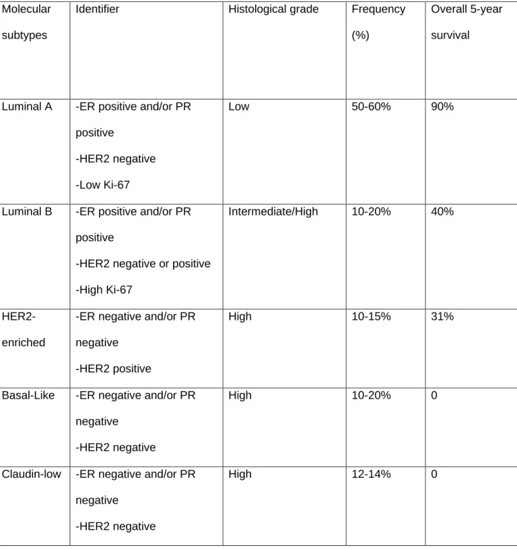
Luminal B, Her2-enriched and Triple negative/basal-like (16). These subtypes are described based on their surface protein expression as detailed in Table 1.1. Effective targeted treatment options for ER+/PR+ and HER2+ breast cancers have been developed. ER+ and /or PR+
tumors make up approximately 60% of breast cancer diagnosis and are treated with hormone modulation therapies (e.g tamoxifen and fulvestrant), aromatase inhibitors (e.g. letrozole, exemestane, and anastrozole), and CDK 4/6 inhibitors (eg, palbociclib, ribociclib, abemaciclib) (used in metastasis disease) (17–19). HER2+ tumors represent about 20% of invasive breast cancers and overexpress the HER2 receptor. Treatment of HER2+ tumors involves targeting the HER2 receptor and preventing downstream signaling using monoclonal antibodies (e.g trastuzumab, pertuzumab) (20–22); antibody-drug conjugates (e.g ado-trastuzumab-emtansine, and trastuzumab derutecan); or tyrosine kinase inhibitors (e.g tucatinib, neratinib) (23).
Triple negative breast cancer (TNBC) does not express any of the above receptors hence treatment options are limited with chemotherapy as the main standard of care. In searching for vulnerability in this aggressive subtype, TNBCs are found to be addicted to glutamine
metabolism both in pre-clinical and clinical settings (24–26). While breast cancer in general is not very immunogenic, TNBC has been found to be responsive to immune checkpoint inhibitors.
Indeed, atezolizumab and pembrolizumab in combination with chemotherapy are well tolerated in TNBC patients with high PD-L1 expression (27), broadening the therapeutic option for TNBC patients. Despite the advances in cancer research in recent years, limited efficacy and drug resistance remains a challenge.
5
Table 1.1: Molecular subtypes of breast cancer and biomarker expression (summarized from (16))
Molecular subtypes
Identifier Histological grade Frequency (%)
Overall 5-year survival
Luminal A -ER positive and/or PR positive
-HER2 negative -Low Ki-67
Low 50-60% 90%
Luminal B -ER positive and/or PR positive
-HER2 negative or positive -High Ki-67
Intermediate/High 10-20% 40%
HER2- enriched
-ER negative and/or PR negative
-HER2 positive
High 10-15% 31%
Basal-Like -ER negative and/or PR negative
-HER2 negative
High 10-20% 0
Claudin-low -ER negative and/or PR negative
-HER2 negative
High 12-14% 0
6 Metabolic Reprograming in Cancer
Altered metabolism is an important hallmark of cancer growth and progression. Metabolic reprogramming provides tumor cells the energy and materials to support their large-scale biosynthesis and rapid proliferation. The concept of altered metabolism started in the 1920s when Otto Warburg observed that tumor cells use glucose to generate lactate despite oxygen- rich environment (aerobic glycolysis) (28,29). Initially, Warburg’s theory was based on the fact that neoplastic cells had a dysfunctional mitochondrion. However, studies later revealed that cancer cells were able to use glucose and fatty acids at the same rate as normal cells (30,31).
In contrast to normal cells, cancer cells utilize glycolysis to generate glycolytic intermediates for biosynthesis while reducing the production of reactive oxygen species from oxidative
phosphorylation (32–34). Advances in metabolic research has made it evident that in addition to glucose, tumor cells also require lipids and amino acids for biomass and proliferation (35).
Altered metabolism depends on the dysregulation of several enzymes in the different metabolic pathways, indicating that appropriate intervention against these key enzymes may be leveraged to inhibit tumor growth and metastasis (36). Given the in-depth study in the field of tumor metabolism, I will now focus on reviewing the contributions of glutamine metabolism in supporting cancer growth and progression.
Glutamine Metabolism and Transporters in Cancer
Glutamine is an abundant non-essential amino acid in the human body. It is largely used for energy production (bioenergetics) by replenishing the TCA cycle and feeding in carbon and nitrogen for the synthesis of nucleotides, glutathione (GSH), amino acids and fatty acids (4,37).
In the cell, glutaminase (GLS) converts glutamine into glutamate, which acts as a precursor for the antioxidant glutathione. Glutamate is further metabolized to α-ketoglutarate either by
7
glutamine dehydrogenase (GLUD) or aminotransferases. Through α-ketoglutarate, glutamine enters the TCA cycle as an anaplerotic source of carbons (4,38). In this capacity, glutamine provides nitrogen for biosynthesis of nucleotides, amino acids, and hexosamines, but also serves as a mitochondrial substrate. Glutamine-derived α-ketoglutarate is reduced to citrate by isocitrate dehydrogenase (IDH) enzymes through reductive carboxylation thereby fueling lipid biogenesis. In addition to its role in the reductive carboxylation reaction, IDH mutations generate the oncometabolite, 2-hydroxyglutarate (2HG), which regulates epigenetics by inhibiting
demethylase enzymes that are members of the α-ketoglutarate-dependent dioxygenase family (39,40) (Figure 1.1). The accumulation of 2HG in breast cancer is associated with c-MYC activation contributing to an increase in glutamine metabolism through the expression of glutaminase, hence poor prognosis in breast cancer patients (40). Cancer cells can exploit these multiple functions of glutamine to drive tumor growth.
Amino-acid transport systems are essential for the growth of cancer cells, not only because they provide the amino acids required for protein synthesis but also due to their capability of
activating signaling pathways involved in cell growth, such as the mammalian target of rapamycin complex 1 (mTORC1). Importantly, glutamine transporters are required to sustain
“glutamine addiction” in tumor cells (41,42). Several glutamine transporters exist in mammalian cells, the best studied of which are the alanine-serine-cysteine transporter 2 encoded by SLC1A5 (ASCT2) gene and the L-type amino acid transporter 1 encoded by the SLC7A5 (LAT1) gene (43). These two transporters are highly expressed in aggressive forms of breast cancer, including triple-negative and HER2-positive, suggesting that these transporters
cooperate to promote tumor growth and progression (44–46). In addition to other neutral amino acids, SLC1A5 /ASCT2 mediates both the Na+-coupled influx/efflux of glutamine while
SLC7A5/LAT1 mediates the efflux of glutamine in exchange of leucine influx into the cells and therefore regulates the activation of mTORC1 (43). Pre-clinical studies showed that ASCT2 is
8
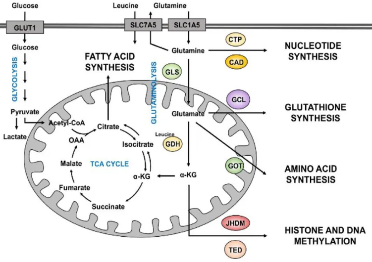
Figure 1.1: Different uses of glutamine metabolism in cancer cells. Glutamine enters the cells through the transporters SLC1A5 or SLC7A5. Once inside the cell, glutamine can contribute to nucleotide
biosynthesis directly (through CAD for example) or is converted to glutamate by GLS. It can also be exported outside of the cell in exchange of leucine, a coactivator of GDH. Glutamate can further be converted to α-KG by GDH. Glutamate can also contribute to the synthesis of glutathione through the activity of different enzymes, including GCL. Amino acid synthesis is supported by the aminotransferases (GOT) which converts glutamate to α-KG. Glutamine-derived α-KG enters the TCA cycle to produce energy for the cell or proceed backwards via the reductive carboxylation to provide an alternative source of lipid synthesis. Additionally, α-KG is a co-substrate of dioxygenase enzymes (JHMD and TED) require in the regulation of histone and DNA methylation. (Review in Nguyen T and Duran R.V., Cancer Drug Resist.
2018)
9
responsible for glutamine-dependent cell growth in basal-like breast cancer and showed that ASCT2 loss is sufficient to significantly reduce basal-like breast cancer cell growth in vitro and in vivo (47). In another study, the role of SLC7A5/LAT1 was investigated in breast cancer growth and progression. The authors showed that applying an SLC7A5/LAT1 inhibitor, 2-aminobicyclo- (2,2,1)-heptane-2-carboxylic acid (BCH) or knocking down the transporter in triple-negative and HER2-positive human breast cancers cell lines significantly inhibited cell growth (46). The expression of these transporters have emerged as major pro-tumoral transporters with increased expression levels correlating with poor patient prognosis in different cancer types (48–51) including breast cancer (45,52), suggesting the potential of glutamine transporters as prognostic biomarkers for breast cancer.
Once inside the cell, glutamine is catabolized to glutamate by glutaminase (GLS), which
represents the rate-limiting step of glutaminolysis (53). Two isoforms of GLS exist in mammals:
the kidney-type glutaminase (GLS1 or GLS) and liver-type glutaminase (GLS2) (54). GLS1 is up-regulated in some human cancers including breast and is associated with a higher disease stage and poor prognosis (24). Additionally, GLS exists as two alternative splice variants, known as glutaminase C (GAC) and kidney glutaminase (KGA). These two isoforms have the same N- terminal and catalytic domains but different C-termini with unknown function. The KGA isoform is expressed in most human tissues except liver, while GAC is only expressed in specific
tissues, such as heart and kidneys (24,55), making GAC a more selective target for tumor cells.
Oligomerization is both sufficient and necessary for KGA and GAC activation with the formation of tetramers from dimers, enhanced by inorganic phosphate, to encompass full enzymatic activity (56,57). The GAC isoform, the more catalytically active form of GLS, is often
overexpressed in human cancers especially in breast cancers and is associated with sensitivity to glutamine withdrawal (58,59). Furthermore, glutamine-dependent breast cancers are less efficient of synthesizing glutamine from glutamate using glutamine synthetase within the cells
10
(58) enabling glutamine addicted breast cancer cells to rely heavily on exogenous glutamine.
The resulting glutamate produced by the tumor cells is exported out of the cell by Xc-
cystine/glutamate antiporter (SLC7A11), which promotes aggressiveness and invasiveness of breast cancer (60), suggesting that GLS (specifically GAC) is an important drug target.
In breast cancer, GLS has been shown to be highly expressed in HER2-positive and triple negative breast cancers (TNBC). Not surprisingly, Kim et al showed that patient samples representing these breast cancer subtypes have higher levels of glutamine consumption and glutaminolysis compared to luminal subtypes (44). To further validate the importance of
glutaminolysis in breast cancer cell viability, Lampa and colleagues investigated the role of GLS as a therapeutic target in TNBC using GLS specific shRNA constructs and glutaminase inhibitor CB-839 as a pharmacological tool. They demonstrated that GLS knockdown in glutamine- dependent human TNBC cell lines led to a decrease in downstream metabolites and significant cell growth inhibition (24). Additional metabolomics analysis of 270 clinical breast cancer tissues and 97 normal breast tissues revealed an elevated glutamate to glutamine ratio in breast cancer samples due to increase GLS1 expression (61). Increased glutamate levels in breast tumors have been associated with invasiveness and drug resistance, correlating with increased risk of recurrence. For this reason, the increased ratio of glutamate to glutamine may represent another biomarker that could help to stratify patients’ treatment with specific glutamine inhibitors.
While much has been shown about GLS in different cancers, the role of GLS2 is still not fully understood. Some studies have enumerated the role of GLS2 as a tumor suppressor while others have suggested GLS2 as a tumor promoter whose upregulation contributes to cancer cell survival. Lui Juan and colleagues showed that GLS2 is a downstream target gene of p53 and increased expression of GLS2 significantly reduced liver tumorigenesis by inhibiting anchorage-
11
independent growth of hepatocellular carcinoma cells (HCC) and the growth of HCC xenograft tumors (62). However, GLS2 has also been shown to support tumor growth and promote ionizing radiation resistance in cervical cancer (63), indicating the role of GLS2 in cancer may be context dependent and requires further studies.
Regulation of Glutaminase in Cancer
Like most biological processes, glutaminolysis is well regulated in normal cells. The MYC oncogene is a transcription factor that is frequently dysregulated in human cancers. MYC regulates the expression of multiple genes involve in diverse processes including cellular metabolism, differentiation, vasculogenisis, cell adhesion, cell growth, apoptosis, and DNA damage responses (64). MYC regulates glutamine metabolism by upregulating GLS, glutamine synthase (GLUL), GLUD and aminotransferases and the glutamine transporters SLC1A5 and SLC38A5 (65–67) (Figure 1.3). MYC promotes GLS expression indirectly by repressing the transcription of miR-23a and miR-23b. Other signaling pathways like c-JUN, GSK3α/β, and mTORC1/S6K1 upregulate GLS directly or indirectly (reviewed in (67)). GLS can also be regulated by hypoxia-inducible factor 1 (HIF1) both at the mRNA and protein expression (68). Studies from our lab provide evidence that the receptor tyrosine kinase (RTK) EphA2 regulates glutamine metabolism through the EphA2-Rho-glutaminase pathway in a breast cancer mouse model driven by activated HER2/ErbB2 (MMTV-NeuT). EphA2 RTK promotes glutamine metabolism by activating the transcriptional coactivators YAP and TAZ.
Once activated, YAP/TAZ binds to TEAD4 transcription factor to upregulate GLS and SLC1A5 genes thereby promoting glutaminolysis in cancer (69,70).
Alternatively, GLS2 is regulated differently from GLS. Studies have shown that GLS2 is
regulated by the tumor suppressor p53 (71,72). Members of the p53 family, TAp63 and TAp73
12
also regulate GLS2 (73,74). Taking together, targeting glutamine metabolism is warranted for therapeutic drug development.
Targeting Glutaminolysis in Cancer
The diverse roles played by glutamine in tumor metabolism present an opportunity for targeting glutamine metabolism for cancer therapy, especially in the glutamine-addicted HER2-positive and triple negative breast cancers. Although advances in targeted therapy have improved the survival of ER+/PR+ and HER2 patients, long-term response rates are limited due to acquired resistance. Because no oncogenic drivers have been identified in TNBC, therapeutic options for most of these patients are very limited and prognosis remains poor. , Immunotherapies are beginning to be used in TNBCs patients, however, strategies are needed to identify patients who will benefit from these therapies (75). Additionally, efforts are needed to reduce immune- related toxicity, and cost on these patients.
The first strategy to target glutamine metabolism would involve inhibiting the transporters to block the import of glutamine. The small molecule inhibitor of ASCT2, gamma-l-glutamyl-p- nitroanilide (GPNA), has been proven to be effective in glutamine-dependent cancers (51).
However, due to toxicity to other healthy cells that rely on glutamine and lack of specificity, GPNA could not be used in the clinic (76,77). Recently, a new ASCT2 inhibitor, V-9302, was discovered and has proven potent in blocking glutamine transport. Using the V-9302 inhibitor in murine models resulted in decreased cancer cell growth and proliferation, increased cell death, and increased oxidative stress, thereby contributing to anti-tumor responses both in vitro and in vivo (78).
13
In addition, as the rate-limiting step in glutaminolysis, GLS is a suitable target in cancer therapy.
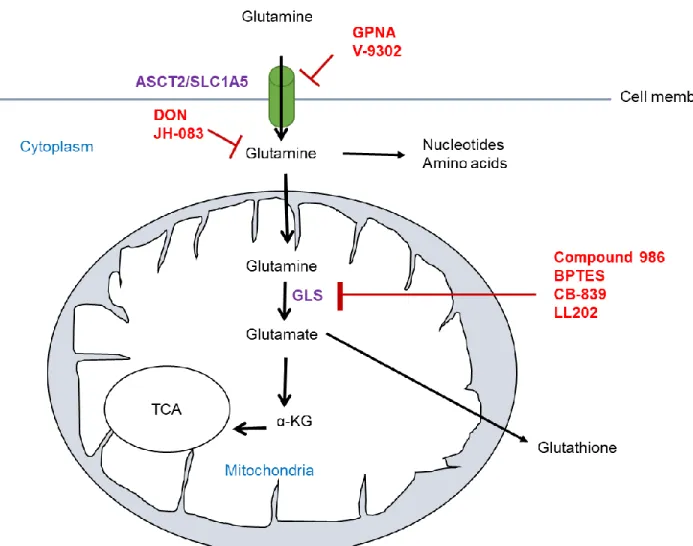
Indeed, several approaches have been employed to target glutaminase (Figure 1.2) including small molecule analogues, such as, 6-diazo-5-oxo-L-norleucine (DON), compound 968, BPTES, CB-839, JHU-083 and LL202. DON is a glutamine antagonist which binds covalently to the enzyme active site and blocks glutamine-dependent enzymes (GLS and glutamine amido- transferases) involved in de novo nucleotide biosynthesis, amino acid synthesis and
hexosamine production in tumor cells (67,79–81). DON exhibited a degree of anti-proliferative effects in pre-clinical studies involving in vitro assays and xenograft animal models. However, DON use and development was discontinued due to dose-limiting neurotoxicity, gastrointestinal toxicity and myelosuppression (82). To diminished the toxicity of DON, a novel prodrug, JHU- 083, was discovered which selectively blocks glutaminase activity (83). Being a pro-drug, JHU- 083 itself is inactive. JHU-083 is converted either in the plasma by plasma esterases or
intracellularly by cathepsin, which is more highly expressed in the tumor compared to normal tissues (84). Overall, DON and JHU-083 are pleiotropic and have effects on other metabolic pathways such as glycolysis.
Unlike DON and JHU-083, Compound-968, BPTES, and CB-839 target glutaminase without affecting other glutamine metabolism reactions. Compound-968 belongs to the family of benzophenanthridinone and it can allosterically inhibit GAC. This drug binds to the monomeric forms of GAC and prevents the formation of the active GAC tetramer (85). Compound-968 is specific to GLS however, it shows limited potency in the presence of the inorganic phosphates that promote GLS activation by tetramerization (85). BPTES blocks GLS in an uncompetitive fashion by causing a conformational change upon binding to KGA and GAC, trapping these isoforms in an inactive tetramer form (86,87). BPTES suppresses tumor growth both in vitro and in vivo in various cancers including breast cancer. However, BPTES is not the appropriate candidate for GLS inhibition because of its poor solubility and bioavailability (88).
14
Due to the limited potency of Compound-968 and the low solubility of BPTES, the efforts of developing new pharmacological inhibitors for GLS, and especially GAC, is necessary. CB-839 is an analogue of BPTES that inhibits both the GAC and KGA glutaminase splice variants but not GLS2 (88). Unlike the other glutaminase inhibitors, CB-839 exhibits low nanomolar potency in biochemical and cellular assays and has good oral bioavailability (89). In pre-clinical studies, CB-839 has been shown to inhibit the growth of several cancers including lymphoma and non- small cell lung cancer (90). Furthermore, CB-839 is able to inhibit proliferation of triple negative breast cancer cells but not ER+ cells both in vitro and in vivo (88,89). Although CB-839
monotherapy has been well tolerated and demonstrated evidence of efficacy in a subset of patients with solid tumors, CB-839 has only yielded 15% of stable disease in TNBC patients (91). To achieve a durable effect of CB-839, clinical trials combination with chemotherapies in different solid cancers including TNBC are underway (ClinicalTrials.gov identifiers
NCT03428217, NCT04265534, NCT03875313, NCT03057600).
Most recently, a novel macrocyclic inhibitor, LL202, was developed which binds GLS with high affinity and targets GLS allosterically with an IC50 value of 6nM. LL202 blocks glutamine metabolism by increasing ROS level and have a similar in vivo antitumor activity as CB-839 (92).
15
Figure 1.2: Summary of glutaminolysis inhibition in cancer cells. Glutamine is imported through the transporters ASCT2 to be used in the glutaminolysis pathway. The inhibitors in red indicates their various targets.
16 Blood Vessel Formation
The inner walls of blood vessels are coated by endothelial cells. These cells enable the exchange of nutrients and oxygen between the bloodstream and the surrounding tissues.
Blood vessels form from de novo vasculogenesis, which requires precursor cells
(angioblasts) to differentiate into endothelial cells, or angiogenesis, which is sprouting of pre- existing blood vessels (93,94). Angiogenesis is a complex, well-regulated process. Both processes of blood vessel formation are observed in the embryo; however, in adults, new blood vessels primarily develop in response to physiological stimulus through angiogenesis.
Physiological situations during which new blood vessels form include the cycling of the ovary, wound healing and in the placenta during pregnancy. Dysregulation of angiogenesis can lead to pathological conditions including ocular diseases, inflammatory disorders and cancer (95,96).
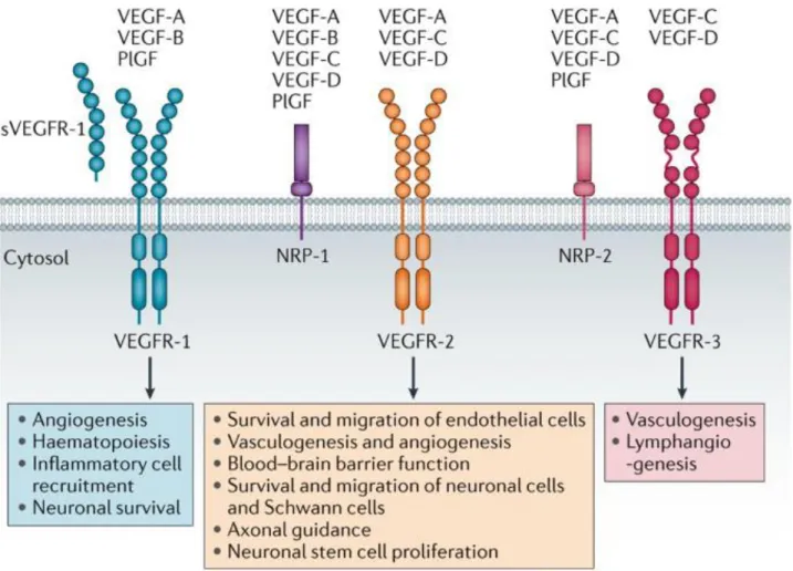
One important stimuli of angiogenesis which is required in both normal and tumor blood vessels is the vascular endothelial growth factor (VEGF). The VEGF gene is upregulated by other growth factors including platelet-derived growth factor (PDGF), fibroblast growth factor (FGF), epidermal growth factor (EGF), tumor necrosis factor (TNF), transforming growth factor- beta (TGF-β) and interleukin-1 (IL-1) (97). In addition to the above growth factors, hypoxia is also an inducer of VEGF. The VEGF protein family comprises of five members: VEGF-A, VEGF- B, VEGF-C, VEGF-D and placental growth factor (PlGF). These proteins signal through three receptor tyrosine kinase; VEGFR-1 (FLT1), VEGFR-2 (KDR/FLK1) and VEGFR-3 (FLT4) (98) (Figure 1.3) that are located on the surface of endothelial cells. VEGF-A, which preferentially binds to VEGFR2, is the main inducer of angiogenesis and activates pro-angiogenic signaling resulting in migration and survival of the endothelial cells (97,98).
17 Angiogenesis
Angiogenic sprout is promoted by the protrusion of highly motile tip cells and continues with stalk cells lagging behind as the sprout continue to elongate (99). Tip cells are highly polarized and rich in cell surface receptors and molecules that destroy the basement membrane and extracellular matrix degradation. Stalk cells however, are highly proliferative. They trail behind tip cells ensuring tube and branches, and lumen formation (100). Crosstalk between tip and stalk cells depends on VEGF and Notch signaling, where VEGF induces tip cell migration while Notch controls tip cell selection (101–103). Binding of VEGF to VEGFR-2 or VEGF-C/D to VEGFR-3 induce a signal transduction cascade which promotes expression of the Notch receptor ligand DLL4 resulting in high Notch signaling activation. This activated Notch signaling contributes to sprouting angiogenesis by differentially regulating the levels of VEGFR-1 and VEGFR-2 in stalk cells (104–106).
Once the new blood vessels are formed, they undergo a maturation phase which involves the enhancement of tight junctions and the recruitment of perivascular cells (107). Endothelial cells secrete Platelet-derived growth factor (PDGF) which signal through the PDGFR to recruit pericytes to the newly formed blood vessels enabling stability to the vessels. Vascular maturation by pericytes coverage have being associated with the Ang-Tie receptor system (107,108). The Tie/Ang signaling system is made up of Angiopoietin ligands 1-4 and two Tie tyrosine kinase receptors (Tie-1 and -2). Pericytes express Ang-1 which bind to Tie-2 on endothelial cells. Ang-2 is expressed both by endothelia and smooth muscle cells and has an antagonistic effect which can cause loss of pericytes to the ECs.
Another interesting signaling pathway in angiogenesis is the Eph receptor tyrosine kinase pathway. Eph proteins belong to a superfamily of receptor tyrosine kinases which are
18
subdivided into either subclass- A or subclass-B. They were first identified in neural
development guiding axons to their targets, and subsequently found to be involved in human cancers (109) and other biological processes. The ligands for Eph proteins, ephrins, are tethered to neighboring/adjacent cell membranes and could either signal through forward or reverse signaling (110). Amidst the plethora roles of Ephs and ephrins in vascular development, tissue-border formation, cell migration, axon guidance, their roles have also been described in tumor growth and neovascularization. For example, blockade of EphA2 using soluble
recombinant fusion proteins inhibited in vivo tumor angiogenesis and progression (111–113).
EphA receptors promote tumor growth and also have a role in the endothelium, thereby making them a potential tumor therapeutic targets (112,114).
19
Figure 1.3: Different vascular endothelial growth factor isoforms and their binding
receptors. Ligands: VEGF (also known as VEGF-A), VEGF-B, VEGF-C, VEGF-D and placental growth factor (PlGF). Receptors: VEGFR-1, VEGFR-2 and VEGFR-3 and the co-receptors neuropilin-1 (NRP-1) and NRP-2 (Reviewed in Lange et al. Nature Reviews Neurology, 2016
20 Tumor Angiogenesis
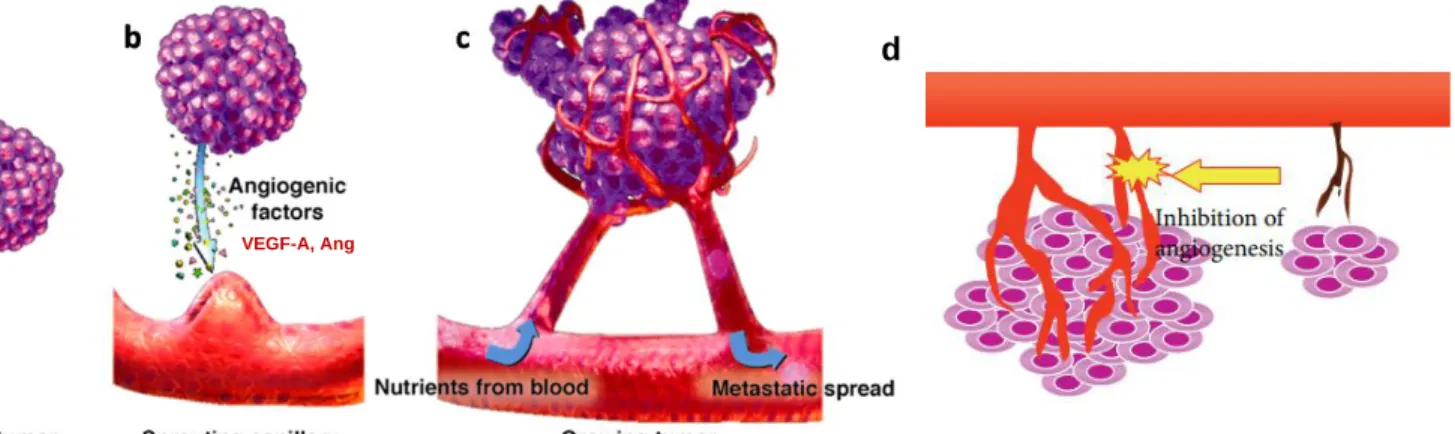
Growing tumors acquire nutrients and oxygen through diffusion. However, for solid tumors to grow beyond 2mm3 in diameter, they require a vascular system (115). Judah Folkman, also known as "The Father of Angiogenesis", in 1971 hypothesized that tumor growth was
angiogenesis dependent. He stipulated that inhibition of angiogenesis could be a therapeutic strategy for solid malignant tumors (6). This suggested that anti-angiogenesis could hold the tumor in a non-vascularized state to restrict the tumors from nutrients, oxygen, and promote shrinkage and eventually death of the tumor (Figure 1.4). Tumor blood vessels are
characterized by being morphologically abnormal and structurally dysfunctional. Compare to normal blood vessels, tumor vessels are tortuous, leaky, irregular and form chaotic networks (116). The endothelia cells that line tumor blood vessels are irregular in shape and are
disorganized. These ECs also have weak junctions promoting trans-migration. In addition, pericytes coverage is lost in tumor vasculature hence resulting in poor stability.
Figure 1.4: Formation of tumor angiogenesis from a small tumor (a), sprouting vessels (b), to growing tumor (c). Tumor blood vessels can be targeted using anti-angiogenesis (d). (Adapted from Loizzi et al. Int. J. Mol.
Sci. 2017)
d
VEGF-A, Ang
21
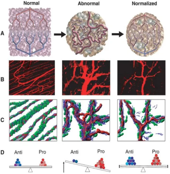
The chaotic nature of tumor blood vessels promotes a hostile tumor microenvironment due to poor oxygen delivery and the removal of waste resulting to a hostile hypoxic milieu. The hypoxic medium promotes cells that are in cable to withstand the harsh environment to escape hence promoting tumor progression and metastasis (117,118). Additionally, the lack of mural cell coverage on tumor blood vessels have been associated with metastasis in human cancers (119). Furthermore, abnormal tumor blood vessels prevent the delivery of drugs into the tumor parenchyma. Based on Folkman’s hypothesis, antiangiogenic drugs inhibit the blood vessels and at the same time renders drug delivery difficult. In addition, the hypoxic milieu has been linked to radiation and chemotherapy resistance in breast cancer (120). Understanding the concept that abnormal tumor blood vessels result when pro-angiogenic factors outweighs the anti-angiogenic factors, Rakesh Jain hypothesized that using judicious dose of anti-angiogenic drugs would “normalize” tumor vasculature (121) (Figure1.5).
Tumor Vessel Normalization
The concept of tumor vessel normalization can be defined as re-establishing the balance between pro- and anti-angiogenic factors created during angiogenic switch. This new paradigm has indeed been validated in mouse studies which have shown that blocking VEGF/VEGFR2 using either bevacizumab (a monoclonal antibody targeted against VEGF) or DC101 (a rat monoclonal antibody targeted against mouse VEGFR2) normalized the vasculature and hence decreased interstitial fluid pressure, microvessel density and improved intratumoral perfusion (122–124). Other genetic and pharmacological strategies have been shown to promote vessel normalization to improve drug delivery and immune cell infiltration (125–128).
22
Figure 1.5: Proposed concept of tumor vessel normalization in response to
antiangiogenic therapy. A and D Steps from normal to abnormal and normalized bloods vessel maintained by the perfect balance of pro-and antiangiogenic molecules. B and C showing vascular normalization induced by antiangiogenic treatment and pericytes (green) coverage from normal to normalized vessels. (Adapted from references 11 and 12)
23
Because leukocytes circulate through the hematogenous vasculature (129) abnormal tumor blood vessels often impair tumor infiltration of lymphocytes, which may contribute to tumor immune evasion. Increasing data suggest that tumor vessel normalization may enhance the efficacy of immunotherapy, not only by improving delivery of immune checkpoint inhibitors (ICIs) to tumors, but also by promoting new lymphocyte infiltration (130–136). Therefore, therapeutic strategies that improve vessel normalization may synergize with ICIs to improve treatment response and patient outcome. Indeed, anti-VEGF antibody increased lymphocyte infiltration and enhanced the effectiveness of adoptive immunotherapy in a B16 tumor model (137) (29).
Subsets of infiltrating immune cells, including Th1 cells and eosinophils have been shown to enhance blood vessel normalization (128,138) and further attraction of infiltrating lymphocytes.
Thus, immune-vascular crosstalk may mediate a feedback loop of vascular normalization and reprogramming of anti-tumor immunity.
Metabolism in Endothelial and Tumor Endothelial Cells
Glucose Metabolism
Like cancer cells, vascular ECs also reprogram their metabolism for rapid proliferation (99). The main energy provider is through the glycolysis pathway. This results in ~75-85% of ATP
production (139,140). Similar to cancer cells, ECs utilize glucose under aerobic conditions resulting to lactate production (140–142). Studies have confirmed that glucose metabolism is essential for EC functionality and upkeep as inhibiting glycolysis using 2-deoxy-D-glucose promotes ECs cytotoxicity. Again, pharmacological inhibition or genetic loss of
phosphofructokinase-2/fructose-2,6-biphosphatase 3 (PFKFB3) hinders ECs proliferation,
24
migration and sprouting (140,142,143). In addition to energy production, ECs use glucose metabolism in the pentose phosphate pathway to yield nicotinamide adenine dinucleotide phosphate (NADPH) and ribose-5-phosphate (R5P). These molecules are necessary for antioxidant defense and nucleotide biosynthesis (35).
Tumor endothelial cells (TECs) line the inner walls of tumor blood vessels. Like normal endothelial cells (NECs), TECs are hyper-glycolytic for ATP production and still maintains functional mitochondria. The hyper-glycolytic phenotype is observed due to increased expression of glucose transporter GLUT1 and the glycolytic activator PFKFB3. Inhibition of PFKFB3 in TECs promoted tumor vessel normalization and decrease tumor metastasis while promoting chemotherapy drug delivery (13,144).
Fatty Acid Metabolism
ECs utilize fatty acid metabolism to support their proliferation, differentiation and permeability.
Like other cells where the mitochondria serve as the energy powerhouse, ECs use the
mitochondria as a biosynthetic hub. Fatty acid derived carbons are source for production of the amino acid aspartate and deoxyribonucleotides use in DNA synthesis (145). Carnitine
palmitoyltransferase 1a (CPT1a) is the rate-controlling enzyme of fatty acid oxidation. This enzyme links carnitine to long chain fatty acyls so they can be imported into the mitochondria.
Once in the mitochondria, fatty acids are metabolized through beta-oxidation to yield acetyl-CoA which in turns enters the TCA cycle for energy production (146). Endothelial-specific inhibition of CPT1a has resulted to vascular sprouting defects in vivo and ablation of EC proliferation in vitro (145). Fatty acid metabolism regulates EC membrane stiffness by modulating the lipid
25
composition. Unlike glycolysis, which controls EC proliferation and migration, EC fatty acid metabolism controls only EC proliferation (139,140,145,146).
Compared to NECs, TECs also increase lipid production by upregulating fatty acid synthase (FASN), the rate-limiting enzyme in the FA synthesis pathway (147). Additionally,
pharmacological inhibition of FASN with cerulenin and orlistat impairs lymphatic ECs viability, proliferation and migration which contributed to decrease melanoma cancer spread (148).
Lymphatic ECs are not necessarily TECs, but these studies suggest that targeting fatty acid metabolism in TECs is a therapeutic potential which remains to be exploited.
Glutamine Metabolism
In addition to glucose and FA metabolism, a growing body of evidence shows that glutamine metabolism also contributes to EC proliferation, providing carbons for biomass production that is required for EC proliferation (9,14,141). Glutamine metabolism is also required for angiogenesis by contributing to the TCA cycle anaplerosis and redox homeostasis. Pharmacological inhibition or EC-specific deletion of glutaminase (GLS), blocked EC proliferation and migration (9,14). The withdrawal of glutamine from culture medium also had a similar effect. However, with the
addition of asparagine in glutamine-depleted medium, EC function and protein synthesis was restored (14,32).
Glutaminolysis is important for vessel development and homeostasis in vivo. For example, pharmacological blockade of glutaminase using CB‐839 in vivo suppressed pathological ocular angiogenesis (14). One mechanism of regulating glutamine metabolism in ECs is through the transforming growth factor-β (TGF-β) and Raf-MEK-ERK signaling pathways (149).
Glutaminolysis is enhanced within NECs upon infection with Kaposi’s sarcoma virus, rendering
26
these cells dependent on glutamine metabolism for survival, suggesting that glutamine may play an important role in tumor endothelial cells (150). However, there is a gap of knowledge on the role of glutamine metabolism in TECs. In this dissertation I sought to examine the role of glutaminnolysis in TECs on mammary tumor growth and progression.
Anti-angiogenic Drugs for Cancer Therapy
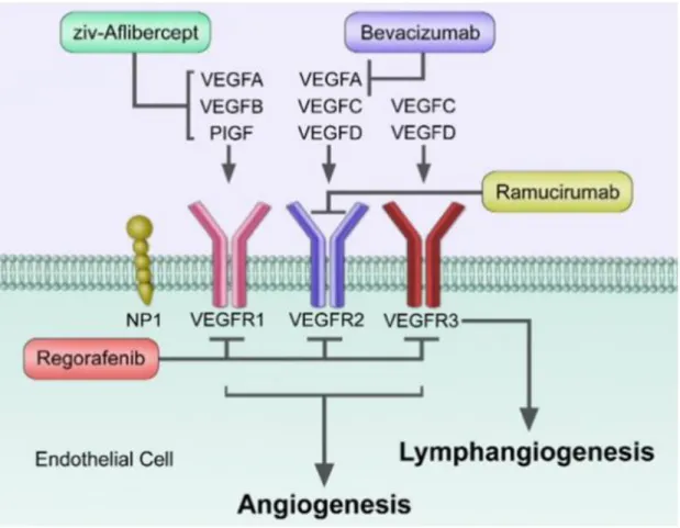
Disrupting the process required by tumors to grow is critical thereby making tumor angiogenesis an attractive target for cancer treatment. Cancer patients have benefited from Aanti-angiogenic drugs, however, because of resistance, insufficient efficacy and even toxicity, the responses from these drugs are not durable. In this section, the use of targeted angiogenic drugs in a clinical setting will be discussed (Figure 1.6) as well as how EC metabolism can be leveraged as an anti-angiogenic target.
Bevacizumab (Avastin) is a humanized anti-VEGF-A monoclonal antibody. Binding of
bevacizumab blocks VEGF-A from binding to VEGFR1 and VEGFR2. When used as a single therapy, bevacizumab failed to improve patients’ overall survival (OS) (121); however, in combination with chemotherapy, it was able to increase progression free survival and/or OS in several solids tumors including metastatic colorectal cancer, non-small cell lung cancer
(NSCLC), Glioblastoma multiforme (GBM), and breast cancer (151–153).
Ziv-aflibercept (Zaltrap) is a recombinant fusion protein where key domains of human VEGFR1 and VEGFR2 are fused to the Fc portion of human IgG1. It blocks VEGF-A, B and PIGF ligands from binding to VEGFR1 and VEGFR2 receptors. It is being used in combination with 5-
27
fluorouracil, leucovorin, and irinotecan (FOLFIRI) in metastatic colorectal cancer patients after progression with oxaliplatin-containing regimen (154–156).
Ramucirumab (IMC-1121B) is a monoclonal antibody which targets the extracellular domain of VEGFR2 blocking VEGF-A from binding to its receptor. The regimen either as a single dose or in combination with paclitaxel is use in patients with metastatic gastric, metastatic colorectal cancer, and gastroesophageal junction cancer after progression on fluoropyrimidine or platinum- containing protocols. It is also used in addition with erlotinib as a first-line metastatic EGFR- mutated NSCLC regimen (154,156–158)
In addition to monoclonal antibodies targeting either the VEGF or the VEGF receptors,
preclinical and clinical studies with small molecule inhibitors to block angiogenesis are ongoing.
Sorafenib and sunitinib are tyrosine kinase inhibitors that target primarily the VEGFR2 receptor (159) . Sorafenib is used in renal cell cancer (RCC), hepatocellular cancer (HCC) and thyroid cancer while sunitinib is used in RCC, pancreatic neuroendocrine tumors, and gastrointestinal stromal tumors (GIST). Regorafenib is a TKI that blocks VEGFR1-3, KIT (platelet-derived growth factor receptor-β) and fibroblast growth factor receptor (FGFR) and is used in colorectal cancer (CRC), HCC and GIST tumors (160). Additional small molecule inhibitors are described in Table 1.2.
28
Figure 1.6: Mechanism of action of antiangiogenic drugs tarting either the ligands (VEGF-A, B, PIGF) or the receptors VEGFR-1-3 (Reviewed in Clarke et al. Cancer Treatment Reviews 2014)
29
Drug Cancer Mechanism Company Approval date
Bevacizumab (Avastin)
mCRC,
NSCLC, RCC, GBM, Ovarian cancer,
cervical cancer
Monoclonal anti- VEGF antibody
Genentech Feb 26, 2004
Ziv-Aflibercept (Zaltrap)
mCRC Recombinant fusion protein again VEGF
Sanofi and Regeneron Pharmaceuticals
Aug 3, 2012
Ramucirumab (Cyramza)
mCRC, gastric cancer,
NSCLC, and HCC
Monoclonal anti- VEGFR2 antibody
Eli Lily and Company April 21, 2014
Sorafenib (Nexavar)
RCC, HCC and thyroid cancer
TKI against
VEGFRs, PDGFRs, RAF, KIT, FLT3 and RET
Bayer December 20,
2005 Sunitinib Malate
(Sutent)
RCC,
pancreatic and GIST
TKI against
VEGFRs, PDGFRs, CSFR1, FLT3 and RET
Pfizer Jan 26, 2006; May
20, 2011; Nov 16, 2017
Regorafenib (Stivarga)
GIST, CRC, and HCC
TKI against
VEGFRs, PDGFRs, RAF, FGFR1 KIT and RET
Bayer September 27,
2012 Table 1.2: Anti-angiogenic Drugs Use in Cancer Treatment
30 Limitations of Anti-angiogenic Therapy
Targeting VEGF/VEGFRs represented a great breakthrough in cancer patients. However, durable and complete response are rare with this treatment option. However, therapeutic resistance remains the major challenge, due in part to the heterogenous nature of tumor vasculature. Another mechanism of resistance results from several different pathways being activated upon the inhibition of VEGF/VEGFR signaling pathway trying to compensate for the blockade (161,162). Additionally, tumors are able to become vascularized using non-angiogenic methods, including vascular mimicry (formation of vascular-like structures by non-vascular cells), vessel co-option (hijacking existing vasculature of non-tumor cells in the surrounding and migrate along these vessels) and intussusception (splitting of preexisting vessels into daughter vessels)(162–164).
Vandetanib (Caprelsa)
Medullary thyroid cancer
TKI against VEGFRs, EGFR and RET
AstraZeneca April 6, 2011
Axitinib (Inlyta) Advanced RCC TKI against
VEGFRs, and c-Kit
Pfizer Jan 27, 2012
Cabozantinib (Cometriq)
Metastatic medullary thyroid cancer
TKI against VEGFRs, KIT, TRKB, FLT-3, AXL, RET
Exelixis Nov 29, 2012
31
In addition to resistance mechanism, use of anti-angiogenic therapies are limited by adverse effects. Compared to conventional chemotherapy, these side effects are very different because the drugs function differently. The most common toxicity observed are hypertension,
proteinuria, headaches, intestinal bleeding, clots in the arteries and poor wound healing (154,165–167). With toxicity and safety issues, it can be speculated that targeting the
VEGF/VEGFR signaling pathway is not the best approach for future development of targeted tumor vasculature therapies. Instead, targeting the mechanisms exploited by tumor endothelial cells may be preferential. This dissertation will explore if tumor endothelial cell glutamine metabolism may represent a therapeutic option to target angiogenesis in tumors.
Summary and Thesis Project
Breast cancer and metastatic disease continue to be a significant cause of cancer mortality in women worldwide. In spite of advances in cancer research and treatment options, resistance to targeted therapies is increasing in cancer patients. Thus, it is imperative to investigate molecular mechanisms that promote breast cancer progression and metastasis as measures to better target the disease and improve patient outcomes. Deregulation of cellular energetics and tumor angiogenesis are hallmarks of cancer in which extensive research has been performed to improve therapeutic options. Targeting tumor angiogenesis was initially considered to be a sound treatment strategy aimed to block tumor blood vessels and shrink tumors by restricting nutrient delivery. Anti-angiogenic molecules have been successful in pre-clinical studies;
however, limited or no efficacy has been observed clinically. This is, in part, due to tumors switching to alternative angiogenic routes or increased metastatic progression in patients.
32
The tumor vasculature is morphologically and functionally abnormal. These blood vessels are leaky, tortuous, irregular, heterogeneous in shape and size, and form chaotic network. Tumor blood vessels are lined by endothelial cells, but these are disorganized, irregular in shape and are associated with fewer pericytes compared to normal vessels. With the shortcomings of anti- angiogenic drugs in the clinic, exploration of other mechanisms to better target tumor
angiogenesis are necessary. One such concept that has evolved is tumor vessel normalization (TVN), which aims to remodel the chaotic tumor blood vessels to restore their structure and function. This dissertation aims to explore targeting glutamine metabolism of endothelial cells as a new way to induce tumor vessel normalization and reduce breast tumor growth and
metastasis.
In Chapter II, I describe the material and methods used in the studies presented in Chapter III and Chapter IV. Chapter III presents data showing the effect of vascular endothelial GLS deletion in primary tumor growth and metastasis in a model of breast cancer. This chapter further shows the role of endothelial GLS in tumor angiogenesis and in vascular integrity and function. In Chapter IV, I discus implications, future directions and limitations of this thesis.
Chapters III and IV are primarily data modified from a publication in Cancer Research
Communication, with some additional data not included in the manuscript (Ngwa et al. 2022).