I thank my undergraduate advisor, Louis Kuo, for pushing me to succeed when I lacked courage. Every text and phone call cheered me up and pushed me to keep going.
Caenorhabditis elegans
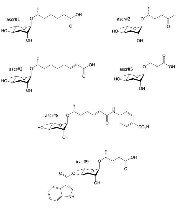
The term ascaroside was first coined to describe a new lipid discovered in the intestinal parasite Ascaris lumbricoides over 100 years ago.15 These lipophilic molecules containing long aliphatic side chains formed a layer around Ascaris eggs to protect them from harsh environmental conditions.16 V recently, a larger collection of more hydrophilic ascarosides was identified in C.
Ascaroside Biosynthesis
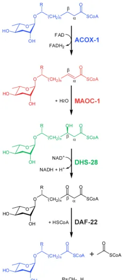
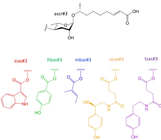
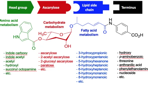
The b-hydroxyacyl-CoA intermediate is then converted into a b-ketoacyl-CoA ester by DHS-28, a homolog of the dehydrogenase domain of mammalian peroxisomal multifunctional protein MFE-2. Many of the headgroups that modify the 4' position of the ascarylose sugar moieties, including indole-3-carboxylic acid (icas), p-hydroxylenisoic acid (hbas) and (E)-2-methyl-2-butenoic acid (mbas) have been derived of amino acid metabolism (Figure 1.4).
Ascarosides in Dauer Formation
To expand the search for new dauer-inducing ascarosides, Pungaliya et al conducted NMR spectroscopy-based comparative metabolomic studies that revealed the additional dauer pheromone component called ascr#8, which contains a p-aminobenzoic acid group at the terminal end of the unsaturated fat. acid side chain.32 In addition, Butcher et al discovered. an ascaroside containing an unusual indole-3-carbonyl group attached in the fourth position of the ascarylose core called icas#9.33 Unlike other ascarosides that induce dauer formation in a manner positively correlated to pheromone concentration, icas#9 dauer formation activity decreases at higher concentrations . The synergistic abilities and different activity-concentration relationships between ascarosides may suggest that pheromone perception may involve multiple receptors.
Ascarosides in Mate Attracting Behavior
In addition, ascr#4, a glucosylated derivative of ascr#2, was found to synergize with ascr#2 and ascr#3 and increase male attraction, but does not attract only males (Figure 1.8). Additional NMR-based metabolomics studies have shown that ascr#8, another component of the dauer pheromone, also synergizes with ascr#2 and ascr#3 to enhance male attraction activity.
Ascarosides in Social Behaviors
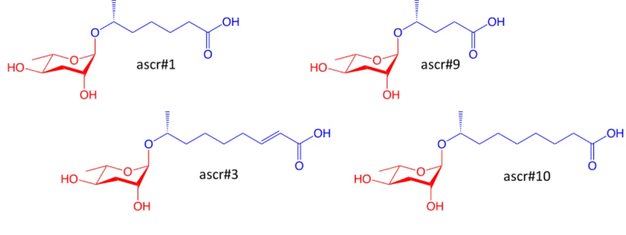
While ascr#3 demonstrates strong male attractant activity and repels hermaphrodite worms, ascr#10 is highly attractive to hermaphrodites. At concentrations as low as 100 fM, both solitary and social hermaphrodites demonstrate significant levels of attraction, thus indicating icas#3 and icas#9 as effective aggregation pheromones.40 The p-hydroxybenzoyl modified ascaroside hbas#3 was was shown to attract hermaphrodites at concentrations up to 10 fM and is therefore the most potent hermaphrodite aggregation signal reported.18.
Ascaroside Perception
Furthermore, it was found that in the presence of ascr#2 srbc-64; gpa-3 and srbc-66; gpa-3 double loss-of-function mutant worms show similar defects in dauer formation compared to their single-mutant counterparts. Together, these results support the model that ascaroside signals promote dauer formation via SRBC-64 and SRBC-66 chemoreceptors and the GPA-3 Ga protein in the ASK neuron.
Mass Spectrometry-Based Ascaroside Profiling
Thesis Summary
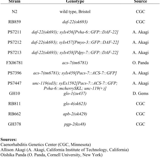
In Chapter 2, we found that by selectively directing the expression of DAF-22, an enzyme required for the production of biologically active ascarosides, within specific tissues, we were able to assess ascarosides produced within the gut, hypodermis, and wall muscle. of the worm's body. Our results revealed that while the intestine is the main site of ascaroside biosynthesis, modest amounts of ascarosides are produced in the hypodermis and body wall muscle, which are sufficient in quantity to rescue various C regulated by ascarosides.
Figures
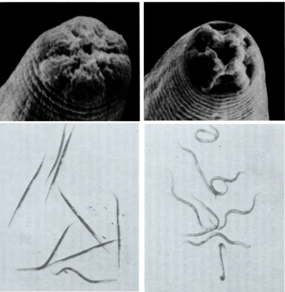
R; Kim, E.; Clardy, J., A potent dauer pheromone component in Caenorhabditis elegans that acts synergistically with other components. G.; Brenner, S., Electron microscopic reconstruction of the anterior sensory anatomy of the nematode Caenorhabditis elegans.?2UU.
Abstract
Introduction
Although much of the biosynthetic pathway of ascarosides remains to be elucidated, previous studies have shown that DAF-22, a thiolase responsible for the final step of peroxisomal b-oxidation of the lipid moiety attached to the 1'-position of the ascarylose sugar is attached. nucleus, is required for the production of biologically active short-chain ascarosides (Figure 2.1).4-5 Transgenic expression of an N-terminally tagged GFP DAF-22 fusion protein (GFP::DAF-22) under control of the native daf -22 gene promoter revealed that the protein is expressed in the gut, the hypodermis and the body wall muscle of the worm.4,. 6 Furthermore, when expressed exclusively in the intestine under the control of the vha-6 promoter, GFP::DAF-22 rescue was able to restore some dauer-inducing activity. It has therefore been hypothesized that the intestine is the main site of ascaroside biosynthesis; however, the possible roles of DAF-22, which is expressed in the hypodermis and body wall muscle, in the biosynthesis of ascarosides remained uninvestigated.4 An additional hypothesis can be made that ascarosides production can occur in different places within the worm and that the ascarosides that are produced may vary from tissue to tissue.
To investigate this issue, we created three types of worms that drive the expression of GFP::DAF-22 specifically in the intestines, hypodermis, or body wall muscle. Pheromone signals collected in each strain were then tested for activity in dauer formation, male attraction, and hermaphrodite repulsion.
Results and Discussion
As expected, exogenous pheromone collected from daf-22 ( ok693 ) loss-of-function mutant worms induced no dauer formation in N2 worms, whereas wild-type dauer pheromone induced dauer formation in SD) larvae. The pheromone generated by intestinal expression of GFP::DAF-22 restored approximately half of the dauer-forming activity compared to the N2 control (35 ± 4%). GFP::DAF-22 rescue within body wall muscle and hypodermis produced dauer pheromone that induced much lower levels of dauer formation at 6 ± 2% and 4 ± 2%, respectively.
Although very modest, GFP::DAF-22 expression in both the body wall muscle and hypodermis does restore dauer formation compared to the daf-22(ok693) mutant. Furthermore, ascr#5 was found in the supernatant of intestinal and hypodermal GFP::DAF-22 rescue cultures, as well as icas#9.
Conclusions
Moreover, the amounts of ascr#10 derivatives produced by hypodermal rescue of GFP::DAF-22 exceed the levels shown by intestinal rescue. Fluorescence microscopy can then be used to test for colocalization of GFP::DAF-22 with potential intestinal granule-like structures within the hypodermis. DIC and GFP fluorescence microscopy images of larval L4 expressing a GFP::DAF-22 transgene at 20X and 100X magnification.
The upper focal plane is in focus, showing the alae (red arrow). I-L) Pmyo-3::gfp::daf-22 is expressed within body wall muscle (white arrows). HPLC-MS analysis of liquid culture supernatant reveals ascaroside profiles of worms expressing GFP::DAF-22 in gut (Pvha-6::gfp::daf-22), hypodermis (Pdpy-7::gfp::daf - 22), and body wall muscle (Pmyo-3::gfp::daf-22).

Abstract
Introduction
Using HPLC-MS analyzes of the worm excreta, we found that ACS-7, a putative acyl-CoA synthetase, plays a role in the attachment of different headgroups to the 4'-position of the ascarylose sugar scaffold of the simple ascaroside ascr #9. Up to this point, the only enzymes known to play a role in the production of ascarosides are those involved in the peroxisomal b-oxidation of the fatty acid side chain.13-. This suggests that oac-50 may play a role in the biosynthesis of mbas#3, but that the change in mbas#3 production observed in the oac-38(gk648702) is most likely due to a change in tigyl-CoA metabolism.
This result suggests that acs-7 may be important in modifying the simple ascaroside ascr#9 at the 4' position. To confirm the role of ACS-7 in the biosynthesis of the modular ascarosides icas#9 and osas#9, as indicated by our initial screen, we determined the expression of a C-terminal green fluorescent protein (GFP)-tagged ACS-7 protein fusion (ACS-7::GFP) under the native acs-7 promoter (Pacs-7:acs-7::gfp) within an acs-7(tm6781) loss-of-function mutant background.
Conclusions
The million mutation strain VC20784 contained 219 mutations, including the oac-50(gk402144) allele, which contained a nonsense mutation in the first exon of the gene. HPLC-MS ion chromatograms (ESI-) for icas#9 and osas#9 show rescue of icas#9 and osas#9 production in Pacs-7::acs-7::gfp transgenic worms (green) compared to acs-7 (tm6781) loss-of-function mutant (red). As determined by HPLC-MS analysis of liquid worm culture supernatant, production of icas#9, tsas#9, and osas#9 is restored in worms expressing the ACS-7::GFP fusion protein.
DIC image of the midsection of a young adult worm expressing the ACS-7::GFP fusion protein under the acs-7 promoter, the peroxisomal marker mCherry, and the unc-119(+) rescue construct within the unc-119(- ) mutant background. DIC image of the midsection of a young adult worm expressing ACS-7::GFP under the native acs-7 promoter.
Materials and Methods
Mak.15 The mCherrySKL sequence was placed downstream of the vha-6 promoter and upstream of the 3'UTR region of the unc-54 gene. Isolated injected animals were grown at room temperature and F1 progeny of the roller phenotype were individually selected and plated. Ascarosides were detected as [M-H]- ions in negative ionization mode (spray voltage 3.5 kV, cone voltage -40 V) and confirmed based on comparison of retention times with those of synthetic standards.
C., 2D NMR-based metabolomics uncovers interactions between conserved biochemical pathways in the model organism Caenorhabditis elegans. Maduro, M.; Pilgrim, D., Identification and cloning of unc-119, a gene expressed in the Caenorhabditis elegans nervous system.
Introduction
Results and Discussion
Figures
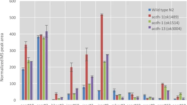
The acdh genes are mitochondrial paralogs of acox-1, the gene that encodes the protein responsible for the first peroxisomal oxidation site b by introducing an a,b-unsaturation site. Loss of function in the acdh-1 and acdh-13 genes results in higher levels of the ascarosides hbas and icas, with the acdh-1 (ok1489) allele being more penetrant than the acdh-1 (ok1514) allele.
Introduction
Results and Discussion
HPLC-MS analysis of mutant secretions did not show a decrease in ascaroside ica biosynthesis (Figure B.3). HPLC-MS analysis of liquid culture supernatant showed that none of the three mutants examined caused a significant decrease in icas production, indicating that none of the tested enzymes is involved in the biosynthesis of the indole-3-carboxylic acid precursor (Fig. B .4) . The elo genes encode elongase proteins that catalyze the elongation of the carbon chain of fatty acids.
We found that elo-1(gk48) and elo-5(gk183) mutants produced greater amounts of the longer chain ascaroside ascr#18 and oscr#18 but did not significantly reduce the production of any specific ascaroside (Figure B .5). In some bacteria and plants, decarboxylase has been shown to play a role in the biosynthesis of indole-3-carboxylic acid.

Ho Yi Mak (H.Y. Mak, Hong Kong University of Science and Technology, Hong Kong) Henry Le (H. Le, Cornell University, New York). Cornelia Bargmann Lab (Bargmann Lab, The Rockefeller University, New York) Paul Sternberg Lab (Sternberg Lab, California Institute of Technology, Californië) David Pilgrim (D. Pilgrim, Universiteit van Alberta, Edmonton, Canada). Andy Fire Lab (Fire Lab, Stanford University, Californië) Joshua Arribere (J. Arribere, Stanford University, Californië) Addgene (Massachusetts).