1!>04) reports Do/'t/icht/tt/s hoaja andJJuinatiUs in the rivers of the Mahij^an Peninsuhi. Rondelet studied the fish alive in the water and his observations are very accurate, except for the one error regarding the sex of the pouch-bearing fish. They ])reed in the summer, the females throwing their spawn in the false ))ellie of the male.
This caused the lips of the bag to open and the young came out and swam in the water. Therefore, he concludes that the maturation of the eggs and the formation of the pouch keep pace with each other. He obtained these results: (1) by "stripping" the tissues and noting the white fluid containing spermatocytes; (2) by dissecting the ovaries and testes and noting the golden-red eggs shining through the walls of the ovary; (3) by making microscopic examinations of the products in 1 and 2.
They are the first to describe a slotted opening at the anterior end of the niarsupiunium. The following observations on the breeding habits of Sipliostoma Jloridx were made at the laboratory of the United States Bureau of Fisheries at Beaufort, North Carolina, on July 17, 1903. Their bodies touch in three places — in the anterior region, just behind the sternum; in pos.
463. teriorrog-ion, at a point about two-thirds of the way from the anus to the caudal; and on the anal openings.
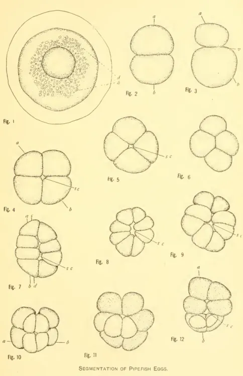
THE SEGMENTATION OF THE EGG OF THE PIPEFISH—
- THE OVARIAN EGG
- THE METHOD OF DEPOSITION
- FERTILIZATION
- MATURATION
- FOLIATION OF THE GERM DIRK
This has already been described in the first part of this article, but . it may be good to emphasize the fact that the process is such that it prevents absolutely any contact of the eggs and sperms with the sea water. In the description of the copulation and accompanying phenomena, attention was called to similar sexual excitement in an Amphibia, a (lanoid, and a cephalopod, which is preparatory to the secretion of sperms as well as of. In this connection Brook (1887 ) that the eggs of the herring vary in size in the same fish or in fish from different places, but consider that this in no way affects their development.
Reighard (1893) found that sea pike sperm die after one minute in water. In the case of tubes, fertilization is not necessary for the formation of the germinal plate. Brook (1887) confirms this for herring, but I found them to be the roe of the sargassum fish, Pterophrijne hlxtrlo.
In pelagic eggs, generally, the oil is in a large globule near the center of the yolk, or in pipefish. 31 on the same plate shows a blastodisc taken from many eggs in the stage of invagination (forty to fortj^-eight hours).
YI. SEGMENTATION
THE PERIBLAST
The origin of this layer, together with the peculiarities of its structure, is mentioned in the descriptions of the plates. The modes of its formation in other Teleosts, and to show under what modes of its formation in other Teleosts, and to show under which of these locks the pipefish egg falls, and finally to give references to a. In eggs, with the first furrow cutting through to the yolk, the periblast is formed by a thin protoplasmic sheet extending inward.
In eggs in which there are no oil droplets beneath the germinal disk, or in which the protoplasmic mass separates sharply from the yolk, the periblast is formed when the inner ends of the cells at the four- and eight-cell stages are excised and lifted from below. - lying thin protoplasmic sheet. In eggs in which there is an imperfect separation between the germinal disc and the yolk, or in which there is a layer of oil droplets beneath the blastodisc, the central periblast has a very peculiar mode of origin. Cells are cut from the protoplasmic disk in successive layers from top to bottom and the central periblast is the remnant of blastodisk on the left.
The explanation for this is that the protoplasm continues to flow from the germinal disk to the germinal disk until the segmentation has progressed some distance. The central periblast nuclei, in types 1 and 2, arise from division of the Edge nuclei and migrate centrally into this layer. In Sijdio.sfo/ita forldpe the two methods of central periblast formation described in Type 2 and 3 above are found.
It is well to note here that the migration of the nuclei to the peripheral region and the formation of a "wreath" with the disappearance of the cell walls due to the turbidity of Qg^^ was not observed in the tubes. The difficult (question whether in the pipehsh egg cells bud from the central periblast and add to the blastomeres cannot be discussed here. For a fuller discussion of the origin of the periblast and its nuclei and the fate of the latter, the reader is referred to Brooke (1887).
It has been the writer's intention to carry it forward, at least to the closure of the blast hole, and so the sections have been cut across, but the difficulties have caused so many. The Q,gg of the pipefish is very different from most other teleost eggs in terms of its segmentation and the dual origin of its peri-blast, together with the "post-segmentation" of cells therefrom. These differences are so marked that it seems correct to say that the figures in this paper are representative of the sections of a thousand or more eggs obtained from third-three fish during three summers.
BIBLIOGRAPHY
EXPLANATION OF PLATES
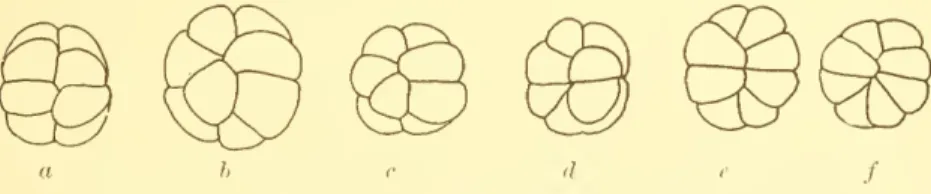
Bottom view of a high-curved 16-cell scene showing 9 cells in the first layer, 6 in the second, and 1 keystone along with the large segmentation cavity. Below the dotted line yolk and protoplasm mixed.. 29. Section through germ disc, 11 hours and 20 minutes in water, showing formation of buds. Eight-celled stage, section in right angle and longitudinal axis of such stages as fig. .. cell stage, section through a-h in fig. 12 showing a protoplasmic bridge.
Sixteen-celled stage, section through blastoderm as fig.. arc 16-celled stage, section through l)lastoderm as fig. 22, with large segmentation cavity. Same stage, third type, showing cells forming in the .. curved type of this stage, with solid-stained nuclei-periblast completely free of cells. Many nudei and vacuoles are found in the periblast, of which a number of cells are 76. SamTstage; other cloth.
The periblast is immersed in ^^e yolk,^^ th« . the blastonu^res only sparingly fills such an enlarged segmentation cavity.