The Ostracode Family Cypridinidae and the
Genus Pterocypridina
LOUIS S. KORNICKER
SMITHSONIAN CONTRIBUTIONS TO ZOOLOGY • NUMBER 379
Emphasis upon publication as a means of "diffusing knowledge" was expressed by the first Secretary of the Smithsonian. In his formal plan for the Institution, Joseph Henry outlined a program that included the following statement: "It is proposed to publish a series of reports, giving an account of the new discoveries in science, and of the changes made from year to year in all branches of knowledge." This theme of basic research has been adhered to through the years by thousands of titles issued in series publications under the Smithsonian imprint, commencing with Smithsonian Contributions to Knowledge in 1848 and continuing with the following active series:
Smithsonian Contributions to Anthropology Smithsonian Contributions to Astrophysics
Smithsonian Contributions to Botany Smithsonian Contributions to the Earth Sciences Smithsonian Contributions to the Marine Sciences
Smithsonian Contributions to Paleobiology Smithsonian Contributions to Zoo/ogy Smithsonian Studies in Air and Space Smithsonian Studies in History and Technology
In these series, the Institution publishes small papers and full-scale monographs that report the research and collections of its various museums and bureaux or of professional colleagues in the world of science and scholarship. The publications are distributed by mailing lists to libraries, universities, and similar institutions throughout the world.
Papers or monographs submitted for series publication are received by the Smithsonian Institution Press, subject to its own review for format and style, only through departments of the various Smithsonian museums or bureaux, where the manuscripts are given substantive review. Press requirements for manuscript and art preparation are outlined on the inside back cover.
S. Dillon Ripley Secretary
Smithsonian Institution
The Ostracode Family Cypridinidae and the Genus Pterocypridina
Louis S. Kornicker
SMITHSONIAN INSTITUTION PRESS City of Washington
1983
Kornicker, Louis S. The Ostracode Family Cypridinidae and the Genus
Pterocypridina. Smithsonian Contributions to Zoology, n u m b e r 379, 29 pages, 9 figures, 4 plates, 2 tables, 1983.—A classification of the Cypridinidae is proposed based on the morphology of the suckers on the b- and c-bristles of the 1st antenna of the adult male. Two new species of Pterocypridina are described and illustrated, one from off southeastern Australia and the other from off southeastern North America. The latter species is the first record of the genus in the Atlantic. In addition to a key to species of Pterocypridina, keys are presented to subfamilies, tribes, groups, and genera of the Cypridinidae.
OFFICIAL PUBLICATION DATE is handstamped in a limited number of initial copies and is recorded in the Institution's annual report, Smithsonian Year. SERIES COVER DESIGN: The coral Montastrea cavernosa (Linnaeus).
Library of Congress Cataloging in Publication Data Kornicker, Louis S., 1919-
The Ostracode family Cypridinidae and the genus Pterocypridina.
(Smithsonian contributions to zoology ; no. 379) Bibliography: p.
1. Cypridinidae—Classification. 2. Pterocypridina—Classification. 3. Crustacea—Classifi- cation. I. Title. II. Series.
QL1.S54 no. 379 JQL444.085] 591s [595.3'3] 82-19266
Contents
Page
Introduction 1 Acknowledgments 1 CYPRIDINIDAE Baird, 1850 2 Pterocypridina Poulsen, 1962 5 Key to Species of Pterocypridina 6 Pterocypridina dedeckkeri, new species 6 Pterocypridina sex, new species 11 Appendix: Keys to Subfamilies, Tribes, Groups, and Genera of the
Cypridinidae 18 Literature Cited 21 Plates 22
The Ostracode Family Gypridinidae and the Genus Pterocypridina
Louis S. Kornicker
Introduction
The genus Pterocypridina in the family Cypridi- nidae was proposed by Poulsen (1962:234) for three new species: P. alata Poulsen, 1962:236 based on 21 males, females, and juveniles col- lected at depths of 10-20 m off Thailand, P.
excreta Poulsen, 1962:240 based on a single female collected in a trawl at 60-90 m off SE Australia, and P. birostrata Poulsen, 1962:243 based on a single female collected off Singapore in shallow water.
A single specimen, an ovigerous female, which I have proposed as a new species, P. dedeckkeri, herein, collected off Long Reef, Sydney, New South Wales, Australia, and sent to me by Patrick De Deckker, provided the opportunity to study the shell of a member of the genus with a Scan- ning Electron Microscope. Later, another new species of the genus (P. sex) was identified from collections off southeastern North America (from North Carolina to Florida) at depths of 5.5-40 m.
The collection contained two males in addition to adult females and juveniles. Previously, the male was known from only P. alata. The suckers on the b- and c-bristles of the 8th joint of the 1st antenna of the adult males of the two species differ considerably from those of other genera of the Cypridinidae. Examination of the suckers of
Louis S. Kornicker, Department of Invertebrate Zoology, National Museum of Natural History, Smithsonian Institution, Washington, D.C. 20560.
P. sex stimulated my reviewing the morphology of the suckers on other members of the Cypridi- nidae, and to propose a classification of the Cy- pridinidae in which five informal groups are in- cluded in the Tribe Cypridinini.
ACKNOWLEDGMENTS.—I wish to thank the fol-
lowing individuals and Institutions for the speci- mens upon which this study was based: Donald Weston, Virginia Institute of Marine Science; Dr.
Willis E. Pequegnat, TerEco Corporation, Col- lege Station, Texas; John Miller, Harbor Branch Foundation, Fort Pierce, Florida; and Patrick De Deckker for a specimen collected by personnel of the Australian Museum Benthic Survey. My thanks also to Mrs. Carolyn Bartlett Gast for rendering shaded drawings of carapaces; Kathryn Schroeder Brown for assisting in preparation and inking of appendage drawings; Walter R. Brown and Mary J. Mann, Smithsonian Institution, who operated the Scanning Electron Microscope; Dr.
Roland Hower, Smithsonian Institution, who as- sisted in freeze-drying of specimens in preparation for the SEM; and Jack Korytowski for final ed- iting and preparation of the manuscript for pub- lication. My appreciation also to Dr. K.G.
McKenzie, Dr. R. B. Manning and Mrs. Anne C. Cohen for reviewing all, or part of the manu- script.
DISPOSITION OF SPECIMENS.—Specimens have been deposited at the National Museum of Nat- ural History, Smithsonian Institution. These have been given USNM numbers in the text.
CYPRIDINIDAE Baird, 1850
Skogsberg (1920:168) considered the family Cypridinidae to consist of two subfamilies: the Cypridininae and Philomedinae. That concept has been followed by Poulsen (1962:14), Hart- mann (1965:552), Hartmann and Puri (1974:11), and Hartmann (1975:672, 673). Previously, Miiller (1912:7) had considered the Cypridininae and Philomedinae to have equal status with the subfamilies Sarsiellinae and Asteropinae (= Cy- lindroleberidinae), all in the family Cypridinidae.
Skogsberg (1920:168) raised the Sarsiellinae and Asteropinae to family rank, but retained the Cy- pridininae and Philomedinae as subfamilies of Cypridinidae. Kornicker (1968:448, 1975:83) considered the Cypridininae and Philomedinae to have family status equivalent to the families Cylindroleberididae, Sarsiellidae, and Rutider- matidae, all members of the Cypridinacea. I con- tinue to adhere to that classification. Therefore, the category Cypridininae of Skogsberg, Poulsen, Hartmann, and Puri, is equivalent to my concept of Cypridinidae. I have indicated this in the text by following Cypridininae by (= Cypridinidae) where warranted.
After consideration of many of the morpholog- ical characters to be found in the Cypridininae (= Cypridinidae), Skogsberg (1920:194) consid- ered the equipment of the b- and c-bristles of the 1st antenna of the adult male, and also the mor- phology of the endopodite of the 2nd antenna of the adult male to be "the most noteworthy" in classifying the Cypridininae.
The b- and c-bristles of the adult male Cypri- dinidae have discs that have been considered by many to be suctorial organs used by the adult male for grasping the female during copulation (Figure 1). Discs similar in morphology to some on the Cypridinidae were described and illus- trated on the cladocopid ostracode Metapolycope hartmanni Kornicker and van Morkhoven,
1976:16, fig. 14. The discs on that species form a row on the two ventral bristles of the 4th joint of the male 2nd antenna. The two bristles were termed sensory bristles by Kornicker and van Morkhoven (1976: fig. 14). Several halocyprid
species have a single "sucker" on the e-bristle of the male 1st antenna (see Poulsen, 1973:11, item 4 in key; Martens, 1979:341). I consider the pres- ence of suckers on cladocopes and halocyprids in comparison with those on the Cypridinidae to be the result of convergence. In this paper, following Skogsberg (1920:195), the discs on the Cypridi- nidae are termed suckers, although it is quite possible that some may have a sensory function.
Skogsberg (1920:195) thought that the cypri- dinid genus Crossophorus (= Azygocypridina) was without suckers. There is no evidence that Skogs- berg had examined males of the genus; however, Miiller (1906:29, fig. 4: 8) observed broad tips on filaments of bristles of the male 1st antenna of Crossophorus gibber Miiller, 1906, and speculated (p. 29) that they might be olfactory organs
"(Riechborsten?)." Similar discs were observed on adult males of Azygocypridina rudjakovi Kor- nicker, 1970:10 by Kornicker (1970; fig. 5h), Iso- cypridina quatuorsetae Kornicker, 1975:206, by Kor- nicker (1975, fig. 121d-f) and Azygocypridina im- periahs (Stebbing, 1901:100) by Athersuch (1980,
FIGURE 1.—Tips of 1st antenna of adult males of the Cypri- dinidae illustrating differences in types of "suckers" on b- and c-bristles of 7th joint: a, Azygocypridininae: Isocypridina quatuorsetae (8th joint not shown), from Kornicker, 1975; fig.
118a. b-n, Cypridininae: b, Gigantocypridinini: Gigantocypris muelleri base of b-bristle, from Skogsberg, 1920, fig. xxvi: 6.
c-n, Cypridinini: c-e, Cypridina Group: c, Paradoloria australis, tip of 1st antenna, from Poulsen, 1962, fig. 78d; d, Cypridinodes acuminata, b-bristle, from Skogsberg, 1920, fig. LXIII: 13; e, detail of proximal sucker at base of b-bristle, from Skogsberg,
1920, fig. LXIII: 14. f-h, Monopia Group:/, Monopia flaveola, 1st antenna, from Poulsen, 1962, fig. 126e; g,h, side and medial views of 2nd filament of b-bristle, from Poulsen, 1962, fig.
126f. i-l, Codonocera Group: Codonocera polygonia, i, tip of antenna, from Poulsen, 1962, fig. 143b; j , proximal part of c-bristle, from Poulsen, 1962, fig. 143c. Codonocera couenta, k, proximal part of c-bristle, from Poulsen, 1962, fig. 145d'; /, detail of suckers forming cluster on c-bristle, from Poulsen, 1962, fig. 145d". m,n, Pterocypridina Group: Pterocypridina alata, m, 1st antenna (not all filaments off- and g-bristles shown), from Poulsen, 1962, fig. H i d ; n, detail of suckers of b- and c-bristles, from Poulsen, 1962, fig. l l l d ' , d " . (The small letters b and c on illustrations refer to the b- and c-bristles commonly used to designate specifically located bristles on the 7th joint of members of the Myodocopina; (see Skogs- berg, 1920:188).
AZYGOCYPRIDININAE
CYPRIDININAE
Gigantocypridinini
Codonocera Group
Cypridinini
Cypridina Group Monopia Group
Ptemcypridina Group
TABLE 1.—Classification of the Cypridinidae and diagnoses of taxa based on distribution of suckers on b- and c-bristles of adult males
Taxon Diagnosis
Family CYPRIDINIDAE Baird, 1850
Subfamily AZYGOCYPRIDININAE Kornicker, 1970
Subfamily CYPRIDININAE Baird, 1850
Tribe GIGANTOCYPRIDININI Hartmann, 1974 (in Hartmann and Puri, 1974) Tribe CYPRIDININI Baird, 1850
Cypridina Group
Monopia Group
Codonocera Group
Plerocypridina Group
Group ?
Suckers present on b- and c-bristles of adult male (Figure 1).
Both bristles with numerous (about 20) short, marginal, me- dial filaments bearing near tip a single oval sucker facing medially (Figure la).
Both bristles bearing at least 1 short, stout, proximal fila- ment with large sucker or cluster of suckers (Figure \b-n).
b-bristle with 3 or 4 short, stout, proximal filaments, each with large sucker near middle facing medially (Figure Ib); c- bristle with 1, or at most 2, similar filaments.
b- and c-bristles with not more than 1 short, stout, proximal filament with large sucker near middle facing medially, or with 1 or more large terminal suckers; long additional fila- ments bearing small marginal suckers may or may not be present (Figures \c-n).
The short, stout, proximal filament on b- and c-bristles bear- ing large marginal sucker, followed by generally 2 (rarely none or 1) long slender filaments with single row of 2 to 13 small, round, marginal suckers (Figure \c-e).
The short, stout, proximal filament on b- and c-bristles fol- lowed by 2 long slender filaments, each with double row of 20-40 small marginal suckers, each consisting of narrow shaft and a broader, flattened tip (Figure \f-h).
b- and c-bristles each with 1 (rarely 2) stout proximal fila- ments, each with cluster of 6-10 terminal branches, each branch bearing single terminal sucker (Figure 1 i-l).
b- and c-bristles each with stout proximal filament with single terminal sucker (Figure \m,n).
This group is necessary because adult males are unknown for 4 recognized genera. (See Table 2 for names of genera.) fig. 6f). I interpret the discs to be equivalent to
suckers on other cypridinids. The fact that they occur on the b- and c-bristles makes this interpre- tation probable.
The male Monopia was not known until 1962 when it was described (Poulsen, 1962:273, fig.
126e,f). The distribution of suckers on the b- and c-bristles differed from those previously known.
Discovery of the new genus, Pterocypridina Poulsen, 1962:234, also revealed a previously unknown type of sucker on the b- and c-bristles (in 1962 the male of only P. alata Poulsen, 1962:236, was known, but the male of a new species, P. sex, is described herein). The adult males of four
genera remain unknown: Paracypridina Poulsen, 1962:245; Amphisiphonostra Poulsen, 1962:249;
Hadacypridina Poulsen, 1962:230; a n d Rugosidoloria Kornicker, 1975:197.
Whether or not the endopodite of the male 2nd antenna is developed as a clasping organ was apparently considered by Skogsberg (1920:195) to be of secondary importance in the classification of the Cypridininae (= Cypridinidae). I concur with that view.
A classification based on morphology and dis- tribution of the suctorial organs on adult males of the Cypridinidae is listed along with diagnoses of the taxa in Table 1 and illustrated on Figure
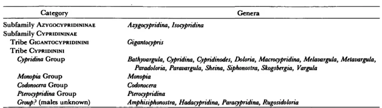
TABLE 2.—Genera referred to higher categories
Category Genera
Subfamily AZYGOCYPRIDININAE Subfamily CYPRIDININAE
Tribe GIGANTOCYPRIDININI Tribe CYPRIDININI
Cypridina Group Monopia Group Codonocera Group Pterocypridina Group Group? (males unknown)
Azygocypridina, Isocypridina Gigantocypris
Bathyvargula, Cypridina, Cypridinodes, Doloria, Macrocypridina, Melavargula, Metavargula, Paradoloria, Paravargula, Sheina, Siphonostra, Skogsbergia, Vargula
Monopia Codonocera Pterocypridina
Amphisiphonostra, Hadacypridina, Paracypridina, Rugosidoloria
1. Informal group names are given for divisions within the Cypridinini; no new formal names are proposed.
This classification is useful in understanding relationships among members of the Cypridini- dae. Unfortunately, the groups into which the Cypridinini have been subdivided are of very limited value in practice for two reasons: (1) adult males are unknown for four genera {Paracypridina, Amphisiphonostra, Hadacypridina, a n d Rugosidoloria) and (2) adult males are unknown for many of the remaining species of Cypridinini. A reason for this is that adult males are relatively sparse in collections. For example, of the five described species of Pterocypridina, adult males are known for only two; there is no assurance that when the adult males of the remaining three species are identified, those species will not have to be re- ferred to a different genus. Similar taxonomic uncertainty also applies to species currently re- ferred to many other genera of Cypridinini. The genera referred to the taxa in the present classi- fication are listed in Table 2; referrals are tenta- tive for those genera in which the male is un- known.
Females of species are generally referred to higher taxa based on the morphology of the carapace, upper lip, endopodite of the 2nd an- tenna, furca, and for some genera, the mandible, maxilla, 5th, 6th, and 7th limbs as well as eyes and organ of bellonci are useful. There is rela- tively little difficulty in separating females of the subfamilies Azygocypridininae and Cypridini-
nae, because of the flap-like, hirsute appendage in place of the lateral eye found only on members of the Azygocypridininae. Within the Cypridini- nae, the tribes Cypridinini and Gigantocypridi- nini are also relatively easy to identify because of the globose thin shell and large medial eye of the latter. Within the Cypridinini, the small number of claws (4) on the furca of members of the Codonocera Group permits its separation from other groups, but identification of members of many remaining groups cannot always be made with a high degree of certainty if adult males are lacking.
To assist identification an Appendix has been included presenting keys to subfamilies, tribes, groups, and genera of the Cypridinidae. My ini- tial drafts of the keys were tested by Anne C.
Cohen, mainly by working with descriptions in the literature. This resulted in many revisions.
Therefore, I consider the keys, especially the "Key to Genera of the Cypridina Group and Group ?"
to be a joint effort with Mrs. Cohen.
Pterocypridina Poulsen, 1962
TYPE-SPECIES.—Pterocypridina excreta Poulsen, 1962 (subsequent designation, Kornicker, 1975:
142).
COMPOSITION.—This genus contains 5 species:
P. alata Poulsen 1962:236, from off Thailand at depths of 10-20 m; P. birostrata Poulsen, 1962:243, from shallow water off Singapore; P. excreta Poul- sen, 1962:240, from off SE Australia, at trawl depth of 60-90 m; P. dedeckkeri, new species, from
off SE Australia at a depth of 43 m; and P. sex, new species, from off the SE coast of North America (North Carolina to Florida) at depths of 5.5-40 m.
A species described by Brady (1897:89, pi. XV:
figs. 20, 21) from a single specimen collected in Flinders Passage, Australia, at a depth of 12.8 m,
and named by him Cypridina (?) armata, bears 2 lateral processes on each valve, suggesting that it might belong in the genus Pterocypridina. Until the appendages are known the species should be re- tained in the category to which it was referred by Miiller (1912:50), "Cypridinidarum genera dubia et species dubiae."
Key to Species of Pterocypridina
1. Lateral process on the posterodorsal part of each valve 2 Without lateral posterodorsal processes 3 2. Furcal claw 2 fused to lamella 4 Furcal claws 2 and 4 fused to lamella P. alata Poulsen 3. Furcal claw 2 fused to lamella P. birostrata Poulsen Furcal claws 2 and 4 fused to lamella P. sex, new species 4. Posterodorsal process on each valve located near middle of posterodorsal
margin; 6th limb with 11-12 epipodial bristles
P. dedeckkeri, new species Posterodorsal process on each valve close to tip of caudal process; 6th limb with 7-8 epipodial bristles P. excreta Poulsen
Pterocypridina dedeckkeri, new species
FIGURES 2-4; PLATES 1-4
ETYMOLOGY.—The species is named for Patrick De Deckker, from whom I received the holotype.
HOLOTYPE.—Ovigerous female on slides and in alcohol; unique specimen, USNM 158240.
TYPE-LOCALITY.—Off Long Reef, Sydney, New South Wales, Australia, depth 43 m. Col- lected 27 Apr 1972 at a diving station of the Australian Museum Shelf Benthic Survey.
DISTRIBUTION.—Collected only at type-locality.
DESCRIPTION OF ADULT FEMALE (Figures 2-4;
Plates 1-4).—Carapace oval in lateral view with well-developed rostrum and incisur; caudal proc- ess longer on right valve than on left; each valve with small triangular process near middle of pos- terodorsal margin, and with very low bulge on anterodorsal quarter of valve at about same level as triangular process (Figure 2; Plates la,b, 2a);
left valve only with small bulge at posterodorsal corner (Figure 2); lateral process present on ros- trum and near anteroventral margin of each valve (Figure 2; Plate la,b).
Ornamentation (Figures 2, 3d; Plates la,b, 2a,b, 3a,b): Valve surface with ridges forming reticu- lations; upper edge of ridges bearing 1 or 2 rows of minute tubercles. (These tubercles are more clearly visible using a compound microscope and transmitted light than on the SEM micrographs;
see Figure 3d.) Surface with sparsely distributed bristles emerging from open pore encircled by rim bearing a single process (Plate 3a,b).
Infold (Figure 3a,b; Plates lc,d, 3c,d, 4a,b):
Rostral infold with 30-40 bristles (Figure 3a;
Plate \c,d); 2 bristles present at edge of valve at inner end of incisur; anteroventral and anterior half of ventral margin with about 39 divided bristles, each consisting of a short bare part and a long branching part (Figure 3a), these bristles forming row close to valve edge; anteroventral infold also with about 15 short branching bristles proximal to outer row of divided bristles; poste- rior half of ventral infold, extending from middle of ventral margin to point where list curves away from valve edge anterior to caudal process with about 15 short bristles; edge of list anterior to caudal process with minute protuberances, each
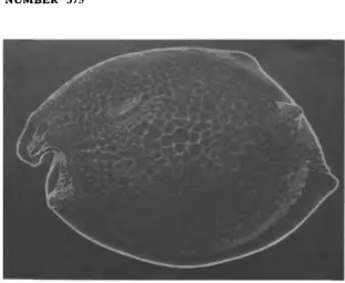
FIGURE 2.—Pterocypridina dedeckkeri, new species, adult female, holotype, USNM 158240, lateral view of complete specimen, length 1.57 mm.
bearing a pore with small bristle (Figure 3b;
Plates U, 3c,d, 4a,b).
Selvage: Anterior and ventral margin with broad lamella prolongation with smooth outer edge; prolongation divided at inner end of incisur, and absent, or very narrow, along edge of caudal process and posterior margin; prolongation along anterior half of ventral margin with second pro- longation reaching middle of wider prolongation;
prolongations with closely spaced striations per- pendicular to valve edge; the striations tending to be wavy along anterior half of ventral margin.
Central Adductor Muscle Attachments (Plates la, 2b, 4c-/): Consisting of round and oval attach- ments located just anterior and ventral to valve middle (Plates la, 2b, 4c,d); remnants of muscles where pulled away from shell appearing as closely spaced irregular tufts (Plate 4*,/).
Dorsal Muscle Attachments (Figure 3c; Plates \c, 2c,d): Consisting of several muscles near middle of dorsal margin.
Size: Holotype, length 1.57 mm, height 1.08 mm.
First Antenna (Figure 3*): 1st joint bare. 2nd joint with medial and ventral spines. 3rd joint short, with 2 bristles (1 ventral, short; 1 dorsal, longer than ventral bristle). 4th joint elongate with well-defined proximal and distal margins, and with 2 bristles (1 ventral, 1 dorsal). Sensory
bristle of 5th joint with 7 stout proximal filaments and 4 slender, shorter, distal, marginal filaments.
6th joint with slender medial bristle. 7th joint: a- bristle longer and stouter than bristle of 6th joint;
b-bristle about 1/3 longer than a-bristle, with 4 short proximal filaments; c-bristle about 1/4 longer than sensory bristle of 5th joint, with about 7 marginal filaments. 8th joint: d- and e-bristles bare with blunt tips; d-bristle about 1/4 longer than b-bristle; e-bristle slightly shorter (tip broken off on illustrated limb); f-bristle shorter than c- bristle, with about 7 marginal filaments; tip of g- bristle missing on both appendages of holotype, with 8 marginal filaments on remaining part on left limb. Some marginal filaments of c-, f-, and g-bristles with faint spines.
Second Antenna (Figure 3/): Protopodite with short, spinous, medial bristle (Figure 3f). Endo- podite 1-jointed, with 3 proximal bristles (1 short, 2 longer), and 1 long terminal bristle (broad base of terminal bristle could be interpreted as being a minute 2nd joint). Exopodite: 1st joint without minute medial bristle on distal margin; bristle of 2nd joint reaching to 9th joint, with 7 ventral spines, no hairs; bristles of 3rd and 4th joints long, with elongate ventral spines near middle, and natatory hairs; bristles of joints 5-8 long, with natatory hairs; 9th joint with 3 bristles (1 short with long, slender, marginal spines or hairs;
2 long with natatory hairs); joints 3-8 with basal spines increasing in length on distal joints; basal spine of 8th joint slightly longer than 9th joint;
lateral spine of 9th joint about same length as spine of 8th joint; joints 3-8 with faint lateral spines forming row along distal margin.
Mandible (Figure 3g): Coxale endite spinous, with 2 stout spines at tip having peg between them. Basale: ventral margin with 3 medial a- bristles, 1 lateral b-bristle quite close to a-bristles,
1 minute b-bristle with base on ventral margin about half-way between a- and c-bristles, 2 short c-bristles of similar length, and 1 long spinous d- bristle; dorsal margin with 1 midbristle and 2 subterminal bristles; a few faint spines forming row present just proximal to midbristle of dorsal margin. Exopodite about 3/4 length of dorsal
margin of 1st endopodial joint, hirsute distally, and with 2 subterminal bristles. 1st endopodial joint with 4 ventral bristles (2 long, 2 short). 2nd endopodial joint: ventral margin with bristles forming 3 groups (1 bristle in each of proximal groups, 2 in distal group); dorsal margin with 7 or 8 bristles (1 of these short, spinous); spines present on medial surface and proximally on ventral and dorsal margins. End joint with 3 short claws (shortest about 3/4 length of longest) and 4 bristles.
Maxilla (Figures 3h, 4a): Endite I (proximal endite) with 9 claws and bristles; endite II with 7 bristles; endite III with 5 distal bristles. Precox- ale and coxale with hairs forming dorsal fringe.
Goxale with spinous dorsal bristle. Basale with 3 bristles (1 dorsal, 2 ventral; 2 latter bristles shown beneath endite I on illustrated left limb). Exo- podite well developed, with 1 short proximal and 2 long distal bristles (short bristle and middle long bristle hirsute proximally and with small spines distally). 1st endopodial joint with 2 long bare alpha-bristles and 2 beta-bristles (outer of these pectinate); cutting edge of large tooth with small indentation terminally; surface of joint with spines forming rows. End joint with 3 bare a- bristles, 2 pectinate b-bristles, 2 pectinate c-bris- tles, and 3 pectinate d-bristles (anterior of these with fewer teeth than other 2).
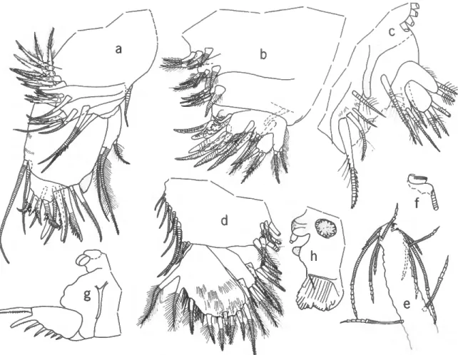
Fifth Limb (Figure Ab,c): Endite I with 5 bris- tles; endite II also with 5 bristles; endite III with 7 bristles, including a small, triangular, tooth-like FIGURE 3.—Pterocypridina dedeckkeri, new species, adult female, holotype, USNM 158240: a, rostrum and incisur of right valve, inside view; b, caudal process and posteroventral infold of right valve, inside view; c, inside view of mid-dorsal part of right valve showing attachment to valve of distal ends of muscles attached to body and appendages, anterior towards left; d, outside view of mid-area of right valve viewed with transmitted light showing reticulated ornamentation formed of ridges bearing minute tubercules, anterior towards right; e, right 1st antenna, medial view; /, distal part of protopodite, endopodite, and proximal part of 1st joint of exopodite of left 2nd antenna, medial view; g, right mandi- ble, medial view; h, part of left maxilla showing dorsal bristle of coxale, 3 bristles of basale, dorsal spines and terminal ventral tooth of 1st endopodial joint, anterior view.
bristle. Protopodite with long, slender, anterior, finger-like process. 1st exopodial joint: anterior side with 1 proximal bristle with long proximal hairs and distal group of 3 bristles (larger of these pectinate and with stout proximal spines) (Figure Ac); main tooth consisting of 5 pectinate teeth and proximal small broad peg; bristle with long proximal hairs near proximal peg (Figure Ab).
2nd exopodial joint with 3 pectinate, unringed a- bristles, a total of 5 pectinate, ringed b- and b'- bristles, 1 posterior c-bristle with long proximal hairs, and 1 anterior d-bristle with long proximal hairs. 3rd exopodial joint with 3 bristles (2 with long hairs, 1 bare, unringed) on inner lobe, and 2 bristles (with long proximal hairs) on outer lobe. Fused 4th and 5th joints with 3 bristles.
Sixth Limb (Figure Ad): Endite I with 1 ter- minal and 2 medial bristles; endite II with 2 or 3 terminal and 2 medial bristles; endite III with 3 terminal and 1 medial bristle; endite IV with 4 terminal and 1 medial bristle. End joint with 9 bristles (posterior 2 hirsute, remaining bristles with only short spines, or with long proximal and short distal spines); ventral margin with long hairs, some spine-like; 11 or 12 bare bristles in place of epipodial appendage.
Seventh Limb (Figure Ae,f): 10 bristles (5 on each side) in terminal group; 2 bristles in proxi- mal group (1 on each side); each bristle with up to 7 bells. Comb consisting of about 4 short teeth on each side of a longer recurved tooth. Tooth opposite comb obscure but consisting of sclero- tized ridge having numerous spines (Figure 4/).
(One of the 7th limbs on holotype lost.)
Furca (Figure 4^): Each lamella with 7 or 8 claws; claw 2 not separated from lamella by suture; claw 3 more slender than claw 4 and slightly longer; all claws with teeth along poste- rior margin; right lamella slightly anterior to left lamella.
Bellonci Organ (Figure Ah): Small, oval, with button-like process at tip.
Eyes: Medial eye with brown pigment (Figure Ah). Lateral eye about same diameter as medial eye, with black pigment and about 20 ommatidia (Figure Ah; not all ommatidia shown).
Upper Lip (Figure Ah): Anterior unpaired part
FIGURE 4.—Pterocypridina dedeckkeri, new species, adult female, holotype, USNM 158240: a, right maxilla, anterior view; b, right 5th limb, posterior view; c, part of left 5th limb, anterior view;
d, right 6th limb, medial view; e, distal part of 7th limb (all marginal bristles shown);/, detail of ridge opposite comb of 7th limb shown in e; g, left lamella of furca, left Y-sclerite, left genitalia; h, lateral eye, medial eye and bellonci organ, anterior process, and upper lip, anterior towards left.
with both anterior and ventral glandular proc- esses and projecting ventrally past paired poste- rior part. Paired posterior part with ventral glan- dular processes; each paired part followed by 2 small lateral processes.
Genitalia (Figure 4g): Consisting of rounded process with attached spermatophore on each side of body anterior to furca.
Anterior of Body (Figure Ah): A single sclero- tized process with rounded tip present between upper lip and medial eye.
Posterior of Body: Bare, evenly rounded poster- odorsal corner.
Y-Sclerite (Figure 4g): Normal for family.
Eggs: Holotype with 7 eggs in marsupium.
COMPARISONS.—Pterocypridina dedeckkeri resem-
bles closely P. excreta Poulsen, 1962:240, and both species were collected in the same general area off southeast Australia. Because only 1 specimen of each species is known, their intraspecific variabil- ity is unknown. Both specimens are adult females.
Because they differ in so many minor characters,
proposing P. dedeckkeri as a new species seems warranted. The length of the carapace of P. de- deckkeri is 1.57 mm compared to 1.87 mm for P.
excreta. The posterodorsal process on each valve of P. dedeckkeri is smaller and more dorsally lo- cated than that of P. excreta. The ridges on valves of P. excreta are formed by rows of minute tuber- cles (Poulsen, 1962:241, fig. 113d) whereas the tubercles project from a ridge on P. dedeckkeri (Figure 3d; Plate 2b). The end joint of the 6th limb extends much farther past the end of the 4th endite on P. excreta than it does on P. dedeckkeri.
The 9th exopodial joint of the 2nd antenna of P.
dedeckkeri bears 3 bristles. Although Poulsen indi- cates in a key and table 24 (1962:235, 236) that P. excreta bears 4 bristles, in the text (p. 242) he ambiguously writes: ". . . the end-joint has 3 bris- tles, also the somewhat shorter dorsal bristle has natatory hairs." The endopodite of the 2nd an- tenna of P. excreta bears 5 bristles compared to 4 on P. dedeckkeri. Needless to say, if additional collections produce intermediate forms, the 2 spe- cies should be synonomized.
Pterocypridina sex, new species
FIGURES 5-9
ETYMOLOGY.—The name of the species from the Latin sex (six), in reference to the presence of 6 claws on each lamella of the furca of the holotype.
HOLOTYPE.—USNM 157760, ovigerous female, on slide and in alcohol.
TYPE-LOCALITY.—BLM sta 6E, on continental shelf off Jacksonville, Florida, 30°23'N, 80°26'W, depth 39 m, collected on 1 Mar 1977.
PARATYPES.—North Carolina continental shelf:
USNM 159078, 1 adult male, sta 016-1, R / V Eastward, cruise E-2-77, 34°37'54"N, 76°08'00"- W, depth 40 m, collected in June 1977. South Carolina continental shelf: USNM 158257, 1 fe- male (probably adult), BLM sta 2C, 32°50'N, 79°04'W, depth 22 m, collected on 13 Feb 1977.
East Florida continental shelf: USNM 158256, 3 juveniles, USNM 158255, 1 juvenile, same station
data as holotype; USNM 158254, 1 early instar,
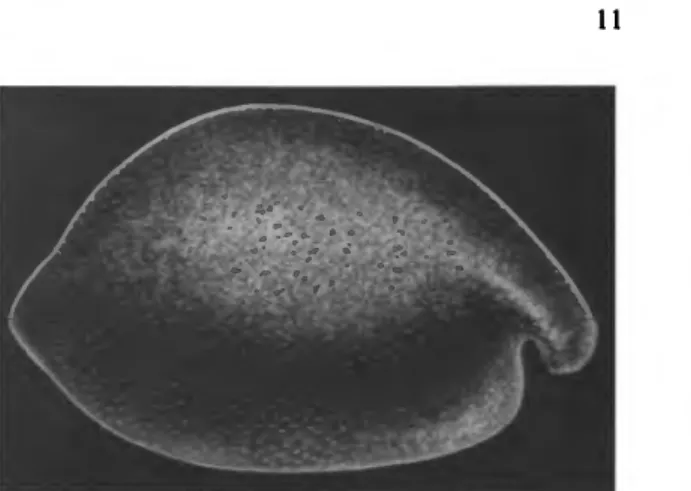
FIGURE 5.—Pterocypridina sex, new species, adult female, hol- otype, USNM 157760, lateral view of complete specimen, length 1.66 mm.
BLM sta 6C, 30°23'N, 80°51'W, depth 29 m, collected on 28 Feb 1977; USNM 158364, 1 adult male, sta LD-19B, 27°29'36"N, 80°17'18"W, depth 5.5 m, collected on 28 Jun 1977.
DISTRIBUTION.—Continental shelf off SE North America. Known depth range 5.5-40 m.
DESCRIPTION OF ADULT FEMALE (Figures 5-
8).—Carapace in lateral view more convex dor- sally than ventrally; posterior margin with pro- jecting caudal process (Figure 5); incisur fairly
deep.
Ornamentation: Surface with well-developed punctae; outer edge of rostrum with minute scal- lop-like processes (Figure 6a); small, low, lateral process present on rostrum of each valve.
Infold (Figure 6a-c): Infold of rostrum with 11 or 12 divided bristles paralleling outer ridge of rostrum and 1 divided proximal bristle (Figure 6a); infold posterior to incisur with 1 proximal bristle and 2 longer bifurcate bristles near outer edge; infold ventral to incisur with 1 short bristle followed by space and then 14-17 divided bristles forming row parallel to anteroventral edge of valve; a few small bristles present proximal to the row of anteroventral bristles; ventral infold bare or with only 2 or 3 small bristles; infold of caudal process complex; that of left valve with large process (Figure 6c), that of right valve with long narrow process overlapping pocket (Figure 66);
processes with few small bristles; minute bristles
present along outer edge of caudal process.
Selvage: Wide lamellar prolongation with smooth outer edge present along anterior and ventral valve margins; prolongation divided at inner end of incisur.
Size: USNM 157760, length 1.66 mm, height 1.13 mm; USNM 158257, length 1.52 mm, height 1.02 (probably adult, contains fairly large unex- truded eggs).
First Antenna (Figure 6af): 1st joint bare. 2nd joint with few lateral, ventral, and dorsal spines
and many medial spines forming rows. 3rd joint short with 2 bristles (1 ventral, 1 dorsal). 4th joint long with 2 bristles (1 short, ventral, subterminal;
1 longer, dorsal terminal). Sensory bristle of squarish 5th joint with 5 long proximal filaments (a minute filament present adjacent to each of 2 long proximal filaments), 2 short filaments with bases just distal to 5th proximal filament, and 1 small filament near tip. 6th joint squarish with 1 short medial bristle. 7th joint: a-bristle spinous, slightly longer than bristle of 6th joint; b-bristle about twice length of a-bristle, with 3 marginal filaments; c-bristle reaching past tip of sensory bristle of 5th joint, with about 8 marginal fila- ments. 8th joint: d- and e-bristles bare with blunt tips, about twice length of b-bristle; f-bristle longer than d- and e-bristles, with about 9 mar- ginal filaments; g-bristle longer than f-bristle, with about 8 marginal filaments.
Second Antenna (Figure 6^): Protopodite with short, spinous, medial bristle, otherwise bare. En- dopodite with minute 2nd joint fused to 1st joint:
1st joint with 4 bristles (2 of these much shorter than others); minute 2nd joint with 1 long fila- ment-like bristle. Exopodite: bristle of 2nd joint about same length as combined joints 2-9, with
FIGURE 6.—Pterocypridina sex, new species, adult female, hol- otype, USNM 157760: a, rostrum and incisur of right valve, inside view; b, c, caudal process and infold of right and left valves, inside view; d, right 1st antenna, lateral view; e, distal part of protopodite and endopodite of left 2nd antenna, medial view; /, left lamella of furca, lateral view; g, right side of anterior of body showing right lateral eye, medial eye and bellonci organ, anterior process, and upper lip, anterior of body towards right.
about 11 stout ventral spines but no natatory hairs; bristles of joints 3 and 4 with proximal ventral spines and both proximal and distal na- tatory hairs; bristles of joints 5-8 with natatory hairs; 9th joint with 2 bristles with natatory hairs and stout dorsal spines, and 1 shorter dorsal bristle with only natatory hairs; joints 3-8 with basal spines increasing in length on distal joints;
basal spine of 8th joint and lateral spine of 9th joint about same length as 9th joint; joints 2-8 with minute spines forming row along distal mar- gin.
Mandible (Figure 7a): Coxale endite spinous with bifurcate tip; small bristle present near base of endite on left limb of USNM 157760 but not observed on right limb. Basale: medial side near ventral margin with 2 short a-bristles, 1 minute b-bristle, 2 small c-bristles, and 1 long spinous d- bristle; dorsal margin with 3 bristles (1 near middle, 2 subterminal). Exopodite hirsute, reach- ing to about middle of dorsal margin of 1st endopodial joint, with 2 spinous bristles (distal outer bristle less than 1/2 length of proximal bristle). First endopodial joint with 4 ventral bristles (2 long, 1 short, 1 minute). Second endo- podial joint: ventral margin spinous and with 3 groups of short slender bristles containing 1, 1, and 2 bristles; dorsal margin with 9 spinous bris- tles; medial surface with short spines forming rows. End joint with 4 bristles and 3 claws (all claws with few proximal teeth along ventral mar- gin)-
Maxilla (Figure 7b,c): Endites (not all bristles shown on illustrated limb): endite I with about 8 spinous or pectinate bristles; endite II with about 6 spinous or pectinate bristles; endite III with 1 proximal bristle and about 6 terminal spinous or pectinate bristles. Precoxale with hairs forming dorsal fringe. Usual plumose bristle of coxale not observed on either limb, but both limbs are ob- scure on slide; small bare bristle present either on dorsal margin of coxale or basale; basale with 2 additional longer bristles. Exopodite broad with 1 short, spinous, proximal bristle and 2 long, spinous, terminal bristles (middle bristle and proximal bristle with long marginal hairs) (Figure
7 c). First endopodial joint with triangular cutting tooth with bifurcate tip, 2 alpha-bristles with short marginal spines, and 2 beta-bristles (1 slen- der with short marginal spines; 1 stout, pectinate).
End joint with total of about 10 bristles, some claw-like, pectinate, others ringed and with either marginal spines or both marginal spines and few marginal teeth.
Fifth Limb (Figure ld,e): Epipodial appendage with 54 bristles. Endites I to III with up to 6 bristles (not all shown in illustrated limb). Pro- topodite with small, sclerotized, anterior process on distal margin (Figure le). First exopodial joint with 3 anterior bristles (the inner of these stout, pectinate) (Figure 7e); main tooth consisting of 5 pectinate teeth and proximal peg (Figure Id); a short bristle present proximal to peg. Second exopodial joint with 1 posterior c-bristle, 1 ante- rior d-bristle, 3 a-bristles, and 6 b-bristles. Third exopodial joint: inner lobe with 1 proximal and 1 terminal bristle, outer lobe with 2 spinous bris- tles; 4th and 5th joints divided on left limb of USNM 157760, each joint with 2 bristles, fused on right limb, with total of 4 bristles.
Sixth Limb (Figure 8): 4 short bristles in place of epipodial appendage. Endites I and II some- what fragmented on USNM 157760, but each apparently with 2 spinous bristles; endites III and IV each with 4 spinous bristles. End joint with 13-15 bristles (2 posterior bristles plumose, others either with short distal spines and long proximal hairs, or just short spines).
Seventh Limb (Figure 7/^): Each limb with 2 proximal bristles (1 on each side) and 8 distal (4 on each side); each bristle with 3-7 bells. Termi- nal ventral comb with 4 short blunt teeth on either side of 3 elongate teeth; sclerotized ridge
FIGURE 7.—Pterocypridina sex, new species, adult female, hol- otype, USNM 157760: a, right mandible, medial view; b, left maxilla, inside view; c, right maxilla showing exopodite, distal tooth and proximal parts of alpha-bristles of 1st en- dopodial joint; d, distal part of left 5th limb, posterior view;
e, distal part of right 5th limb, not all bristles and claws shown; / , 7th limb; g, detail of tip o f / Female (probably adult), paratype, USNM 158257, A, upper lip, anterior towards left.
FIGURE 8.—Pterocypridina sex, new species, adult female, hol- otype, USNM 157760, left 6th limb, medial view.
opposite comb with curved tip bearing 5 or 6 minute teeth along edge (not all teeth shown on illustrated limb).
Furca (Figure 6/): Each lamella with 6 claws;
claws 2 and 4 fused to lamella, remaining claws separated from lamella by suture; claw 3 weaker than claw 4; posterior edge of claws with teeth forming 1 or 2 rows.
Bellonci Organ (Figure 6g): Short with small process at tip.
Eyes (Figure 6g): Lateral eyes pigmented black, with about 19 ommatidia. Medial eye about same size as lateral eye, with some black pigment.
Upper Lip (Figures 6g, Ih): Consisting of nar- row, anterior, unpaired part and broader, poste- rior, paired part; short proximal tusk present laterally near posterior edge of paired part; spines present along posterior edge of lip just in front of mouth (see illustrations of male upper lip for details).
Posterior of Body: Without process.
Eggs: USNM 157760 with 8 eggs in marsu- pium.
DESCRIPTION OF ADULT MALE (Figure 9).—Car- apace similar in shape to that of adult female (Figure 9a).
Ornamentation: Surface with well-developed punctae (Figure 9c); outer edge of rostrum with minute scallop-like processes; small, low, lateral process present on rostrum of each valve (Figure 9b).
Infold: Not examined from the inside, but ap-
pearing similar to that of female when viewed from outside in transmitted light.
Selvage: Similar to that of female.
Size: USNM 158364, length 1.41 mm, height 0.95 mm.
First Antenna (Figure 9d,e): 1st and 2nd joints bare. 3rd joint short, with 2 small bristles (1 ventral, 1 dorsal). 4th joint with 2 small terminal bristles (1 ventral, 1 dorsal). Sensory bristle of 5th joint with 7 long and 3 small proximal filaments and 3 slender, short, distal filaments (all bare except for spine at tip of small proximal and short distal filaments). 6th joint with small, bare, me-
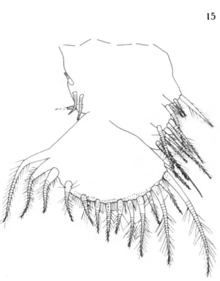
FIGURE 9.—Pterocypridma sex, new species, adult male, paratype, USNM 158364: a, lateral view of complete specimen, length 1.41 mm; b, anterior of carapace showing rostrum bearing lateral process; c, detail of surface punctae, from specimen shown in a; d, left 1st antenna, medial view;
e, tip of right 1st antenna showing a-bristle and proximal parts of b- and c-bristles, medial view drawn with specimen not under cover slip;/, right lamella of furca and copulatory process; g, anterior of body from left showing left lateral eye, medial eye and bellonci organ, anterior process, upper lip, and opening of mouth with part of esophagous dashed; h, upper lip, posterior view, ventral edge towards bottom; i, upper lip, ventral view, anterior towards bottom.
dial bristle. 7th joint: a-bristle slightly longer than bristle of 6th joint, with short marginal spines; b- bristle with stout proximal filament bearing thin disc at distal end; b-bristle also with 3 slender marginal filaments; c-bristle almost twice length of b-bristle and with proximal filament bearing terminal disc similar to that of b-bristle, but filament slightly shorter; c-bristle also with 9 marginal filaments (2 or 3 of the distal filaments much longer than others; all filaments bare ex- cept for spine at tip). 8th joint: d- and e-bristle bare with blunt tips (both bristles longer than b- bristle but shorter than c-bristle); f-bristle slightly shorter than c-bristle, with stout proximal part bearing abundant, long, slender filaments, and slender distal part with about 6 shorter filaments;
g-bristle about same length as c-bristle, with broad proximal part bearing abundant, long, slender filaments, and slender distal part with 6 or 7 shorter filaments.
Second Antenna: Similar to that of adult female except for 9th exopodial joint having only middle bristle with proximal dorsal spines.
Mandible: Similar to that of adult female, ex- cept exopodite almost reaching distal end of dor- sal margin of 1st endopodial joint.
Maxilla: Similar to that of adult female.
Fifth Limb: Anterior tooth of protopodite
about twice length of that of adult female and with constriction near middle. Left limb of USMN 158634 with 5 bristles on fused 4th and 5th exopodial joints; right limb with 4 bristles.
Limb otherwise similar to that of adult female.
Sixth Limb: Endite I with 1 long terminal and 3 short medial bristles; endite II with 1 long terminal and 2 short medial bristles; endite III with 3 terminal bristles and 1 long medial bristle;
endite IV with 3 terminal bristles and 1 fairly long medial bristle. Distal margin of end joint with 7 anterior spinous bristles followed by space and then 3 stout hirsute bristles. 4 small bristles present in place of epipodial appendage.
Seventh Limb, Bellonci Organ (Figure 9g), Upper Lip (Figure 9g-i), Posterior of Body: Similar to those of adult female.
Furca (Figure 9/): Each lamella with 7 claws, otherwise similar to that of female.
Copulatory Organ: Elongate, terminating in several lobes, smaller lobes with sclerotized ridges (Figure 9/).
COMPARISONS.—The new species, Pterocypridina sex, resembles P. birostrata Poulsen in not having lateral posterodorsal processes. Furcal claws 2 and 4 are fused to the lamella of P. sex, whereas, only claw 2 is fused to the lamella of P. birostrata. Each furcal lamella off. birostrata bears 10 claws com- pared to only 6 or 7 for P. sex.
Appendix
Keys to Subfamilies, Tribes, Groups, and Genera of the Cypridinidae
CYPRIDINIDAE Baird, 1850
Key to Subfamilies of the Cypridinidae
(adult males and females)
Each lamella of furca with 18-29 claws; lateral eyes flaplike, hirsute, without
ommatidia AZYGOCYPRIDININAE
Each lamella of furca with 4-15 claws; lateral eyes absent, or bare, oval with ommatidia CYPRIDININAE
AZYGOCYPRIDININAE Kornicker, 1970 Key to Genera of the Subfamily Azygocypridininae
(adult males and females)
Outer lobe of 3rd joint of 5th limb with 2 bristles; endopodite of 2nd antenna of male formed as clasper Azygocypridina Outer lobe of 3rd joint of 5th limb with 4 bristles; endopodite of 2nd antenna of male similar to that of female Isocypridina
CYPRIDININAE Baird, 1850
Key to Tribes of the Subfamily Cypridininae
(adult males and females)
Shell halves united for 1/2 to 2/3 of margin, carapace globose, thin-shelled, large, length 9 mm or more, evenly rounded posteriorly, or at most with minute caudal process GiGANTOCYPRlDiNiNi Shell halves united only along dorsal and posterodorsal margin, carapace either thin-shelled or well calcified, length less than 8 mm, generally elongate, if rounded bearing prominent caudal process CYPRIDININI
GIGANTOCYPRIDININI Hartmann, 1974 The tribe contains only the genus Gigantocypris.
18
Cypridinini Baird, 1850
Key to Groups of the Tribe Cypridinini (adult males and females; * = genera in Group ?)
1. Each lamella of furca with 4 claws Codonocera Group Each lamella of furca with 5 claws Monopia Group Each lamella of furca with more than 5 claws 2 2. Shell or rostrum without lateral processes
Cypridina Group (part), Hadacypridina * Shell or rostrum with lateral processes 3 3. Rostrum with dorsal opening Amphisiphonostra * Rostrum without dorsal opening 4 4. Endopodite of 2nd antenna, elongate, 3-jointed
Cypridina Group (part), Rugosidoloria * Endopodite of 2nd antenna, short, 1- or 2-jointed 5 5. Proximal filaments of sensory bristle of 5th joint of 1st antenna about same length or shorter than most distal filaments Paracypridina * Most proximal filaments longer than most distal filaments
Pterocypridina Group
Codonocera Group (new group) This group contains only the genus Codonocera.
Monopia Group (new group) This group contains only the genus Monopia.
Pterocypridina Group (new group) The group contains only the genus Pterocypridina.
Cypridina Group (new group) and Group ? See Table 1 for diagnosis.
Key to Genera of the Cypridina Group and Group ? (adult males and females; * = genera in Group ?)
1. Carapace yellow to dark brown with round uncolored area in vicinity of lateral eye Macrocypridina Carapace unpigmented or with black pigment 2 2. Carapace with round opening on dorsal end of rostrum
Amphisiphonostra * Carapace without opening on dorsal end of rostrum 3
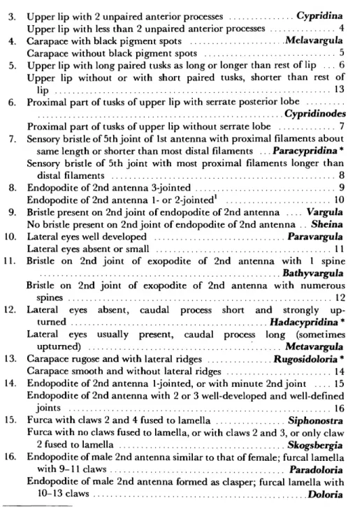
3. Upper lip with 2 unpaired anterior processes Cypridina Upper lip with less than 2 unpaired anterior processes 4 4. Carapace with black pigment spots Melavargula
Carapace without black pigment spots 5 5. Upper lip with long paired tusks as long or longer than rest of lip . 6 Upper lip without or with short paired tusks, shorter than rest of lip 13 6. Proximal part of tusks of upper lip with serrate posterior lobe
Cypridinodes Proximal part of tusks of upper lip without serrate lobe 7 7. Sensory bristle of 5th joint of 1st antenna with proximal filaments about same length or shorter than most distal filaments ... Paracypridina * Sensory bristle of 5th joint with most proximal filaments longer than distal filaments 8 8. Endopodite of 2nd antenna 3-jointed 9 Endopodite of 2nd antenna 1- or 2-jointed1 10 9. Bristle present on 2nd joint of endopodite of 2nd antenna . . . . Vargula
No bristle present on 2nd joint of endopodite of 2nd antenna .. Sheina 10. Lateral eyes well developed Paravargula Lateral eyes absent or small 11 11. Bristle on 2nd joint of exopodite of 2nd antenna with 1 spine Bathyvargula Bristle on 2nd joint of exopodite of 2nd antenna with numerous
spines 12 12. Lateral eyes absent, caudal process short and strongly up- turned Hadacypridina * Lateral eyes usually present, caudal process long (sometimes upturned) Metavargula 13. Carapace rugose and with lateral ridges Rugosidoloria * Carapace smooth and without lateral ridges 14 14. Endopodite of 2nd antenna 1-jointed, or with minute 2nd joint . . . . 15 Endopodite of 2nd antenna with 2 or 3 well-developed and well-defined joints 16 15. Furca with claws 2 and 4 fused to lamella Siphonostra Furca with no claws fused to lamella, or with claws 2 and 3, or only claw 2 fused to lamella Skogsbergia 16. Endopodite of male 2nd antenna similar to that of female; furcal lamella with 9-11 claws Paradoloria Endopodite of male 2nd antenna formed as clasper; furcal lamella with 10-13 claws Doloria
1 Some females of Metavargula ampla Kornicker, 1970, figure 3f with small 3rd joint on endopodite of 2nd antenna.
Literature Cited
Athersuch, John
1980. The Genus Azygocypridina Sylvester-Bradley (Crus- tacea: Ostracoda) with Particular Reference to A.
imperialis (Stebbing, 1901). Bulletin of the British Museum (Natural History), Zoology, 39(3): 139-160, 11 figures.
Baird, W.
1850. The Natural History of the British Entomostraca. 364 pages, 36 plates. London. [Printed for the Ray Society.]
Brady, G.S.
1897. A Supplementary Report on the Crustaceans of the Group Myodocopa Obtained during the Chal- lenger Expedition, with Notes on Other New or Imperfectly Known Species. Transactions of the Zo- ological Society of London, 14(3)7:85-100, plates 15-
17.
Hartmann, Gerd
1965. Neontological and Paleontological Classification of Ostracoda. Pubblicazioni delta Stazione Zoologica di Napoli, supplement 33:550-587.
1975. Ostracoda. In H.G. Bronns, editor, Klassen und Ordnungen des Tierreichs, 5(4):569-786, figures 374- 474.
Hartmann, Gerd, and Harbans S. Puri
1974. Summary of Neontological and Paleontological Classification of Ostracoda. Mitteilungen aus dem Hamburgischen Zoologischen Museum und Institut, 70:7-
73.
Kornicker, Louis S.
1968. Bathyal Myodocopid Ostracoda from the North- eastern Gulf of Mexico. Proceedings of the Biological Society of Washington, 81:439-472, 10 figures, 2 plates.
1970. Ostracoda (Myodocopina) from the Peru-Chile
Trench and the Antarctic Ocean. Smithsonian Con- tributions to Zoology, 32: 42 pages, 25 figures.
1975. Antarctic Ostracoda (Myodocopina). Smithsonian Contributions to Zoology, 163:720 pages, 432 figures, 9 plates.
Kornicker, Louis S., and F.P.C.M. van Morkhoven
1976. Metapolycope, a New Genus of Bathyal Ostracoda from the Atlantic (Suborder Cladocopina). Smith- sonian Contributions to Zoology, 225: 19 pages, 24 figures.
Martens, J.M.
1979. Die pelagischen Ostracoden der Expedition Mar- chile I (Siidost-Pazifik), II: Systematik und Vor- kommen (Crustacea: Ostracoda: Myodocopida).
Milteilingen aus dem Hamburgischen Zoologischen Mu- seum und Institut, 76:303-366.
Miiller, G.W.
1906. Die Ostracoden der Siboga-Expedition. In Uitkom- sten op Zoologisch, Botanisch, Oceanographischen on Geo- logische Gebeid versameld in Nederlandsch Oost-Indie, 1899-1900, 30: 40 pages, 9 plates.
1912. Ostracoda. In Das Tierreich, 31: 434 pages, 92 figures.
Poulsen, E.M.
1962. Ostracoda-Myodocopa, 1: Cypridiniformes-Cy- pridinidae. In Dana Report, 57:1-414, 181 figures.
1973. Ostracoda-Myodocopa, IIIB: Halocypriformes- Halocypridae, Conchoecinae. In Dana Report, 84:1-234, 113 figures.
Skogsberg, T.
1920. Studies on Marine Ostracods, 1: Cypridinids, Hal- ocyprids, and Polycopids. Zoologiska Bidrag fran Uppsala, supplement, 1: 784 pages, 153 figures.
Stebbing, Thomas R.
1901. Giant Ostracoda: Old and New. Knowledge, 24:100.
21
PLATE 1
Plerocypridina dedeckken, new species, adult female, holotype, USNM 158240, left valve: a, lateral view of valve with dorsal end tilted about 40° forward, edge of posterior part of dorsal margin missing, stereoscopic pair, X 58; b, lateral view of rostrum and incisur of valve shown in a, lamellar prolongation of selvage at inner edge of incisur torn, stereoscopic pair, X 275; c, inside view of valve, note distal ends of dorsal muscles near upper edge of valve, X 58; d, inside view of rostrum and incisur, lamellar prolongation of selvage at inner end of incisur torn, X 275.
(Micrographs reduced to 79%.)
PLATE 2
Pterocypridina dedeckkeri, new species, adult female, holotype, USNM 158240, left valve: a, triangular process near middle of posterodorsal margin from Plate la, stereoscopic pair, X 400;
b, low mound bearing central adductor muscle attachments, from lower left of Plate la, stereoscopic pair, X 375; c, inside view of valve showing remnants of dorsal muscles, from dorsal part of Plate Ic, X 370; d, detail of left set of muscles in c, X 1500. (Micrographs reduced to 78%.)
PLATE 3
Pterocypridina dedeckkeri, new species, adult female, holotype, USNM 158240, left valve: a, bristle and bristle pore from near top of posterodorsal triangular process, from Plate 2a, stereoscopic pair, X 900; b, bristle and bristle pore adjacent to tip of posterodorsal triangular process, stereoscopic pair, from Plate 2a, X 9000; c, caudal process from inside, from Plate Ic, X 250; d, minute protuberances bearing pore with small bristle along edge of list just anterior to caudal process, from right end of list shown in c, X 2100. (Micrographs reduced to 78%.)
PLATE 4
Plerocypndina dedeckken, new species, adult female, holotype, USNM 158240, left valve: a, minute protuberance, bearing pore with small bristle, along edge of list just anterior to caudal process, from Plate 3d, X 10,000; b, same type pore and bristle from list of caudal process opposite posterior tip of process, from Plate 3c, X 10,000; c, central adductor muscle attachment scars, from Plate \c, X 175; d, same, X 360; e, attachment scar located to upper left of main group of scars, from c, X 1250;/, detail of surface where muscle had been detached, from e, X 10,000.
(Micrographs reduced to 78%.)