Thanks also go to dr. for his invaluable advice on the synthesis of isotopically labeled cysteine. One of the nitric oxide complexes is. complex NO-bridge, demonstrating that the site of oxygen reduction is between two metal centers. These studies represent the first time that the ligands of any metal center in cytochrome c oxidase have been unequivocally elucidated.
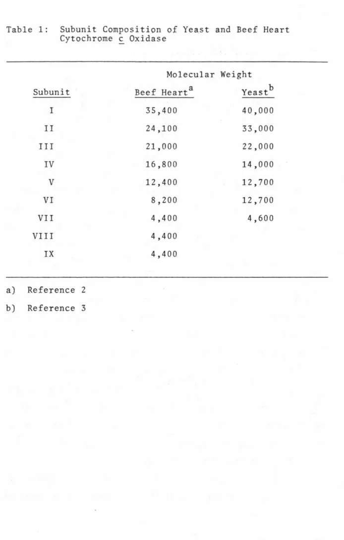
The macroscopic structure of cytochrome £ oxidase was studied on two-dimensional arrays of oxidized substances. The "roots" of the tooth protrude from the matrix side of the inner mitochondrial membrane. Spectroscopic studies have played an important role in understanding the role of metal centers in protein function.
CHAPTER II: STRUCTURE OF THE OXYGEN BINDING SITE 1. INTRODUCTION
MATERIALS AND METHODS
The intensity of the high-spin cytochrome ~3 EPR signals was determined relative to an external myoglobin standard. The binding curve of NO to oxidized cytochrome £ oxidase (Fig. 5) demonstrates that the intensity of the high-spin cytochrome ~3 signal is dependent on the NO pressure. The pressure of NO corresponding to the appearance of SO% of the observed high-spin cytochrome ~3 E?R signal is about 65 mm Hg.
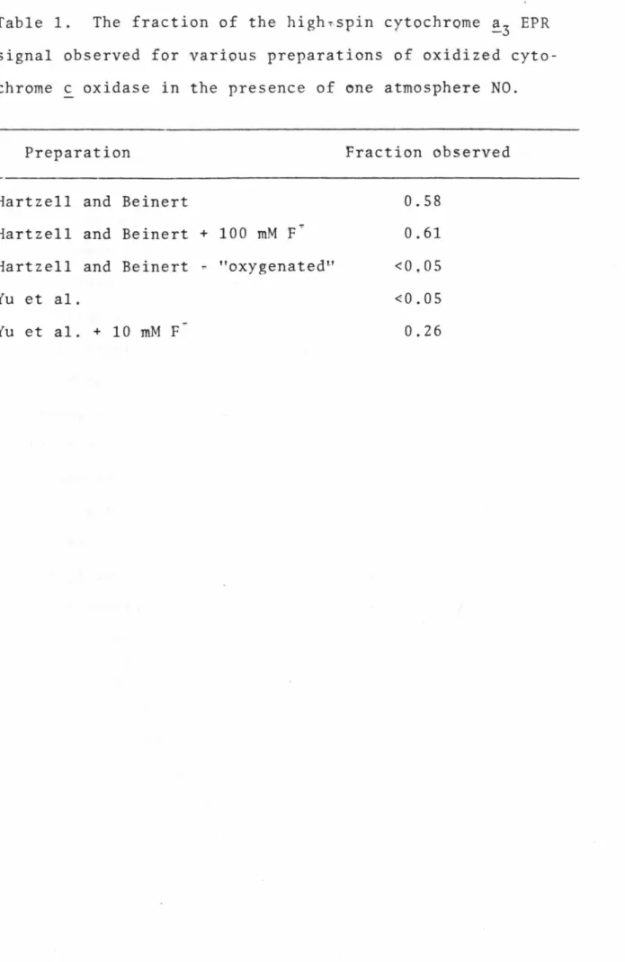
In this respect we have found that the Hartzell and Beinert preparation of the enzyme which. Since the fluoride-bound Hartzell and Beinert preparation of the enzyme exhibits a new high-spin heme EPR signal upon addition of NO, we have also investigated the addition of NO to the fluoride-bound Yu et al. As was observed for the Hartzell and Beinert preparation of the protein, the NO complex in the presence of fluoride is reversible upon removal of NO from the sample.
The intensity of this new EPR signal at g = 3~5 is similar to that of the high-spin cytochrome ~3 EPR signal induced by NO. Finally, the low-spin cytochrome ~;3~.CN~ EPR signal induced by the addition of NO completely disappeared after the removal of the NO.
DISCUSSION
In model A, a tightly bound imidazole bridges the iron and copper metal centers (32), with the ligand binding site being the free axial position of the heme iron. However, since the results of the interaction of NO with oxidized protein in the presence of azide provide the most discriminating evidence for the two models, the relative merits of the two models will be discussed in Section 4.4. However, it is also possible that NO binds to all cytochrome ~ oxidase molecules, but that a conformational heterogeneity of the protein allows only a fraction to be observed by EPR spectroscopy.
It is probably a change of a3. in the conformation of the cytochrome a3~cu site will change. the magnitude of the exchange interaction between these two metal centers. Fluoride and cyanide binding studies to the oxidized protein-NO complex have shown the existence of at least three different conformations of the oxidized protein: i) a conformation that gives rise to rhombic high spin. The sum of these conformations accounts for 100% of the enzyme molecules in both Hartzell and Beinert and Yu et al.
Furthermore, EPR signals typically appear from a triplet species, originating from the thermally accessible S = 1 excited state of the antiferro~. In contrast, the bindins of NO in the case of the fully reduced cytochrome c oxidase-NO complex (Fe +2-NO) and in. This allows a state of the protein to be prepared in which cytochrome ∼3-co is reduced and Cua is oxidized.
Model B also suggests the possibility of the formation of an ~~oxo bridge between these two post-metal centers. If this is the case, then the conformational change normally associated with these two states of the enzyme can be understood. This model explains the results of our EPR studies of the oxidized enzyme bound to NO.
In this regard, it is important to compare the reduction of the protein under aerobic and anaerobic conditions.
INTRODUCTION
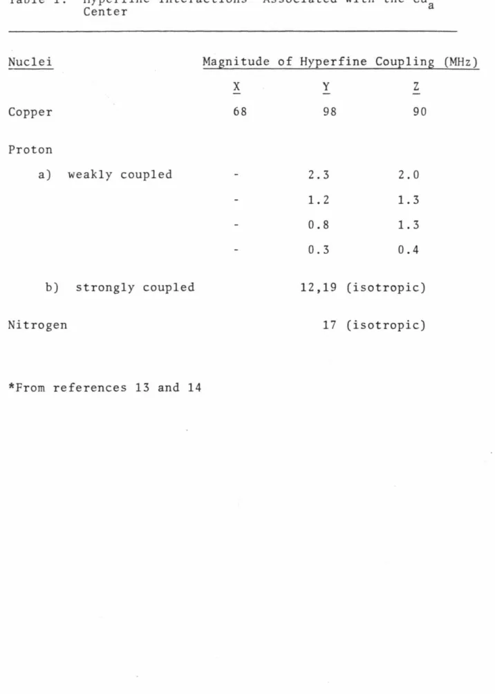
In this model, Cua is bound to two cysteines and two histidines, with electron delocalization from one of the cysteinyl sulfurs to Cua, resulting in a Cua(I)-S spin system. Measurement of the Cu EPR signal in the S-band (3 GHz) allowed the resolution of hyperfine copper along the X and Z orientations (11. Recent ENDOR studies have provided some insight into the origin of this 25 G coupling in the Cu EPR spectrum (13,14).
However, the proton couplings were small, 4 and 6 G, and the ENDOR spectra did not reveal a 25 G-proton coupling that could explain the 25 G coupling observed in the S-band EPR. However, calculations taking into account the g-anisotropy and the very small, almost isotropic copper-hyperfine interaction of the Cu EPR signal (l2) indicate that this is the case. Thus, it appears that the best description of the Cu center is that of a Cu(I)-S complex.
The presence of at least one nitrogen in the Cu center has been implicated in ENDOR studies and in Outline of the conversion of diethylamine malonate to cysteine with an isotopic label introduced as formaldehyde. Cells grown on minimal plus cysteine were then tested for growth in the absence of cysteine and growth on defined medium containing all amino acids except cysteine.
The initial culture of both auxotrophs was added at a cell density of approximately 3x10⁶ cells/mil. Since each liquid nitrogen treatment resulted in breakage of only between 15 and 20% of the yeast cells, this procedure was repeated until almost all cells were broken, as indicated by the size of the light fluff. a layer of broken cell debris in a 2000 xg pellet. A number of published procedures for the isolation and purification of yeast cytochrome oxidase have been attempted, but none have been particularly satisfactory.
EPR spectra were typically recorded on 0.3-.4 ml samples of enzyme at a protein concentration of 0.1-0.2 mM.
RESULTS
13c NMR spectra for 15N-histidine and 12cn2-cysteine were performed on a JEOL FX90 and Varian XL-100 NMR spectrometer, respectively. This finding demonstrates that cysteine has retained the high isotopic enrichment obtained in formaldehyde. EPR spectra of protein fractions eluted from a cytochrome affinity column with 0.5% chelate and with 2%.
The EPR spectrum of the cytochrome c oxidase that eluted with 1M KCl and precipitated at 38% ammonium sulfate saturation is shown in Figure 6. Therefore, the procedure for isolating yeast cytochrome c oxidase described in this work is very satisfactory for the preparation of protein samples for EPR spectroscopy. The total yield of purified oxidase from 500 grams of yeast cells is approximately 15 - 30 mg protein.
The EPR spectra of 15N-his and 12cn2-cys of yeast cytochrome oxidase are shown in Figure 7. The EPR spectra for both isotopically labeled proteins are qualitatively similar to the EPR spectrum of the unlabeled yeast protein. The instrumental conditions were the same as for Fig. 5 except that the microwave power was 0.2 mW.
UNLABELED YEAST OXIDASE
15 N-HIS YEAST OXIDA. SE
The origin of the spectral sharpening is the elimination of small hyperfine couplings to the unpaired electron. So if histidine was a ligand for Cua (or two histidines), then the substitution of 15N-his should cause a narrowing of the EPR spectrum. The narrowing of the 12cn2-cys oxidase EPR spectrum must arise from the elimination of methylene proton hyperfine interactions with Cua.
The resolved hyperfine structure in the 12 CD2-cys yeast oxidase EPR spectrum allows a more detailed analysis of the Cua center EPR signal. The determination of the involvement of cysteine and histidine as ligands to Cu now allows a more detailed. It is therefore necessary to discuss the polarization of the inner shell paired as well as bonding electrons by the outer shell unpaired electron.
The origin of the 17 MHz nitrogen-hyperfine coupling can also be explained by a spin polarization model. Thus, it has been shown that all hyperfine interactions associated with the EPR signal from the Cua center can also be quantitative. The unusual nature of the Cua center as proposed in our model (Figure 1) should certainly be related to the center.
In this regard, it has been shown that the Cua center is probably buried deep within the protein matrix(lO) and thus reduction of this center will result in an isolated negative charge within a region of low dielectric constant. This is expected to lead to a large increase in potential energy of the Cua center which can then be linked to the conservation of energy in cytochrome £oxidase. Upon transfer of an electron away from the Cua center, this proton will now leave an isolated positive charge in a hydrophobic environment.
Therefore, it may be that the unusual nature of the Cua center is directly related to its unique role of coupling electron transfer to proton pumping in cytochrome c oxidase.
CHAPTER IV: SU~~ARY
The oxygen reduction site of cytochrome £ oxidase is known to consist of cytochrome. The model that received the most favor when this work began assumed an imidazole bridge between Cua and cytochrome!3· It did not exist. Nitric oxide binding studies of isotopically labeled reduced yeast protein have conclusively established that histidine is the axial ligand of the cytochrome.
However, these studies did not address the question of the position of the imidazole relative to the Cu center. This one-electron reduced NO-bound protein complex gave rise to a triplet state whose EPR spectrum revealed new information about the oxygen. The EPR parameters together with the chemical stability of this NO-bound complex led to the conclusion that nitric oxide bridges the two metal centers in it.
This conclusion carried the implication that the already identified histidine was axial ligand for cytochrome. In this triplet state, Cu was also found to be square planar or octahedral with strong tetragonal distortion, ruling out the suggestion that Cu. Furthermore, these nitric oxide binding studies demonstrated for the first time that Cu was able to bind exogenous ligands.
The results presented in this work represent a contribution to the understanding of the general role of metal centers in the structure and enzymatic function of cytochrome c-oxidase. Further studies will be necessary to fully elucidate the structure of the metal centers in the absence of high-resolution X-ray crystal data.