DYSREGULATED T-B LYMPHOCYTE COLLABORATION IN AUTOIMMUNITY POSES A BARRIER TO TRANSPLANT TOLERANCE
By
Blair Taylor Stocks
Dissertation
Submitted to the Faculty of the Graduate School of Vanderbilt University
in partial fulfillment of the requirements for the degree of
DOCTOR OF PHILOSOPHY in
Microbiology and Immunology August, 2016
Nashville, Tennessee
Approved By:
Amy Major, PhD, Chair
Daniel Moore, MD, PhD, Thesis Advisor Luc Van Kaer, PhD
Alvin C. Powers, MD Edward Sherwood, MD, PhD
DEDICATION
To my Mother,
who gave everything for her children
and
To my wife Meredith, my rock.
Copyright © 2016 by Blair Taylor Stocks All Rights Reserved
ACKNOWLEDGEMENTS
As so concisely and eloquently stated by Peter Medawar in his 1960 Nobel Lecture, “Far too much is still uncertain.” Although in reference to the scientific community’s understanding of immunologic tolerance, to which I can only hope that my thesis work has contributed to in some way, Dr. Medawar’s brief statement reminds us why science exists in the first place.
Simply, we don’t have all the answers. Luckily, there is no better time to find these answers than right now and no purer way to discover new knowledge than science. And in today’s world, the scientific community is just that, a community. Gone are the days of the lone scientist. Instead, knowledge is now created through the immense and coordinated efforts of scientific teams.
Accordingly, I would like to briefly thank my scientific team, as none of my work would have been possible without their unwavering support.
First and foremost, I am immensely grateful for the many sources of funding that have allowed me to complete my thesis, permitting me to ask any scientific question that my brain could come up with. In order of receipt, personal support for my training has been provided by the Vanderbilt Medical Scientist Training Program Grant (Public Health Service Award T32- GM07347), the Thomas Huggins Winn Memorial Scholarship for MD/PhD students at Vanderbilt, and the Glen and Barbara Merz Scholarship in Transplant Immunology for MD/PhD students at Vanderbilt. Funding for my experimental work has been provided by NIH grants K08-DK090146, R03-DK097410, and R21-AI119224, a JDRF Career Develop Research Award, as well as institutional funds provided by the Vanderbilt Department of Pediatrics.
Words cannot fully express the immense gratitude that I have for my mentor Dan (and for the sake of professionalism, Daniel Moore MD, PhD). Simply put, Dan is the best mentor in the
world. I cannot thank Dan enough for the countless hours he has spent pushing me to be a better scientist. Dan is the smartest guy in the room and I have been extremely lucky to be in that room these past four years. His unwavering perseverance, caring demeanor, and lightning-fast turn- around time are characteristics that I wish to emulate throughout my career. I wholeheartedly dedicate my career as a physician scientist to Dan and look forward to working with him in the future. I can only hope that when I have graduate students of my own, I can provide them with a fraction of what Dan has given me.
And of course, the Moore Lab would not be what it is without my fellow graduate student Chris Wilson and lab manager Andrew Marshall. Walking in to lab everyday was a surprise – but always in a good way! Whether the day was spent working together to complete a 14-hour experiment from which 500 flow samples needed to be later analyzed, or instead, was filled with endless crossword puzzles, 2-hour long lunches, and fruitless efforts to start our own comedy podcast, I will always cherish our time in lab together. I could not imagine completing graduate school without Chris - he is always, always willing to help me get through my experiments, let me bounce experimental ideas off him, and simply be there for moral support. Similarly, I extremely grateful of Andrew, who perfectly managed our ever growing and genetically complex mouse colony, provided beautiful figures for my papers, grants, and dissertation, and was simply always willing to help out no matter what. And although we sometimes fought like 12-year old brothers, I feel extremely lucky to have become such good friends with Chris and Andrew.
I cannot thank the members of my thesis committee enough for their continued support throughout my graduate training. I feel extremely fortunate that my committee chair, Dr. Amy Major, provided not only wonderful scientific guidance, excellent organization of our meetings, and enjoyable scientific camaraderie, but moreover the opportunity to collaborate with the
members of her lab (Jillian Rhoads and Ashley Wilhelm) in exploring the barriers to tolerance in the setting of lupus. I am deeply grateful for Dr. Luc Van Kaer’s thought provoking questions and for providing me with an extremely strong foundation in immunology during my first year of medical school. I thank Dr. Al Powers for his strong career guidance and would further like to thank him for allowing me to work directly with members of his lab and utilize his scientific resources. Finally, I thank Dr. Edward Sherwood for our detailed discussions of IL-15 biology, allowing me to work in collaboration with members of his lab (Yin Guo), and for his valuable perspectives concerning physician-scientist development as a fellow MD/PhD.
I would next like to thank those individuals at Vanderbilt who helped contribute to my work. I am indebted to Dave Flaherty, Britney Matlock, and Chris Warren of the Vanderbilt Flow Cytometry Core for teaching me how to run advanced flow cytometry, a technique I will carry with me for the rest of my scientific career. Learning how to isolate islets and perform islet transplantation would have been impossible if not for the unwavering patience and highly effective teaching skills of the awe-inspiring microsurgeon Greg Poffenberger. I would specifically like to thank Rachel Henry Bonami for her technical expertise, advice in experimental design, and perspective on a career in science. I would further like to thank Cindy Lowe of the Vanderbilt Translational Pathology Shared Resource for preparing beautiful tissue samples, Troy Hutchens and Chris Reissaus of the Piston Lab for teaching me how to perform 2- photon microscopy, Adel Tawfilis of Vanderbilt’s Division of Animal Care for caring for and helping maintain our mouse colony, Miss Virginia for always keeping our lab clean and providing a smile at the end of the day, and Mary Lou Millet in helping me submit 5 NIH fellowship applications and teaching me how to use a fax machine. Finally, a number of these experiments would have gone uncompleted if not for the help of our summer and rotation
students including Elizabeth Ockerman, Stephen Owen, David Meehan, Nicole Putnam, Mabel Seto, Ly Pham, and Lauren Brewer.
I would also like to thank the numerous teachers and mentors who have contributed to my scientific development. I would like to first thank Dr. Shayn Peirce-Cottler, Dr. Adam Katz, and Dr. Alexander Baily for helping me realize my love for scientific discovery during my time as an undergraduate at UVA. Furthermore, I would also like to thank a number of Immunology and Pediatrics Faculty here at Vanderbilt for both their in- and outside of class lessons. Thank you Dr. Bill Russell, Dr. Nathan Bingham, Dr. David Jacobson, Dr. Mark Boothby, Dr. Eric Sebzda, Dr. James “Tom” Thomas, Dr. Louise Rollins Smith, Dr. Sebastian Joyce, Dr. Peggy Kendall, Dr. Jeffrey Rathmell, and Dr. Earl Ruley. Although too many too list, I would also like to thank all of my medical school instructors who provided me with a strong medical background which helped keep my scientific discoveries in perspective.
I am forever indebted to the Vanderbilt MSTP, that for reasons still unknown to me, decided to take a chance on me and offer me admission. Dr. Terry Dermody is one of the hardest working, energetic, and caring individuals I have had the pleasure of working with. I cannot thank Dr. Larry Swift enough for his advice to join Dan’s lab and Dr. Jim Bills for making sure I met all of my coursework requirements in a timely, military-like fashion. I am extremely appreciative of having the opportunity to work with Dr. Michelle Grundy on the MSTP admissions committee these past three years, and look forward to working with our new associate directors Dr. Sally York and Dr. Danny Winders in the coming years. And finally, I would like to thank Melissa Krasnove for not only making the Vanderbilt MSTP the best in the country, but more importantly so, for being such a loyal and caring friend.
And of course, I would not be where I am today without my family and friends. I simply cannot put into words how thankful I am of my mother, Judith Stocks, whose selfless sacrifice has provided me with limitless opportunities to follow my dreams. My sisters Devon Kuehn and Jen Stocks are wonderful role models to whom I will always look up to. I would also like to thank my father-in-law Dr. Scott McKenney and my mother-in-law Joan McKenney for so lovingly welcoming me, a yankee, into their Texas family. I have made some of my best friends here at Vanderbilt - Brian Palmisano, Scott McCall, Walker Plash, and Elliot Kim – and remain 100% confident that even artistic differences won’t break up “the band.” And most importantly, I would like to thank my wife Meredith Stocks for everything. Her love and support push me to be a better person everyday. I truly am the luckiest person in the world in that she chose me to share her life with.
Overall, I cannot imagine a better place to have earned my PhD than at Vanderbilt. The minute I set foot on campus in January of 2010, my gut told me that this school was made for me. And since enrolling here, my reasons for attending Vanderbilt have only been strengthened.
Every individual associated with this institution is an amazing, hard-working, and passionate person who pushes me to be better. I can only hope that future MD/PhD students have similar experiences here, and I look forward to contributing to this amazing environment in the future.
TABLE OF CONTENTS
Page
DEDICATION ... ii
ACKNOWLEDGEMENTS ... iv
LIST OF TABLES ... xii
LIST OF FIGURES ... xiii
Chapter I. INTRODUCTION ... 1
Overview ... 1
Historical Overview of Clinical Transplantation in the United States ... 3
Immunologic Mechanisms of the Transplant Response ... 6
Mechanisms of Transplant Rejection by Alloreactive T and B Cells ... 13
Immunologic Mechanisms of Transplantation Tolerance ... 20
T and B Cell Mediated Regulation of the Transplant Response ... 25
Resistance to Transplantation Tolerance in Autoimmunity ... 31
Significance of the Research and Synopsis of the Data ... 34
II. B LYMPHOCYTES PREVENT TRANSPLANT TOLERANCE IN T1D PRONE NOD MICE BY LIMITING CD4 T REGULATORY CELL FUNCTION ... 37
Scientific Goal ... 37
Introduction ... 37
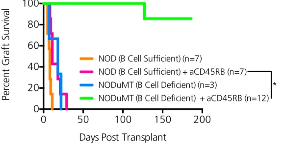
B cell deficient NODμMT mice are permissive to transplant tolerance induction ... 42
Transplant tolerance in NODμMT mice is permanent, antigen specific, and highly robust .... 42
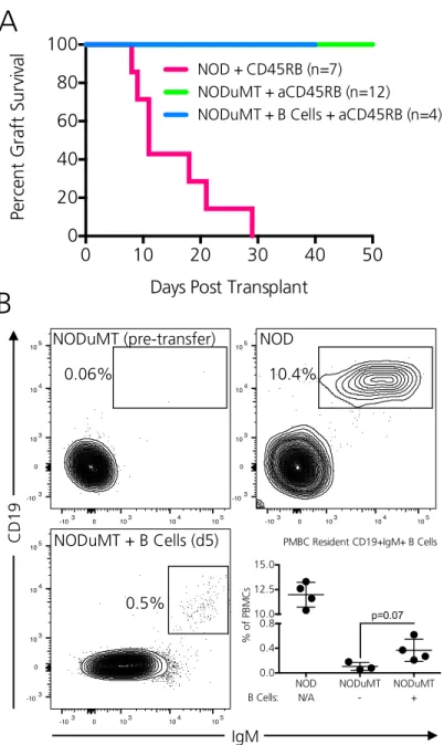
Complementation of NODμMT mice with NOD B cells fails to break tolerance induction ... 46
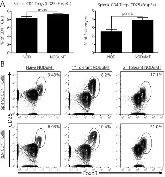
Peripheral CD4 Tregs are expanded in 1’ and 2’ tolerant NODμMT recipients ... 47
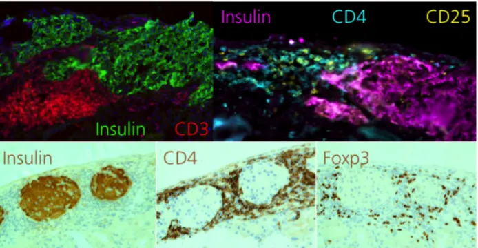
Islet grafts from tolerant NODμMT mice are encased by graft-protective CD4 Tregs ... 49
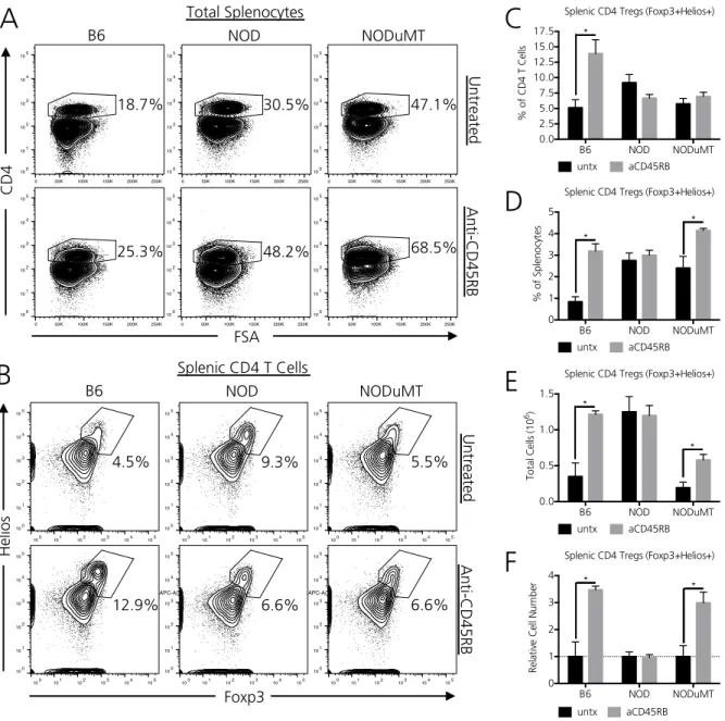
B cells in NOD mice restrain anti-CD45RB mediated CD4 Treg expansion ... 51
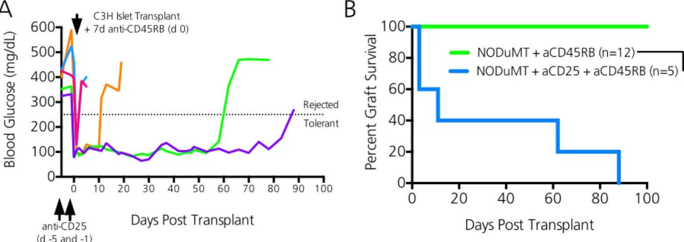
Early CD4 Treg depletion in NODμMT mice impedes transplant tolerance induction ... 53
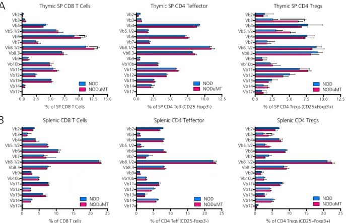
B cells limit Vβ3+ CD4 Tregs in NOD mice ... 55
Discussion ... 58
III. CD8 T REGULATORY CELL CONTROL OF THE TFH-GC B CELL AXIS IS ESSENTIAL IN SUPPRESSING DIABETOGENIC IMMUNITY AND MAINTAINING TRANSPLANT TOLERANCE ... 66
Scientific Goal ... 66
Introduction ... 66
NOD mice possess nonfunctional, classically defined CD8 Tregs and generate exaggerated antibody responses ... 69
Diabetes-prone NOD mice are profoundly deficient of TFH targeting Ly49+ CD8 Tregs ... 71
Macrophages from NOD mice inadequately trans-present and upregulate IL-15 ... 75
NOD Ly49+ CD8 Tregs adequately transduce IL-15 mediated survival signals ... 77
In vivo administration of IL-15C robustly expands NOD Ly49+ CD8 Tregs and partially rescues their suppressive function ... 79 IL-15C activated Ly49+ CD8 Tregs delay diabetes transfer ... 81 IL-15 deficient mice lack CD8 Tregs, mount an enhanced alloantibody response, and resist transplant tolerance induction ... 81 Discussion ... 85 IV. HEMATOPOIETIC STEM CELL MOBILIZATION IS NECESSARY BUT NOT
SUFFICIENT FOR TOLERANCE IN ISLET TRANSPLANTATION ... 93 Scientific Goal ... 93 Introduction ... 93 The tolerance-inducing agent anti-CD45RB promotes HSC mobilization that is necessary to establish transplant tolerance ... 96 The effect of anti-CD45RB is not directly on HSCs but rather on osteoblasts ... 98 Sympathectomy prevents HSC mobilization by anti-CD45RB and abrogates transplantation tolerance ... 101 Blockade of excess CXCR4 enhances NOD HSC mobilization and extends islet transplant survival ... 103 Discussion ... 103 V. LUPUS PRONE MICE RESIST IMMUNE REGULATION AND TRANSPLANT
TOLERANCE INDUCTION... 109 Scientific Goal ... 109 Introduction ... 109 Lupus prone B6.SLE123 mice possess expanded splenic CD4 TFH and Germinal Center B cells, fluctuating CD4 Treg and CD8 Treg populations, and generate an exaggerated
alloantibody response ... 114 Effector T Cells from Lupus prone mice resist CD4 Treg suppression of the IFNγ response 117 Effector CD4 T and B Cells from Lupus prone mice resist CD8 Treg mediated suppression 119 Lupus-prone mice lack ongoing islet immunity yet resist anti-CD45RB mediated tolerance induction to islet allografts ... 119 Discussion ... 123 VI. AN ENHANCED IL-6 RESPONSE AND HEIGHTENED IMMUNOMETABOLISM CONTRIBUTE TO TOLERANCE RESISTANCE IN LUPUS-PRONE MICE ... 128
Scientific Goal ... 128 Introduction ... 128 Single congenic lupus-prone mice resist transplant tolerance induction to varying degrees . 132 Temporary blockade of enhanced IL-6 signaling partially restores transplant tolerance in B6.SLE1 mice but fails to extend allograft survival in B6.SLE123 mice ... 133 Enhanced IL-6 signaling blockade in B6.SLE123 mice fails to extend allograft survival ... 137 Glycolytic blockade via Metformin and 2-DG during anti-CD45RB mediated transplant
tolerance induction significantly extends islet allograft survival in B6.SLE123 mice ... 138 Discussion ... 142
VII. DISCUSSION AND FUTURE DIRECTIONS ... 149
Overview ... 149
The Janus-faced roles of B cells in transplant tolerance in autoimmunity ... 151
Effector T cell resistance to Treg mediated suppression as a barrier to transplant tolerance in autoimmunity ... 156
Targeting a single component of the immune system vs. the network as a whole ... 160
Heterologous immunity as barrier to transplant tolerance in autoimmunity ... 163
Transplant tolerance as a dynamic, ongoing, and highly regulated process ... 165
Innate immune cells: a potential barrier to transplant tolerance in autoimmunity? ... 166
Towards reliably achieving Clinical Operational Tolerance ... 168
Conclusions ... 172
MATERIALS AND METHODS ... 173
Animals ... 173
Lymphoid cell preparation ... 173
Bone marrow preparation ... 175
Peripheral Blood Mononuclear Cell (PBMC) preparation ... 175
Conventional flow cytometry ... 176
Cytokine stimulation assays and phosphoflow cytometry ... 177
Magnetic Activated Cell Sorting (MACS) ... 178
Fluorescence Activated Cell Sorting (FACS) ... 179
Histology ... 179
Anti-Insulin IgG ELISA ... 180
Ex vivo Mixed Lymphocyte Reaction (MLR) ... 180
Cytokine ELISAs ... 181
2-NBDG glucose uptake assay ... 181
Colony Forming Cell assay ... 181
Osteoblast isolation, culture, and functional analysis by flow and RT-PCR ... 182
Ex vivo CD4 Treg suppression assay ... 182
In vivo CD8 Treg suppression assay ... 183
Alloimmunization and alloantibody titer analysis ... 184
Adoptive transfer of diabetes ... 184
Streptozotocin induced diabetes ... 184
Donor islet harvest ... 185
Islet transplantation and anti-CD45RB mediated tolerance induction ... 186
Survival nephrectomy and islet re-transplantation ... 187
Generation of chimeric mice and bone marrow transplantation ... 188
Statistics ... 188
REFERENCES ... 189
LIST OF TABLES
Table Page
1. Onset and immunologic methods of rejection ...11
2. Primers used to evaluate osteoblast gene expression ...100
3. Immunological phenotypes of NZM2410 congenic and subcongenic B6 mice ...130
4. Animal models used in dissertation ...174
LIST OF FIGURES
Figure Page
1.1. Mechanisms of direct and indirect allorecognition by recipient T cells ...9
1.2. Mechanisms of pharmacologically induced transplant tolerance ...24
2.1. B cell deficient NOD mice are permissive to transplant tolerance induction ...43
2.2. Transplant tolerance in NODμMT mice is permanent, antigen specific, and highly robust...45
2.3 Complementation of NODμMT mice with NOD B cells fails to break tolerance induction ...48
2.4. Peripheral CD4 Tregs are expanded in primary and secondary tolerant NODμMT recipients ...50
2.5. Islet grafts from tolerant NODμMT mice are encased by graft-protective CD4 Tregs ...52
2.6. B lymphocytes in NOD mice restrain anti-CD45RB mediated CD4 Treg expansion ...54
2.7. Early CD4 Treg depletion in NODμMT mice impedes transplant tolerance induction ....56
2.8. B lymphocytes limit Vβ3+ CD4 Tregs in NOD mice ...57
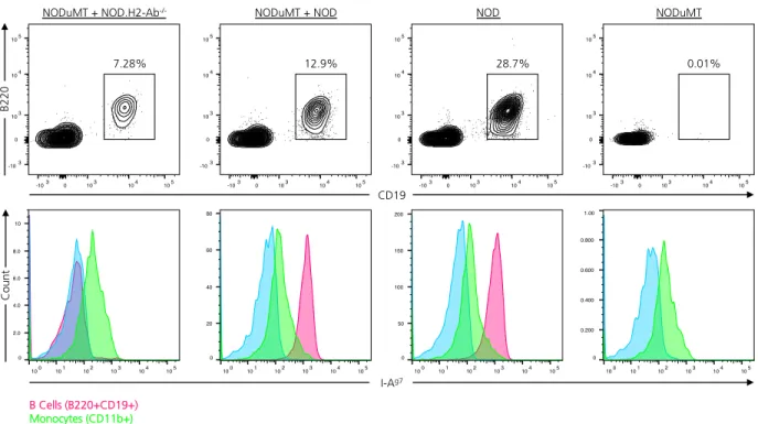
2.9. Generation of chimeric NOD mice in which only B lymphocytes lack MHC Class II expression ...62
2.10. Chapter II – findings and future work ...65
3.1. NOD mice possess nonfunctional, classically defined CD8 Tregs and generate exaggerated antibody responses ...70
3.2 Diabetes-prone NOD mice are profoundly deficient of TFH targeting Ly49+ CD8 Tregs ...72
3.3. The Ly49E/F clone CM4 binds TFH targeting Ly49+ CD8 Tregs in NOD mice ...74
3.4. Macrophages from NOD mice inadequately transpresent and upregulate IL-15 ...76
3.5. NOD Ly49+ CD8 Tregs adequately transduce IL-15 mediated survival signals ...78
3.6. In vivo administration of IL-15C robustly expands NOD Ly49+ CD8 Tregs and
partially rescues their suppressive function ...80
3.7. IL-15C activated Ly49+ CD8 Tregs delay diabetes transfer ...82
3.8. IL-15 deficient mice lack CD8 Tregs, mount an enhanced alloantibody response, and resist transplant tolerance induction ...84
3.9. Chapter III – findings and future work ...92
4.1. Tolerance induction requires stem cell mobilization ...97
4.2. Anti-CD45RB targets osteoblasts ...99
4.3. Sympathectomy blocks HSC mobilization and tolerance induction ...102
4.4. Targeting CXCR4 enhances HSC mobilization and extends islet allograft survival in NOD mice ...104
4.5. Chapter IV – findings and future work ...108
5.1. Lupus-prone B6.SLE123 mice possess expanded splenic CD4 TFH and Germinal Center B cells, fluctuating CD4 Treg and CD8 Treg populations, and generate an exaggerated alloantibody response ...116
5.2. Effector CD4 T Cells from lupus-prone mice resist CD4 Treg mediated suppression of the IFNγ response ...118
5.3. Effector CD4 T and B Cells from lupus-prone mice resist CD8 Treg mediated suppression ...120
5.4. Lupus-prone mice lack ongoing islet immunity yet resist anti-CD45RB mediated tolerance induction to islet allografts ...122
5.5. Chapter V – findings and future work ...127
6.1. Genetic overlap of risk alleles shared between tolerance resistant B6.SLE123 and NOD mice ...131
6.2. Single congenic lupus prone mice resist transplant tolerance induction to varying degrees ...134
6.3. Temporary blockade of enhanced IL-6 signaling partially restores transplant tolerance in B6.SLE1 mice but fails to extend allograft survival in B6.SLE123 mice ...136
6.4. Enhanced IL-6 signaling blockade in B6.SLE123 mice fails to extend allograft
survival ...139 6.5. Glycolytic blockade via Metformin and 2-Deoxy-D-Glucose during anti-CD45RB
transplant tolerance induction significantly extends islet allograft survival in
B6.SLE123 mice ...141 6.6. Chapter VI – findings and future work ...148 7. Summary of findings and future directions ...150
CHAPTER I
INTRODUCTION
Overview
Organ replacement via surgical transplantation represents a potentially life-saving intervention for numerous autoimmune conditions; however, the heightened immunogenicity characteristic of these autoimmune patients remains a significant barrier to long-term graft acceptance. Every year a significant number of autoimmune transplant recipients must undergo re-transplantation directly attributable to both recurrent autoimmunity and heightened allograft immunogenicity. No better is this clinical scenario modeled than the autoimmune non-obese diabetic (NOD) mouse, a model in which no therapy has ever induced permanent transplant tolerance to islet allografts or any other allograft type in the intact NOD immune system, thereby indicating the severity of this immunologic barrier. Overall, determining the immunological barriers to successful organ tolerance in the autoimmune environment would prove invaluable.
Not only would developing a mechanistic understanding of how the autoimmune environment fails to tolerate foreign allografts provide new targets for restoring graft tolerance in autoimmunity, but moreover, this knowledge would enhance the scientific community’s understanding of specific cellular and molecular pathways that govern immunological tolerance.
Autoimmune pathogenesis and failures to maintain graft tolerance are in part driven by insufficiently regulated, antigen-specific T and B effector cells of the adaptive immune system.
Although numerous studies have so far defined specific roles for inadequate immune regulation in driving autoimmunity and perpetuating loss of graft tolerance independently, it remains to be
determined whether the failures in immune regulation that perpetuate loss of self-tolerance in autoimmunity directly contribute to an inability to later “learn” how to tolerate foreign graft tissue as self. Moreover, whether known dysregulations in T-B cell collaboration that drive different forms of autoimmunity directly contribute to an enhanced, inadequately regulated anti- graft response remains to be explored. In my dissertation, I explore the specific hypothesis that dysregulated T-B cell collaboration in autoimmunity poses a stringent barrier to transplant tolerance. Using murine models of Type 1 Diabetes (T1D) and Systemic Lupus Erythematosus (SLE), I define specific cellular and molecular T-B cell aberrancies in these autoimmune environments that lead to a generalized resistance to transplant tolerance.
In setting the stage for my findings, I have provided background information pertinent to the immunologic mechanisms that drive the transplant response. After conducting a brief historical review of clinical transplantation in the United States, I highlight the pioneering work of basic transplant immunologists and their roles in defining the mechanisms of transplant rejection and tolerance. As T and B cells possess the unique ability to recognize graft tissue in an antigen specific manner, I mechanistically address how these cells actively reject or learn to tolerate foreign graft tissue. Finally, I review the key studies in which a generalized resistance to transplant tolerance in autoimmunity was first described while highlighting the outstanding knowledge gaps generated by these bodies of work. Overall, my findings reveal that enhancing graft-protective T Regulatory Cell function is a necessary component of restoring transplantation tolerance in the autoimmune setting. Moreover, interrupting specific pathways that enhance anti- graft effector cell function may further provide a targeted means to enhance transplant tolerance in autoimmunity.
Historical Overview of Clinical Transplantation in the United States
Joseph Murray made clinical organ transplantation a reality when, on December 23rd, 1954 at the Peter Bent Brigham Hospital, he performed the first successful kidney transplant between monozygotic twins1. At 24 years of age, Richard Herrick was dying of renal failure secondary to chronic nephritis. Uniquely however, Richard was blessed with a monozygotic twin brother named Ronald who was willing to donate a healthy kidney to his best friend. After 17 different genetic identity tests (including a skin transplant from Ronald to Richard that showed no signs of rejection after 4 weeks), as well as extensive ethical counseling of the twins, Drs. J.
Hartwell Harrison and Joseph Murray agreed to perform the donor procurement and recipient placement procedures, respectively2. Ronald Herrick’s donated kidney, now anastomosed to his twin brother’s Richard’s iliac vessels, began producing urine just minutes after placement.
Within days, Richard’s renal function and overall health would return to normal. Richard would go on to live another 8 years after his operation, eventually succumbing to a heart attackA. Looking back, Dr. Murray would eloquently reflect in his 1990 Nobel Prize acceptance speech,
“in a way, it was spying into the future because we had achieved our long-term goal by bypassing, but not solving the issue of biological incompatibility3.” Technically, Joseph Murray had proven that solid organ transplantation represented a life-saving surgical intervention.
However, for the majority of patients in need of a functioning organ that were not blessed with a
A Ronald Herrick, aged 79, passed away in 2010 from complications after heart surgery.
Although insurance data at the time suggested that the average life expectancy of those living with one kidney did not differ from those with two, Ronald still agreed to donate his kidney although no data existed for the life expectancy of a living organ donor2.
monozygotic twin donor, there remained the immense need to regulate the intense immune reaction brought on by the introduction of a foreign graft.
Prior to Dr. Murray’s landmark operation, basic immunologists had made some scientific progress in understanding the fundamental biology driving the transplant response (discussed below in detail in the Foundational Studies in Transplantation Immunology section). From these studies, it was apparent to clinicians that successful clinical organ transplantation would require clinical interventions or immunosuppressive agents strong enough to shut down the alloreactive immune response4. Early clinical attempts to quell organ rejection, in part, included recipient immune cell ablation via sublethal Total Body Irradiation (TBI) prior to transplantation5 or use of the anti-leukemic agent 6-mercaptopurine6, its analogue Azathioprine (AZA)7, the immunosuppressive steroid Prednisone8, and the lymphocyte depleting agent Anti-Thymocyte Globulin (ATG)9. Moreover, introduction of clinical Mixed Lymphocyte Reactions (MLRs) in 1964 and Human Leukocyte Antigen (HLA) matching in 1968 helped further reduce the likelihood of the anti-graft response during transplantation10; the process of donor/recipient matching remains in clinical transplantation practice todayB, although this practice now creates the challenge of finding the best match for high need individuals. Although these early trials demonstrated marked improvements in allograft survival in a few instances, overall 1-year graft survival rates continued to hover around 50%. Clearly in its infancy, the medical community
B Today, the required stringency of donor/recipient HLA-matching depends largely on the type of organ graft11. Complete HLA-matching at all 6 loci is critical during bone marrow transplantation [as to avoid Graft Versus Host Disease (GVHD)] and kidney/pancreas allograft survival rates are significantly enhanced with better matching. Due to reduced organ availability, HLA matching for heart and lung transplantation is not necessarily undertaken. HLA matching is
primarily viewed organ transplantation as an experimental procedure in lieu of a clinically adoptable intervention.
Despite a still insufficient arsenal of pharmacologic agents that could adequately, safely, and reproducibly control organ rejection, leading surgeons continued to push the envelope, working to technically master new methods of organ transplantation. In 1963 Dr. Thomas Starzl performed the first successful liver transplant in Denver, CO12; in 1966 Drs. Richard Lillehei and William Kelly performed the first successful pancreas transplant in Minnesota, MN13; and in 1967 Dr. Christiaan Bernard performed the first successful heart transplant in Cape Town, South Africa14. Unfortunately however, clinicians continued to question the ethics of clinical organ transplantation as most of the patients undergoing these pioneering surgeries succumbed to immune mediated rejection and death.
Fortunately, the discovery of Cyclosporine A in 1976 by scientists at Sandoz and the promising results of its efficacy in transplantation15,16 ushered in the modern era of organ transplantation. Cyclosporine A, isolated from the fungus Tolypocladium inflatum, halts T cell proliferation via inhibition of calcineurin17. Spurred by this landmark discovery, research into the development of new anti-rejection drugs exploded. Discovery of FK506/Tacrolimus in 1986 by Goto18, which similarly but more powerfully inhibits T cell activation by blocking calcineurin activity17, further improved clinical graft survival rates. In 1995, the FDA approved Mycophenolate Mofetil for kidney transplantation, an agent that irreversibly blocks requisite purine synthesis in dividing T cells19. Rapamycin/Sirolimus, which blocks IL-2 mediated signaling in T cells by binding the mammalian target of rapamycin complex (mTOR)20, was approved by the FDA in 1999 as an alternative to the often nephrotoxic calcineurin inhibitors (CNIs). Finally, the advent of biologic pharmaceutical era has led to the approval of numerous
monoclonal antibody (mAb) based therapies administered at the time of transplantation. Notable agents and their targets include Basiliximab/Simulect (anti-CD25, which blocks activated T cells21) and Alemtuzumab/Campath (anti-CD52, which targets and destroys mature T and B lymphocytes22). Ultimately, the discovery and introduction of these agents made clinical transplantation the reality that it is today: in 2015, a record 30,973 solid organ transplants were performed in the United States as documented by the Organ Procurement and Transplantation Network.
Immunologic Mechanisms of the Transplant Response Foundational Studies in Transplant Immunology
Our modern understanding of the transplant response is commonly attributed to Peter Medawar who in 1944 demonstrated that rejection of skin allografts in rabbits was mediated by the host immune system in a process termed “actively acquired immune reactions” or host versus graft (HVG) disease23. In this landmark study, Dr. Medawar demonstrated that a skin autograft from the same rabbit (syngeneic) was accepted indefinitely, whereas placement of a skin homograft from a genetically dissimilar rabbit (allogeneic), was rapidly rejected within 3 weeks.
In this same study, Dr. Medawar further demonstrated that placement of a second matched allograft was more quickly rejected than the first, implying, but not yet directly stating, the concept of anti-graft memory. Placement of a second allograft, not matched to allotype of the first graft, was rejected with normal kinetics. The biological principles of the allograft response gleaned from these elegant experiments still hold true today.
Today, it is well appreciated that the Major Histocompatibility Complex (MHC), or Human Leukocyte Antigen (HLA), is the primary driver of the anti-graft response. However, it
would take nearly 30 years to prove this concept. In 1948, Gorer and Snell discovered that the small loci of genes encoding the Major Histocompatibility Complex (MHC) were strong determinants of the alloresponse24. Using a tumor transplant model, Gorer et al demonstrated that a specific set of tumors successfully engrafted in mice that shared similar Antigen II alleles.
However, when transplanted into mice possessing different Antigen II alleles, these tumors were rapidly rejected. Successive crosses and backcrosses between these strains confirmed that the genes controlling the aggressive alloresponse were limited to a small genetic loci, henceforth named the H-2 locus (H for Histocompatibility, 2 for Antigen II). Gorer and Snell named this locus the Major Histocompatibility Complex (MHC) and Snell would receive the Nobel Prize for his findings in 1980. In 1954, Mitchison would demonstrate that allograft immunity was primarily mediated by a cellular, rather than humoral, mechanismC; only the transfer of cells from a tumor draining lymph node from a previously immunized mouse could speed the rejection of an allogeneic sarcoma in an otherwise naïve recipient26. In 1969, McDevitt and colleagues would definitively prove that the MHC locus (H-2 region in mice) was the primary driver of the alloimmune antibody response via a series immunization and antibody mapping experiments among 33 inbred strains of mice possessing 8 different MHC alleles27. Finally, in 1974 Zinkernagel and Doherty introduced the concept of MHC Restriction, a finding that would implicate the MHC as the primary means through which a T lymphocyte is activated. This landmark finding lead to their award of the Nobel Prize in 1996. Using a Cr51 release cell-killing assay, Zinkernagel and Doherty demonstrated that Cytotoxic T Lymphocyte (CTL) function
C At the time, researchers debated whether allograft rejection was primarily a humoral or cell mediated process25. Medawar would strongly contend the former, and Gorer the latter.
Ultimately, researchers would come to appreciate the unique yet often overlapping roles of both
required LCMV infected targets cells to express both the viral antigen as well as an MHC haplotype that matched the CTLs28. In this same report however, Zinkernagel and Doherty referenced previous work by Cerottini and colleagues (1970) providing evidence that T cells alone could robustly mount a cytotoxic response against MHC mismatched lymphocytes29. Thus, despite Zinkernagel and Doherty’s finding that MHC haplotype matching between target and effector cells was an absolute requirement during viral infection, there existed strong evidence that the alloresponse may not be confined by the principles of MHC restriction.
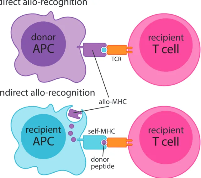
Direct and Indirect Allorecognition by T Lymphocytes
Today, Cerottoni’s findings can be described as the “Direct Alloresponse,” or the ability of a T cell to be activated by engaging with a foreign MHC protein30 (Figure 1.1). MHC Class I expressing graft endothelium represents the primary target of directly alloreactive CD8 T cells whereas MHC Class II expressing donor-derived passenger Macrophages, Dendritic Cells (DCs) and B lymphocytes represent the primary target of directly alloreactive CD4 T cells. Strikingly, research estimates that approximately 5-10% of the polyclonal T cell repertoire is comprised of directly alloreactive T cells31. Two hypotheses exist as to why such a large fraction of one’s T cell repertoire is comprised of directly alloreactive T cells. The High Determinant Density Hypothesis envisions that alloreactive T cells are selected for their ability to bind foreign MHCs regardless of the peptides they are loaded with32. As thymic selection for directly alloreactive T cells would be independent of peptide, a range of low, medium and high affinity T cells would be selected thus contributing to their high precursor frequency. Alternatively, The Multiple Binary Complex Hypothesis posits that a high number of directly alloreactive T cells could instead be selected by specific foreign MHC/peptide structures32. As foreign target cells express
Figure 1.1. Mechanisms of direct and indirect allorecognition by recipient T cells.
TCR
recipient APC
donor APC
donor peptide
allo-MHC
self-MHC
T cell
recipient
T cell
recipient direct allo-recognition
indirect allo-recognition
a variety of different self-peptides within the context of its own MHC haplotype, a host’s directly alloreactive T cell repertoire should be adequately diverse enough to engage with a variety of different foreign MHC/peptide complexes. Regardless of how the alloreactive repertoire is biologically shaped, it is generally appreciated that the acute transplant response, characterized by vascular endothelial destruction 1 to 12 weeks after transplantation, is perpetuated mainly by directly alloreactive T cells30 (Table 1). Conclusive evidence for direct allorecognition was demonstrated in 200033; transplantation of heart allografts into MHC Class II deficient / Severely Combined Immunodeficient (SCID mice, which possess a nonfunctional VDJ recombination element Protein Kinase, DNA activated, catalytic polypeptide [Prkdc]) recipients alongside adoptive transfer of CD4 T cells resulted in rejection only when the heart allografts expressed MHC Class II. Rejection was solely attributable to directly alloreactive CD4 T cells as these cells could not interact with host antigen presenting cells (APCs) to initiate graft rejection.
In addition to Direct Allorecognition, allografts can also be rejected more insidiously through a process termed the “Indirect Alloresponse” (Figure 1.1). Herein, host APCs process graft antigen and subsequently activate alloreactive T cells via presentation of foreign peptide in the context of self-MHC. In contrast to the direct alloresponse, the slowly evolving process of graft fibrosis characteristic of chronic rejection (which occurs anywhere from 3 months to decades after engraftment) is generally attributed to indirect allorecognition30. Initiation and perpetuation of the indirect alloresponse is generally thought to be slower for both anatomical reasons (graft antigen must travel to draining lymph nodes, be processed and presented by host APCs, which then must activate alloreactive T cells, which then must recirculate to the graft itself to initiate destruction) as well as the fact that the precursor frequency of indirectly
Table 1. Onset and immunologic mechanisms of graft rejection.
Type of Rejection Onset Immunologic Mechanism
Hyperacute Minutes to Hours
Caused by preformed antibodies to donor tissue (against mismatched ABO blood type or MHC) Antibodies bind endothelium and activate complement Causes rapid graft intravascular coagulation, thrombosis, and occlusion Acute Weeks to Months
Caused by the recipient’s directly and indirectly alloreactive T cells Alloreactive B cells produce alloantibody that deposits on graft endothelium
Results in graft inflammation of T cell infiltration
Chronic Months to Years Prolonged activation and effector function of alloreactive T and B cells Results in graft vessel intimal thickening, fibrosis, and atrophy
alloreactive T cells is nearly undetectable within a normal polyclonal T cell repertoire34. Conclusive evidence of rejection mediated solely by indirect allorecognition was demonstrated in MHC Class I knockout mice transplanted with skin allografts from MHC Class II deficient mice35. In this setting, graft rejection was restricted to recipient CD4 T cells that could only recognize foreign graft peptides in the context of self-MHC Class II expressing APCs. Despite its delayed initiation however, emerging evidence suggests that in comparison to anti-graft effector T cells generated by direct allorecognition, anti-graft effector T cells generated by indirect allorecognition proliferate more rapidly, take on stronger effector phenotypes, more readily traffic to the allograft, and persist longer as anti-graft memory cells30. Regardless of the primary means through which alloreactive T cells recognize and destroy graft tissue (direct vs.
indirect allorecognition), these endstage effector cells remain the primary barrier to long-term graft survival and function. The molecular means by which alloreactive T cells are activated and exert the their anti-graft effector function are reviewed below (see Mechanisms of Transplant Rejection by Alloreactive T and B cells).
Allorecognition Mediated by B Cells
Complementing the immensely diverse and strongly immunogenic alloreactive T cell repertoire, B lymphocytes can detect alloantigen and perpetuate graft rejection. Similar to T lymphocytes, B lymphocytes undergo VDJ Recombination from which up to 3x10¹¹ unique B Cell Receptors are generated, each possessing its own antigen specificity. Attempts to quantify the endogenous alloreactive B lymphocyte repertoire have proven difficult because of their low precursor frequencies even during active immunization36,37. However, as a single alloreactive B cell can exponentially give rise to numerous antibody-secreting plasma cells capable of secreting
thousands of alloantibodies per second, it is well appreciated that the alloreactive B cell repertoire becomes substantial after foreign MHC sensitization. In a study analyzing 15 HLA- sensitized multiparous women, precursor frequencies of HLA-alloantibody secreting B lymphocytes averaged as high as 43 per 106 B cells38. Although the pathogenic role of alloantibodies during graft rejection has yet to be fully elucidated, alloantibody can induce acute graft rejection via complement activation (C4d deposition) and neutrophil and macrophage recruitment (via Fcγ receptors), as well as chronic rejection via altering gene expression in graft endothelial cells ultimately leading to basement membrane remodeling and fibrosis39. Furthermore, B lymphocytes can present alloantigen indirectly to alloreactive CD4 T cells via MHC Class II40. A detailed description of the alloreactive B lymphocyte response during transplantation is discussed below (see Mechanisms of Transplant Rejection by Alloreactive T and B cells).
Mechanisms of Transplant Rejection by Alloreactive T and B Cells Alloreactive CD8 T Cells in Transplantation
As graft endothelium richly expresses foreign MHC Class I complexes, directly alloreactive CD8 T cells represent some of the earliest responders in the transplant response.
Immediately following transplantation, acute inflammation events result in the release of a panoply of cell attractant chemokines (CCL family) as well as the upregulation of the endothelial cell adhesion molecules (Intercellular Adhesion Molecule 1 [ICAM-1], Lymphocyte Function- Associated Antigen 1 [LFA-1]) 41,42. Subsequently, directly alloreactive CD8 T cells undergo vascular arrest and MHC/T Cell Receptor (TCR) ligation ensues. Whereas blockade of cellular integrins can delay or prevent allograft rejection43,44, blockade of specific chemokines or their
receptors has yet to provide any evidence of graft survival prolongation45-47, perhaps due to the often overlapping and redundant roles of these chemoattractant molecules. However, recent evidence suggests that the cognate interactions between MHCs/TCRs may in fact provide stronger signals for T cell graft arrest than traditional Gai-coupled chemokine receptor signaling pathways48. Specifically, in 2013 Walch and colleagues demonstrated that OT-I CD8 T cells (a transgenic model in which all CD8 T cells react with the Ovalbumin [OVA] derived peptide SIINFEKL loaded in the H-2Kb MHC) strongly arrested in OVA-expressing heart and kidney grafts even when Gai-coupled chemokine receptor signaling was chemically blocked. Moreover, similarly inhibited OT-I cells remained capable of precipitating graft rejection. Elucidating the interplay of cues that guide alloreactive CD8 T cell migration to allografts remains an active area of investigation.
In the event of adequate CD28/B7.1/2 and CD40/CD40L co-stimulation, alloreactive CD8 T cells take on different effector phenotypes49. High avidity TCR/MHC interactions result in the generation of CD8+ Tc1 cells capable of producing IL-2 and IFNγ independent of CD4 T cell help. In contrast, low avidity TCR/MHC interactions result in the generation of CD8+ Tc2 cells skewed toward IL-4 and IL-5 production. In the former case, transfer of Tc1 cells to immunodeficient RAG-/- recipients (Recombination Activating Gene null) receiving cardiac allografts perpetuated graft vasculitis and arteriopathy whereas transfer of Tc2 cells perpetuated increased graft eosinophil infiltration. Despite this dichotomy, characteristic of graft CD8 T cell infiltrate is upregulation of the activation markers CD44/CD69 and downregulation of the cell homing marker CD62L and the Protein Tyrosine Phosphatase (PTP) CD45RB50.
Direct destruction of graft tissue by alloreactive CD8 T cells is largely attributed to their release of inflammatory cytokines and cytolytic molecules. IFNγ production by alloreactive CD8
T cells not only results in graft damage, but also aids in the upregulation of graft MHC Class I expression (leading to enhanced alloantigen presentation), activation of CD4 T Helper cells, and recruitment of Macrophages and NK Cells51,52. In the presence of IFNγ, alloreactive CD8 T cells are capable of producing the chemokines IP-10 and Mig further augmenting neutrophil recruitment53. Although increased levels of granzyme B, perforin, and FasL have been reported in cases of acute kidney rejection54,55, the pathogenic role of these CD8 T cell associated cell- killing molecules remains in question. In a murine model of islet transplantation in which islet allografts were transplanted into immunodeficient SCID recipients, transfer of in vitro primed alloreactive CD8 T cells rapidly led to graft rejection even if they genetically lacked perforin or FasL51. In contrast, CD8 derived IFNγ was absolutely essential for graft rejection51. Moreover, in 2014, Zimmerer and colleagues demonstrated that animals genetically deficient for CD8 T cells (CD8α-/-) generated higher IgG1 alloantibody levels when challenged with fully MHC mismatched splenocytes56. Specifically, CD8 expression of FasL and perforin was required to eliminate alloreactive B cells in an MHC Class I dependent manner. In a broader context, however, the extent and intensity of alloreactive CD8 T cell mediated acute rejection depends largely on the type of organ allograft. Whereas islet and skin allografts appear to be exquisitely sensitive to alloreactive CD8 T cell mediated rejection, the brunt of cardiac and liver allograft rejection is perpetuated by alloreactive CD4 T cells57.
Alloreactive CD4 T Helper Cells in Transplantation
Since the discovery in 1986 that CD4 T cells are composed of unique “Helper Subsets”58, a significant body of research has since described the roles of CD4 T Helper cells in the transplant response. The largest body of investigation has centered on TH1 and TH2 cells.
Whereas the former subset is characterized by its expression of the master transcription factor T- bet which is driven by STAT4 signal transduction, the latter expresses the master transcription factor GATA3 induced by STAT6 signaling59. Immediately following allogeneic stimulation, TH1 cells secrete IL-2 and IFNγ leading to the activation of alloreactive CD8 T cells that are further capable of producing IFNγ60. Alloreactive TH1 cells can also perpetuate graft damage directly through Fas/Fas-L interactions as well as activate B cells to produce alloreactive antibody60. Although other models have associated TH1 cells with enhanced immunogenicity and TH2 cells with protective immunity (IL-4 and IL-10 production)61, this paradigm may not necessarily hold true during the transplant response. As informed by two complementary reports, in vitro differentiated TH1 and TH2 cells can independently reject skin62 or islet63 allografts when transferred to T cell deficient mice. IL-4 production by rejecting TH2 cells was accompanied by enhanced eosinophilic graft infiltrate, an observation consistent with acute rejection events. Further supporting the overlapping roles of CD4 T Helper Cells in allograft rejection, transfer of T-bet deficient T cells to immunodeficient skin allograft recipients failed to delay allograft rejection over the transfer T-bet sufficient cells64. Overall, evidence continues to suggest that both TH1 and TH2 cells are significant participants in acute and chronic rejection.
Nearly a decade prior to the initial discovery of TH17 cells in 200565 (characterized by the master transcription factor RORγt driven by IL-6, IL-23, HMGB-1, and TGFβ mediated STAT3 signaling), a report by Van Kooten and colleagues noted increased IL-17 staining in renal allograft biopsies taken from 6 patients experiencing acute rejection66. In 2007, Snell et al.
found that lung transplant recipients experiencing bronchioloitis obliterans syndrome (BOS) possessed high levels of IL-17 their in bronchoalveolar lavage biopsies67. Transient rises in serum IL-17 were then associated with acute rejection events in liver transplant recipients
(2009)68. However, the direct contribution of IL-17 producing TH17 cells to the transplant response would not be revealed until 2008. In work published by Yuan and colleagues69, the authors demonstrated that T-bet deficient animals displayed accelerated rejection of MHCII mismatched cardiac allografts with increases in IL-17 expressing CD4 TH17 infiltrate. In this setting, antibody mediated depletion of CD4 T cells restored graft tolerance while IL-17 blockade doubled the time to rejection. Further supporting the direct role of TH17 cells in allograft rejection, in 2013 Sabet-Baktach and colleagues found that only 40% of cardiac allografts were rejected when RORγt-/- T cells were transferred to immunodeficient recipients64. Transfer of RORγt sufficient (albeit T-bet deficient) T cells precipitated rejection in 100% of recipients. Although IL-17 itself is a pleotropic cytokine with numerous isoforms, its definitive role in transplantation has been linked to graft neutrophil recruitment59. In fact, depletion of neutrophils can delay cardiac allograft rejection in murine models70. Thus, although it appears that TH17 cells play a significant role in acute rejection, emerging evidence has further implicated these cells in chronic rejection. Of particular note, TH17 derived IL-17 has been noted to induce cardiac fibrosis71 and obliterative airway lesions72 in murine models and cardiac and lung allograft models, respectively. Research in TH17 mediated transplant rejection remains an active area of research, with recent findings implicating Platelet Factor 4 as a strong negative regulator of TH17 cells during cardiac transplantation73.
Finally, in comparison to the vast amount of research describing the roles of TH1, TH2, and TH17 cells in the transplant response, very few reports have so far defined a role for T Follicular Helper (TFH) cells in transplantation. Since the discovery of TFH cells in 200074 (which express the master transcription factor Bcl-6 and produce IL-4 and IL-21) a single report has demonstrated a mechanistic role for TFH cells during the transplant response. In 2012,
Conlon and colleagues demonstrated that only indirectly-alloreactive transgenic CD4 T cells (B6.TCR75, which bind the H-2Kd54-68 derived peptide loaded in self I-Ab) transferred to TCR-/- recipient hosts were capable of differentiating into CXCR5+PD-1+ TFH cells75. In the presence of B cells, these TFH cells perpetuated a long-lasting IgG alloantibody response. Directly- alloreactive transgenic CD4 T cells transferred to TCR deficient hosts receiving cardiac allografts failed to differentiate into TFH cells and no IgG alloantibody response was observed.
Although not directly attributed to attenuation of the TFH cell response, IL-21 Receptor blockade was recently demonstrated to enhance islet allograft tolerance when used in conjugation with anti-CD40L costimulatory blockade + Donor Specific Transfusion (DST) protocols76. Despite a still unclear role of TFH cells in the transplant response, emerging clinical data has found that acute renal rejection episodes are associated with increases in the presence of follicular like structures in kidney graft biopsies containing Bcl-6+ TFH cells and IgG+ B cells77.
Alloreactive B Cells in Transplantation
The pathogenic role of B cell derived alloantibody remains incompletely understood despite clinical data associating chronic rejection events with increased Donor Specific Alloantibody (DSA)39. A direct role for alloantibody in graft rejection was in part defined by Brandle and colleagues, who in 1998 found that when alloreactive T cells were inhibited by Cyclosporine A (CsA), passive transfer of alloantibody-rich serum from B cell sufficient mice rejecting cardiac allografts to B cell deficient hosts hastened graft rejection78. The necessity of CsA administration stemmed from their previous observation that B cell deficient and B cell sufficient hosts rejected cardiac allografts with similar kinetics. Only when T cell mediated rejection was attenuated via CsA did the B cell deficient hosts demonstrate delayed rejection
kinetics. In this setting, whether the absence of alloantibody was the primary determinant of delayed rejection was not directly answered. More recently, B cell rich tertiary lymphoid tissues (TLT) in chronically rejected kidney allografts were found to produce alloantibodies of diverging specificities in comparison to those alloantibodies generated in secondary lymphoid tissues79. The function and pathogenesis of TLT-derived alloantibody vs. secondary lymphoid tissue derived alloantibody remains unknown.
Beyond the perplexing role of DSA in transplantation, alloreactive B cells perpetuate acute and chronic rejection via presentation of alloantigen to T cells. In 2006, Noorchashm and colleagues demonstrated that in the absence B cell MHC Class II expression, median cardiac allograft survival reached > 70 days whereas the presence of MHC Class II expression on B cells rapidly precipitated rapid graft rejection (Median Survival Time 9.5 days)80. Mechanistically, the absence of alloantigen presentation by B cells resulted in attenuated alloreactive CD4 T cell activation and proliferation. In contrast, the absence of MHC Class II on B cells did not delay skin allograft rejection80, thereby bringing to light the diverging roles alloreactive B cells in the context of different organ allografts. Beyond acute rejection events, in 2014 Zeng et al demonstrated that alloreactive B cells promote chronic allograft rejection independent of alloantibody40. Using a model in which B cells were genetically unable to secrete alloantibody, the authors found that B cell alloantigen presentation via MHC Class I and II to alloreactive T cells was required to precipitate chronic cardiac allograft rejection. In the absence of MHC Class I/II mediated alloantigen presentation by B cells, alloreactive T cell IFNγ/TNFα production was attenuated, decreased graft infiltrate was observed, and minimal graft vasculopathy was noted.
Pharmacologic targeting of the B lymphocyte compartment during transplantation represents a new opportunity to abrogate rejection81. However, depletion of specific B cell
subsets leads to different patterns of rejection in murine models depending on the type of organ allograft. Whereas complete B cell, plasmablast, and plasma cell depletion achieved by an anti- CD19 antibody delayed renal allograft rejection and reduced IgG alloantibody levels, partial B cell depletion achieved by an anti-CD20 antibody did not extend renal allograft survival or reduce alloantibody levels82. B cell depletion by either CD19 or CD20 antibodies did not alter cardiac allograft rejection kinetics; however, skin allograft rejection was hastened in the absence of B cells. In the context of murine islet allografting, B cell depletion using a novel anti-CD22 antibody conjugated to the cell toxin calicheamicin (anti-CD22/cal) doubles islet allograft survival83. These results mirror findings that B6 mice genetically deficient of B cells (μMT) are slower to reject islet allografts donated by fully MHC mismatched Balb/c mice84. Alternatively, antibody mediated blockade of the B cell survival factor BAFF/Blys resulted in permanent tolerance of islet allografts when used in conjugation with Rapamycin (Rapamycin alone did not induce permanent tolerance)85. Although still in clinical trials, the B cell depleting anti-CD20 agent Rituximab/Rituxan (indicated for patients receiving HLA mismatched renal transplants) has shown some efficacy in reducing the severity of acute rejection events and preventing chronic alloantibody mediated rejection in high-risk patients86.
Immunologic Mechanisms of Transplantation Tolerance
"Immunological tolerance may be described as a state of indifference or non-reactivity towards a substance that would normally be expected to excite an immunological response.”
– Peter B. Medawar, 1960, Nobel Prize Acceptance Speech87
Foundational Studies in Transplantation Tolerance
Overcoming the aggressive transplant response is a daunting task. In work ahead of its time, however, Billingham, Brent, and Medawar would prove that permanent allogeneic tolerance could in fact be achieved between fully MHC disparate mice. Taking advantage of inbred mouse strains generated by George Snell at the Jackson Laboratories, Medawar and colleagues found that embroynic injection of allogeneic splenocytes (A Strain; today’s A/J H2-a) into d15 pregnant females (CBA Strain; H2-k) would permit permanent acceptance of A Strain skin allografts in these fetally inoculated mice after they reached adult age88. Moreover, CBA mice made tolerant to A-Strain mice were able to rapidly reject non-matched 3rd party skin allografts (AU Strain; today’s B6 H2-b) thereby demonstrating the antigen-specific nature of tolerance. Taking a step back, it is important to reflect on these experimental findings in the context of the time (1953); the function and classification of lymphocytes was essentially unknown, the role of the thymus had yet to be identified, and the identification of the MHC locus as the primary transplant antigen had yet to be made. Justifiably, Medawar received the Nobel Prize in 1960 for his pioneering work in immunological tolerance.
Just two years later, Main and Prehn would develop a radical protocol to render adult mice tolerant to allogeneic tissue. Aptly titled, “Successful skin homografts after the administration of high dosage X radiation and homologous bone marrow,” their work demonstrated that matching graft allotype to immune system allotype could in theory permit graft tolerance89. Such a means to induce graft tolerance did not come without cost however, as Simonsen, Billingham, and Brent quickly recognized the lethality of Graft vs. Host Disease (GVHD) that ensued from allogeneic Bone Marrow Transplantation (1957). From a clinical standpoint, the benefits of graft tolerance did not outweigh the lethality of uncontrollable GVHD.
Thus, developing an alternative means to safely induce graft tolerance was needed; however, it would take nearly 4 decades of research to reach this goal. Researchers would first need to mechanistically understand how the immune system responded to foreign tissue antigen before they were able to control it.
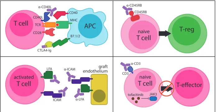
Pharmacologically Induced Transplant Tolerance
In the late 1980s, T cell depletion using a monoclonal antibody directed against CD3 (OKT3) was widely used in clinical transplantation90. Although similar anti-CD3 agents could induce transplant toleranceD in the preclinical murine setting91, the growing number of adverse events associated with total T cell depletion led clinicians to question its safety. Interest in preventing alloreactive lymphocyte homing to graft tissue gave way to the discovery in 1992 that antibody blockade of the cellular adhesion molecules ICAM-1 and LFA-1 could induce transplant tolerance to cardiac allografts in mice92. In 1996, Larsen and colleagues demonstrated that simultaneously blocking T cell co-stimulation using CTLA-4-Ig (which outcompetes B7.1/2 ligation to inhibit CD28 signaling in T cells) and an anti-CD40L antibody (clone MR1, which inhibits the CD40/CD40L co-stimulation axis in T cells) could induce transplant tolerance to skin and heart allografts93. In the same year, Lazarovits and colleagues found that an antibody directed against a splicing variant of the PTP CD45 (anti-CD45RB) could similarly induce
D Reference to induction of “transplant tolerance” herein describes an experimental setting in which the following criteria are met: 1) A short course of the therapy is used around the window of transplantation. Agents given continuously that induce a state of generalized immunosuppression are excluded. 2) Prolonged allograft acceptance is achieved in the majority of animals undergoing therapy (generally accepted as graft survival > 100 days in mice). 3) Placement of a matched allograft is accepted without further treatment whereas a 3rd party non- matched allograft is rejected with normal kinetics.
transplant tolerance to renal allografts in mice and non-human primates94. Although the exact mechanism of tolerance induction by anti-CD45RB remains unknown, its principal mechanism of action is thought to center around the generation of antigen-specific, graft-protective T Regulatory Cells (Tregs – described in detail below)95. More recently, discovery of the JAK-3 specific inhibitor Tofacitinib (CP-690,550), a small molecule that inhibits IL-2 mediated signal transduction in T cells, was demonstrated to induce transplant tolerance to cardiac allografts in mice96. It is important to note that this list of tolerance inducing agents is in no way exhaustive.
However, these selected agents each provide mechanistic insight into how transplant tolerance may be achieved: deletion of effector cells (anti-CD3), prevention of lymphocyte homing to graft tissue (anti-ICAM-1/LFA-1), interruption of T cell co-stimulation (CTLA-4Ig/anti-CD40L), induction of graft-protective T Regulatory Cells (anti-CD45RB), and inhibition of effector T cell activation (Tofacitinib) (Figure 1.2).
Transplantation Tolerance in the Clinic
Clinical Operational Tolerance (COT) is defined as an immunosuppression-free state in which a transplant recipient demonstrates prolonged allograft function in the absence of any signs of acute or chronic rejection after 1 year. It is currently predicted that up to 20% of liver allograft recipients may achieve this state after undergoing immunosuppression-withdrawal protocols97. Potential mechanisms driving COT in liver allograft recipients include 1) increases in peripheral blood resident γδT cells, CD4 Tregs, plasmacytoid and conventional DCs, and B Cells98-100, and 2) evidence that liver allograft endothelium can be replaced by recipient bone- marrow derived cells due to the high vascular turnover of this organ101. In the latter case, the vasculature of liver allografts would be viewed as “self” and consequently protected from
Figure 1.2. Mechanisms of pharmacologically induced transplant tolerance. A variety of preclinical agents can reliably induce permanent transplant tolerance in small animals models.
Mechanistically, these agents act via T cell costimulation blockade (upper left panel – anti- CD40L, CTLA4-Ig), generation of graft-protective T Regulatory Cells (upper right panel – anti- CD45RB), inhibition of activated T cell homing to graft tissue (lower left panel – anti-ICAM-1, anti-LFA-1), or deletion/inhibition of anti-graft T effector cell activation (lower right panel – anti-CD3, Tofacitinib).
T cell
activated
graft endothelium
ICAM LFA
α-LFA α-ICAM
T cell
naiveT-reg
CD45RB α-CD45RB
T cell APC
CD40 CD40L
TCR
MHC
CD28
CTLA4-Ig
B7.1/2 α-CD40L
T cell
naive T-effectortofacitinib JAK3 CD3
α-CD3