Restriction in Drosophila
Thesis By Brian M. Zid
In Partial Fulfillment of the Requirements For the Degree of
Doctor of Philosophy
California Institute of Technology Pasadena, California
2008
(Defended January 17, 2008)
© 2008 Brian M Zid All Rights Reserved
Acknowledgements
I would like to thank Seymour Benzer for taking me into his lab, giving me guidance and at the same time, freedom in pursuing my research. I especially value the insight I have gained from him on pursuing novel ideas in science and hope that this knowledge will guide me in my future research endeavors. I would like to thank Pankaj Kapahi for always being there to bounce ideas off of and argue with, and for bringing excitement to the science, even if the experiments weren’t always going as expected. I would like to thank Eimear Kenny for listening and helping me scientifically as well as non-
scientifically during my time at Caltech. I would like to thank my committee members, Ray Deshaies, Alex Varshavsky, Judy Campbell and, especially, Paul Sternberg for
always having their doors open to answer my questions and to have scientific discussions.
I would also like to thank members of the Benzer and Kapahi Labs as well as other members of the Caltech community for all their help.
Abstract
Aging is characterized by the declining ability of an organism to maintain homeostasis, which eventually leads to death. Dietary restriction (DR), the
reduction of nutrients without malnutrition, extends lifespan in various organisms, yet its molecular underpinnings are poorly understood. We show that in
Drosophila, DR upregulates the translational repressor 4EBP, the eukaryotic
translation initiation factor 4E binding protein, and that this upregulation is
necessary for the full lifespan extension upon DR and sufficient to extend lifespan on a nutrient rich diet. Investigation of the genome-wide translational changes upon DR using translation state array analysis (TSAA) found that translationally downregulated genes tend to have extensive 5’ untranslated regions (UTR) secondary structures, while those that are upregulated have weakly structured 5’UTRs. Among the translationally upregulated genes, mitochondrial ribosomal proteins and electron transport chain components were overrepresented.
Mitochondrial genes were found to have weakly structured 5’UTRs in Drosophila, and this was conserved in Humans. The 5’UTRs of mitochondrial genes were found to be sufficient to confer preferential translation during times of high 4EBP activity in a cap-independent manner to reporter constructs. Upregulation of mitochondrial function was verified and found to be d4EBP dependent, implicating a novel
mechanism for regulating mitochondrial function upon DR. These results implicate mRNA translation initiation in modulating lifespan and mitochondrial function upon DR.
Contents
Acknowledgements iii
Abstract iv Background 1
1 Aging ………. 1
1.1 Introduction ………... 1
1.2 Dietary Restriction ……… 1
1.3 Genetics of Aging ………. 2
1.31 Insulin Pathway ……….. 2
1.32 TOR Pathway ……….. 3
1.4 Genetics of Dietary Restriction ……….… 5
1.41 Sir2 ……….…. 5
1.42 TOR Pathway ……….…. 6
1.43 Insulin Pathway ……….. 6
1.5 Mitochondrial Respiration ……….... 7
2 Translation ……… 8
2.1 Translation Initiation ……….… 8
2.11 Cap Dependent Translation Initiation ……….… 8
2.12 Internal Ribosome Entry ………. 10
2.13 Cancer ………. 10
2.2 Lifespan ………. 11
Results 11
3 Introduction ……….. 11
4 4EBP and Dietary Restriction ………. 13
4.1 Protein Levels ……… 14
4.2 Lifespan ………. 14
4.21 4EBP Null ………….……….. 14
4.22 4EBP Overexpression ………. 20
4.3 Metabolic Changes ……… 22
4.4 Stress Resistance ………... 24
5 Genome Wide Translational Changes ………... 25
5.1 Translation State Array Analysis ……….. 25
5.2 5’UTR Analysis ……… 27
5.3 Mitochondrial Measurements ……… 30
6 5’UTR Analysis ……… 32
6.1 Conserved Gene Ontology Categories ……….. 32
6.2 In vivo Analysis of 5’UTR Function ……… 36
6.21 Monocistronic Reporter ……….. 37
6.22 Bicistronic Reporter ……… 39
Concluding Remarks 40
7 Summary of Results ………. 40
8 Discusion...……….. 42
8.1 IRES Translation ……… 42
8.2 Growth and Lifespan ..……….….…… 44
8.21 Multicellular Organisms ……….……….….….. 44
8.22 Yeast Growth Changes …….……….….……. 45 8.23 Mitochondrial Respiration...….….……… 46
Methods 49
Supplemental Tables 57
References 60
Background Chapter 1 Aging 1.1 Introduction
Aging can be defined as the progressive and irreversible decline in physiological function, which leads to an increased susceptibility to death. While it would seem much simpler for an organism to maintain itself in a healthy adult state compared to the complex development of an adult organism from a single sperm and egg, it is apparent this is not the case. In fact, a human’s chance of dying doubles every 8.9 years after reaching sexual maturity (Arking 1998). From an evolutionary perspective, aging may arise because of a lack of selective forces acting on organisms that have started reproducing.
In most wild populations organisms do not survive to a time in which they have decreased physiological functions because of mortality from environmental factors (Medawar 1952). This leads to a lack of selective pressure against deleterious mutations that may be harmful late in life (Medawar 1952).
1.2 Dietary Restriction
While there is no known way to halt aging, there are ways to delay or slow the aging process. One of the most well studied ways of slowing aging is dietary restriction (DR), the reduction of nutrients without malnutrition. This was first shown to extend the
lifespan of rats in the 1930s (McCay, Crowell et al. 1935). This procedure has since been shown to extend lifespan in yeast (Jiang, Jaruga et al. 2000), C. elegans (Klass 1977), Drosophila (Chapman and Partridge 1996), mice (Weindruch and Walford 1982), and is currently being tested in rhesus monkeys (Roth, Ingram et al. 2001). Along with
extending lifespan, DR slows the progression of many age related diseases including
cancer, diabetes, and cardiovascular disease (Weindruch and Walford 1982; Hursting, Lavigne et al. 2003). Upon DR, organisms are thought to undergo a shift in resource allocation from reproduction to somatic maintenance, which would allow an organism to sustain itself until nutrients are abundant again (Holliday 1989).
In mammals, DR is usually performed by restricting total calories ingested (Masoro 2002). In Drosophila it has been found that the composition of the nutrients restricted is important. While many labs use whole food dilution, the main determinant in lifespan extension from DR in Drosophila is yeast (Mair, Piper et al. 2005), the major amino acid source for the fly. It has also been found that Drosophila compensate their food intake dependent on the food dilution (Carvalho, Kapahi et al. 2005), with the carbohydrate content being a major determinant of this compensation (Kapahi unpublished).
1.3 Genetics of Aging
1.31 Insulin Pathway
Along with DR, single gene mutations extend lifespan in a variety of organisms. The Age-1 mutation in C. elegans was the first single gene mutation found to extend lifespan (Friedman and Johnson 1988). Age-1 is a mutation in the daf-23 gene, a dauer
constitutive gene. The dauer is an alternative form of C. elegans larvae induced during times of stress (Riddle and Albert 1997). While normal larvae progress to adulthood in
~3 days, and live for a couple weeks, dauers can be maintained for more than 70 days and then live a normal adult life (Klass and Hirsh 1976). Age-1 was later cloned and found to be the homolog of mammalian phosphatidylinositol-3-OH kinase (PI(3)K) catalytic subunit (Morris, Tissenbaum et al. 1996). It was found that another lifespan extension
mutant, daf-2, was the insulin/IGF-1 receptor ortholog (Kimura, Tissenbaum et al. 1997).
The dauer constitutive and lifespan phenotypes of daf-2 and age-1 are suppressed by mutations in the dauer defective gene, daf-16, a FOXO family transcription factor (Lin, Dorman et al. 1997; Ogg, Paradis et al. 1997), important for heat and oxidative stress resistance, fat metabolism, fertility, and metabolism (Larsen 1993; Finch and Ruvkun 2001). These, along with mutations in pdk-1, daf-18 (the worm homolog of human PTEN), akt-1, and akt-2, defined the insulin-like signaling pathway (ILSP) as a key regulator of lifespan in C. elegans (Ogg and Ruvkun 1998; Paradis and Ruvkun 1998;
Paradis, Ailion et al. 1999). Reduction of flux through the ILSP has been found to be a conserved lifespan extension pathway. In flies, mutants in the insulin receptor (InR) (Tatar, Kopelman et al. 2001) or the insulin receptor substrate (chico) (Clancy, Gems et al. 2001) extend lifespan, as well as overexpression of the daf-16 homolog (dFOXO) (Giannakou, Goss et al. 2004; Hwangbo, Gershman et al. 2004). In mice, lifespan was extended by both a fat-specific insulin receptor knockout (FIRKO) (Bluher, Kahn et al.
2003) or heterozygote knockout mice for the insulin-like growth factor type 1 receptor (Igfr1) (Holzenberger, Dupont et al. 2003).
1.32 TOR Pathway
Another nutrient sensing growth pathway that is parallel and interacting with the ILSP is the target of rapamycin (TOR) pathway. The Drosophila homologs of human Tsc1 (Hamartin) and Tsc2 (tuberin) function in vivo as a complex that controls growth and size in a cell-autonomous manner (Ito and Rubin 1999; Potter, Huang et al. 2001). Tsc2 acts as a GTPase-activating protein (GAP) for Rheb (Ras homolog enriched in brain), a small, highly conserved guanine triphosphatase (GTPase) (Inoki, Li et al. 2003). Rheb has
been demonstrated through genetic and biochemical analyses to function downstream of the tuberous sclerosis complex (TSC1/TSC2) and activate the kinase activity of TOR (Inoki, Li et al. 2003; Stocker, Radimerski et al. 2003). TOR regulates many processes important for cell growth including protein synthesis (Burnett, Barrow et al. 1998),
ribosome biogenesis (Powers and Walter 1999), and autophagy (Noda and Ohsumi 1998).
Two downstream effectors of TOR which affect protein synthesis are ribosomal S6 kinase (S6K) and the translational inhibitor eIF4E binding protein (4EBP). S6K was originally thought to affect translation by regulating the translation of 5’-tract of
polypyrimidine (TOP) mRNAs, but this has since been found to be untrue (Pende, Um et al. 2004). It has also been seen that S6K1-deficient mice that have a growth defect also have normal phospho-S6 levels, implicating a pathway independent of S6 in S6K’s role in growth regulation (Shima, Pende et al. 1998). A way in which S6K may exert its influence on growth is by phosphorylating the translation initiation factor eIF4B (Raught, Peiretti et al. 2004). Another downstream component of the TOR pathway is 4EBP, which is directly phosphorylated by TOR (Burnett, Barrow et al. 1998; Miron, Lasko et al. 2003). When TOR activity is low, 4EBP is hypophosphorylated and efficiently binds eIF4E, blocking cap-dependent translation (explained below). 4EBP is transcriptionally regulated by many types of stress including hypoxia (Liu, Roy et al. 2006), oxidative stress (Landis, Abdueva et al. 2004), starvation (Zinke, Schutz et al. 2002), and infection (Bernal and Kimbrell 2000). dFOXO is a transcriptional activator of d4EBP (Puig, Marr et al. 2003), and this activation is necessary for the transcriptional upregulation of d4EBP upon starvation and oxidative stress (Teleman, Chen et al. 2005). Increased d4EBP
expression is sufficient to rescue the oxidative stress sensitivity of dFOXO null flies (Tettweiler, Miron et al. 2005).
Similar to the ILSP, reduction of flux through the TOR pathway has been shown to extend lifespan in variety of organisms. Downregulation of the TOR pathway extends both replicative (the measure of how many buds, or daughter cells a single yeast produces) (Kaeberlein, Powers et al. 2005) and chronological lifespan (a measure of the time cells in a stationary phase culture remain viable) (Powers, Kaeberlein et al. 2006) in yeast. In C. elegans, downregulation of TOR, or raptor, a TOR-interacting protein, extended lifespan (Vellai, Takacs-Vellai et al. 2003; Jia, Chen et al. 2004). In Drosophila, overexpression of the negative regulators of TOR, dTsc1, dTsc2, or overexpression of dominant-negative dTOR or dS6K causes lifespan extension (Kapahi, Zid et al. 2004).
1.4 Genetics of Dietary Restriction
1.41 Sir2
There have been several genetic pathways which have been implicated in the lifespan extension due to DR. The first gene that was found to be necessary for DR was the silent information regulator 2 (Sir2) gene in yeast, an NAD-dependent histone deacetylase (Imai, Armstrong et al. 2000; Lin, Defossez et al. 2000). A deletion of Sir2 was
unresponsive to DR, while overexpression of Sir2 extended lifespan, and DR in this long lived strain gave no further benefit (Lin, Defossez et al. 2000). It was also found that increasing Sir2 homologs in C. elegans (Tissenbaum and Guarente 2001) and Drosophila (Rogina and Helfand 2004) also extends lifespan. In Drosophila, the Sir2 homolog dSir2 is necessary for the lifespan extension due to a DR paradigm in which the food was
diluted (Rogina and Helfand 2004). Recently there has been contention about the role of Sir2 in DR. The Kennedy and Kaeberlein groups have shown that in yeast if you delete the fob1 gene, Sir2 is not necessary for the lifespan extension due to DR (Kaeberlein, Kirkland et al. 2004).
1.42 TOR Pathway
The TOR pathway has also been shown to interact with DR in multiple organisms. In Drosophila, downregulation of the TOR pathway extended lifespan on a rich nutrient diet but not upon DR (Kapahi, Zid et al. 2004). Similarly, in yeast DR failed to further
increase the lifespan of the long lived tor1Δ line and sch9Δ (Kaeberlein, Powers et al.
2005). Sch9, a serine/threonine protein kinase involved in cell size and oxidative stress resistance (Fabrizio P 01, Tyers 02), has recently been shown to be a functional S6 kinase (Urban, Soulard et al. 2007).
1.43 Insulin Pathway
While the insulin pathway was originally postulated to be a molecular output for DR, as DR reduces insulin and IGF-1 levels in animals (Sonntag, Lynch et al. 1999; Roth, Lane et al. 2002; Heilbronn and Ravussin 2003), this has failed to be verified experimentally.
In Drosophila, while the DR response of chico is shifted towards higher nutrient
concentrations, it still responds to DR (Clancy, Gems et al. 2002). In C. elegans daf-16 mutants, which completely suppress the lifespan extension due to downregulation of the ILSP, respond normally to DR by nutrient dilution or in combination with the eat-2
mutant, a genetic means of DR (Lakowski and Hekimi 1998; Houthoofd, Braeckman et al.
2003).
1.5 Mitochondrial Respiration
One common, though still controversial proposal, on how DR may be working is by increased respiration. In yeast DR, decreasing the glucose concentration from 2% to 0.5% increases respiration and overexpression of Hap4, which switches the metabolism of the yeast from fermentation to respiration, extends lifespan (Lin, Kaeberlein et al.
2002). Inhibition of the TOR pathway by rapamycin treatment increases respiration by increasing the expression of the TCA cycle and oxidative phosphorylation genes
(Hardwick, Kuruvilla et al. 1999). The tor1Δ also increases respiration by increasing the translation of mitochondrial-encoded oxidative phosphorylation subunits, and this was found to be a primary means by which tor1Δ extends the chronological lifespan of yeast (Bonawitz, Chatenay-Lapointe et al. 2007). Also in yeast, reduction of oxidative
phosphorylation using the Complex III inhibitor antimycin A shortens lifespan, while adding 2,4- dinitrophenol, which uncouples ATP production from electron transport, increases chronological lifespan (Barros, Bandy et al. 2004). C. elegans respire more upon DR (Houthoofd, Braeckman et al. 2002; Bishop and Guarente 2007), and DR was found to upregulate skn-1, a transcription factor, that is necessary for the lifespan extension and increased respiration upon DR (Bishop and Guarente 2007). This upregulation of respiration was shown to be necessary for lifespan extension by administering two different Complex III inhibitiors, antimycin A and myoxithiazol, which reduced respiration and completely rescued the DR lifespan effect. These inhibitors were specific to DR, as there was no effect on the lifespan extension due to daf-2 or on the normal lifespan of the control (Bishop and Guarente 2007). It has also recently been shown that DR increases mitochondrial biogenesis and respiration in mice
by upregulating the expression of eNOS (Nisoli, Tonello et al. 2005), though the necessity of this upregulation on lifespan is still unknown.
Conversely, there is also data that mitochondrial respiration plays no part in the lifespan extension of DR and even that inhibition of mitochondrial respiration can extend lifespan.
In yeast replicative lifespan, it was found that in a more severe form of DR, functional mitochondria were not necessary for lifespan extension upon DR (Kaeberlein, Hu et al.
2005). In C. elegans there are many instances where decreasing mitochondrial function, by mutation and RNAi increase lifespan (Feng, Bussiere et al. 2001; Dillin, Hsu et al.
2002; Lee, Lee et al. 2003). It is interesting that this effect on lifespan is only seen if mitochondrial function is decreased while the worm is still developing (Dillin, Hsu et al.
2002; Rea, Ventura et al. 2007). This effect also depends on the level of inhibition, as high levels of inhibition are detrimental to the organism (Rea, Ventura et al. 2007).
Chapter 2 Translation 2.1 Translation Initiation
Gene expression can be controlled at many levels, including transcription, translation, and protein turnover. The regulation of translation, the conversion of the mRNA to protein by the ribosome and many accessory factors, is a key process for cell growth and proliferation (Jorgensen, Rupes et al. 2004) as well as during times of stress, reviewed in (Holcik and Sonenberg 2005). There are three main steps to translational control;
initiation, elongation, and termination.
2.11 Cap-dependent Translation Initiation
Translation initiation is the rate-limiting step for translation of most mRNAs (Sonenberg, Hershey et al. 2000). This is based on the fact that ribosomes are usually spaced along an mRNA at 80-100nt intervals. If elongation were limiting, they would spaced at 30nt intervals, the limit of putting ribosomes in tandem. Indeed, if you add a translational elongation inhibitor, such as cycloheximide, to make elongation limiting, that is what is seen (Sonenberg, Hershey et al. 2000). During normal conditions, most translation initiation begins with the interaction of the cap-binding complex (eIF4F) with the mRNA
“cap” structure, m7GpppN (where N is any nucleotide, p is phosphate and m is a methyl group), at the 5’ terminus. eIF4F is composed of three initiation factors, eIF4E, the cap- binding protein, eIF4A, an RNA helicase, and eIF4G, a scaffolding protein. eIF4G has binding sites for eIF4E and eIF3, thereby bridging the mRNA to the pre-initiation complex through eIF3’s direct contact with the 40S ribosomal subunit. The 43S pre- initiation complex is composed of the 40S ribosomal subunit which associates with eIF3 and eIF1A, and then is further bound by the ternary complex consisting of eIF2,
methionyl-initiatior tRNA (Met-tRNAiMet), and GTP.
Once the 40S subunit is bound to the mRNA it scans the 5’untranslated region (UTR) from 5’ to 3’, an ATP dependent process, until it finds an initiation codon (AUG) in the right sequence context (Kozak 1980). Upon finding the initiation codon, the 60S
ribosomal subunit joins the complex to form the 80S ribosome and translation elongation commences. Cap-dependent translation is enhanced by circularization of the mRNA and interaction with the poly(A) tail through eIF4G’s interaction with the polyA binding protein (PABP) (Kahvejian, Svitkin et al. 2005).
2.12 Internal Ribosome Entry
An alternative means of translation initiation that is independent of the 5’ cap and eIF4E is internal ribosome entry site (IRES)-mediated translation initiation. While the
mechanism of IRES translation is still unclear, it is known that the cap and eIF4E are not necessary for the translation of a subset of mRNAs. This type of translation was first observed in Picornavirus infections, where the RNAs of the polio and
encephalomyocarditis virus were translated in eukaryotic cells even though they were uncapped (Jang, Krausslich et al. 1988). Viral proteases were found to cleave eIF4G to a form that does not bind eIF4E (Gradi, Svitkin et al. 1998). Other mechanisms that reduce cap-dependent translation initiation include increasing or hypophosphorylating eIF4E binding proteins (4EBPs), which compete with eIF4E for the binding site on eIF4G, as well as hypophosphorylation of eIF4E, which reduces eIF4Es affinity for eIF4G. Along with viral infections, cap-dependent translation is reduced during mitosis (Pyronnet, Dostie et al. 2001) as well as during many types of stress (Holcik and Sonenberg 2005).
2.13 Cancer
Translation initiation plays a key role in growth control and cancer. This first became evident when eIF4E overexpression was shown to cause cellular transformation (Lazaris- Karatzas, Montine et al. 1990). eIF4E is also upregulated in a broad spectrum of cancers (Petroulakis, Mamane et al. 2006), as well promotes tumor formation in vivo (Ruggero, Montanaro et al. 2004). eIF4E is thought to promote carcinogenesis by differential translation of mRNAs. Transcripts with extensive secondary structure in their 5’
untranslated regions (UTRs) are particularly sensitive to the activity of the cap binding complex (Koromilas, Lazaris-Karatzas et al. 1992). Many genes with high 5’UTR
secondary structure are enhanced translationally in vivo upon increased eIF4E expression (Mamane, Petroulakis et al. 2004), and many oncogenes, growth factors and regulatory proteins have 5’UTRs with high secondary structure (Kozak 1991). Recently 4E-BP1 has been postulated as a funnel factor for cancer, as its phosphorylation status is associated with malignant progression in a large variety of cancers regardless of the upstream oncogenic alterations (Armengol, Rojo et al. 2007).
2.2 Lifespan
Recently multiple labs have shown that inhibiting translation can extend lifespan. In yeast, deletion of ribosomal protein subunits are sufficient to extend replicative lifespan (Kaeberlein, Powers et al. 2005). In C. elegans, inhibition of multiple translation initiation and elongation factors as well as many ribosomal protein subunits extend lifespan (Henderson, Bonafe et al. 2006; Curran and Ruvkun 2007; Hansen, Taubert et al.
2007; Pan, Palter et al. 2007). This lifespan extension due to reducing protein synthesis was found to be independent of daf-16 (Curran and Ruvkun 2007; Pan, Palter et al. 2007).
Results
Chapter 3 Introduction
Dietary restriction (DR), the reduction of nutrient intake without malnutrition, is a
method of lifespan extension conserved from yeast to mammals (Masoro 2002). DR also slows the progression of many age-related diseases, including cancer (Hursting, Lavigne et al. 2003). It has been suggested that DR extends lifespan by inducing a shift from growth and reproduction towards somatic maintenance (Holliday 1989). It has become apparent in recent years that nutrient sensing growth pathways are key regulators of
lifespan (Longo and Finch 2003). Two parallel and interacting pathways which affect lifespan in diverse species are the insulin-like signaling pathway (ILSP) and the target of rapamycin (TOR) pathway (Kapahi and Zid 2004; Kenyon 2005). Both of these
pathways integrate nutrient and other environmental signals to mediate growth, and have been implicated in lifespan extension by DR (Clancy, Gems et al. 2002; Kapahi, Zid et al.
2004; Kaeberlein, Powers et al. 2005).
One common downstream component of both the ILSP and the TOR pathways is the translational repressor 4EBP. Decreased ILSP signaling transcriptionally upregulates 4EBP via the forkhead transcription factor FOXO (Puig, Marr et al. 2003), while decreased TOR activity causes hypophosphorylation of 4EBP that increases its affinity for eIF4E (Beretta, Gingras et al. 1996). 4EBP disrupts the interaction between eIF4E and eIF4G, which are components of the eIF4F cap-binding complex that mediates the initiation of mRNA translation (Richter and Sonenberg 2005). Transcripts with extensive secondary structure in their 5’ untranslated regions (UTRs) are particularly sensitive to the activity of the cap binding complex (Koromilas, Lazaris-Karatzas et al. 1992).
Recently, it has been shown in C. elegans that downregulation of components of the cap binding complex extends lifespan (Henderson, Bonafe et al. 2006; Hansen, Taubert et al.
2007; Pan, Palter et al. 2007; Syntichaki, Troulinaki et al. 2007). In mammals, eIF4E overexpression has oncogenic properties (Lazaris-Karatzas, Montine et al. 1990; Ruggero, Montanaro et al. 2004), and upregulation of eIF4E is found in a broad spectrum of
cancers (Petroulakis, Mamane et al. 2006). Since 4EBP and eIF4E are regulated by a
diverse array of nutrient, stress, and mitogenic signals, we investigated the role of 4EBP in lifespan extension by DR in Drosophila.
Chapter 4 4EBP and Dietary Restriction
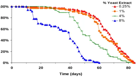
Reducing the concentration of yeast or yeast extract in the fly diet has been shown to extend lifespan (Nusbaum and Rose 1999; Kapahi, Zid et al. 2004; Mair, Piper et al. 2005) (Fig. 1).
0%
20%
40%
60%
80%
100%
0 20 40 60 8
Time (days)
% Survival
0 0.25%
1%
4%
8%
% Yeast Extract
Figure 1 Lifespan of control male flies on diets with varying amounts of yeast extract and constant sucrose.
4.1 4EBP Protein Levels
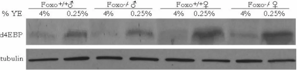
We examined the levels of d4EBP under a paradigm of DR in which the yeast extract (YE) was varied while sucrose, the major carbohydrate source, was constant. As the YE concentration decreased from 4% to 0.25%, both male and female flies showed
upregulation in d4EBP protein levels (Fig. 2). This upregulation was not dependent on FOXO, as dFOXO null flies showed a normal response (Fig. 2).
Figure 2 4EBP protein is induced upon DR. 4EBP protein is induced on 0.25% YE (DR) as measured by western blot using 30μg of protein probed with a polyclonal anti- d4EBP. This induction is not dependent on dFOXO, as dFOXO null flies show a normal response. β-tubulin levels were measured as a loading control.
4.2 Lifespan
4.21 4EBP Null
We next asked whether the upregulation of 4EBP is necessary for lifespan extension upon DR. To do this we used two strains, a d4EBP null line, created by imprecise excision of the P-element insertion Thor1, which is inserted in d4EBP (Bernal and Kimbrell 2000), and a control strain created by precise excision of the same P-element line. Concentrations of YE were varied from 0.1% to 5%, with maximal average lifespan at 0.25% YE for males and 1.5% YE for females (Fig. 3a, b and Table S1). Control flies showed lifespan extension of 27% in males and 32% in females compared to 5% YE (Fig.
3a, 3b). In contrast, d4EBP null flies showed a diminished response across all nutrient concentrations (Fig. 3a, b and Table S1).
a
b
Figure 3 4EBP is necessary for full lifespan extension upon DR in Drosophila.
Lifespan of male and female revertant and d4EBP null flies on various YE concentrations.
a, Male control and d4EBP null flies. b, Female control and d4EBP null flies.
To confirm that lack of d4EBP was causal for the diminished DR response, wild-type d4EBP was ubiquitously expressed with daughterless-Gal4, using the Gal4-UAS system (Brand and Perrimon 1993) in the d4EBP null background. First, the RNA levels of 4EBP were measured using qRT-PCR. Null flies contained very minimal amounts of 4EBP, by qRT-PCR criteria, while flies containing UAS-4EBPwt and the da-Gal4 driver
returned the 4EBP levels to an amount similar to control flies in both males and females.
This expression of 4EBP rescued the DR lifespan response from 8% in 4EBP null males to 37% and from 13% in 4EBP null females to 48%. This was similar to the 35% and 42% DR lifespan extension seen in control males and females, respectively (Fig. 5a, 5b and Table S2).
4EBP RNA Levels
0.0 0.5 1.0 1.5
♂ ♀
Relative 4EBP Levels
+;4EBPwt/+
Null;4EBPwt/+
Null;4EBPwt/da
Figure 4 Rescue of 4EBP RNA levels using the UAS-Gal4 system. qRT-PCR of d4EBP, in male and females, with values normalized to Actin5C, and the background set as control flies with UAS-4EBPwt (n=2).
a
0%
20%
40%
60%
80%
100%
0 20 40 60 8
Time (days)
% Survival
0
null;4EBPw t/+
null;4EBPw t/+
null;4EBPw t/da null;4EBPw t/da +;4EBPw t/+
+;4EBPw t/+
♂
b
0%
20%
40%
60%
80%
100%
0 20 40 60 80
Time (days)
% Survival
null;4EBPwt/+
null;4EBPwt/+
null;4EBPwt/da null;4EBPwt/da +;4EBPwt/+
+;4EBPwt/+
♀
Figure 5 Replacing 4EBP rescues the 4EBP null DR lifespan effect. a,b Rescue of DR effect by ubiquitously expressing d4EBP using da-gal4 in the 4EBP-null background.
Solid symbols DR (0.25% YE), empty symbols control (5% YE) a, Male 4EBP-null flies have an 8% lifespan extension upon DR. Control flies have a lifespan extension of 35%, and putting 4EBP back in the null line gives a lifespan extension of 37%. b, Female 4EBP-null flies have a 13% lifespan extension upon DR. Control flies have a lifespan
extension of 42%, and putting 4EBP back in the null line gives a lifespan extension of 48%.
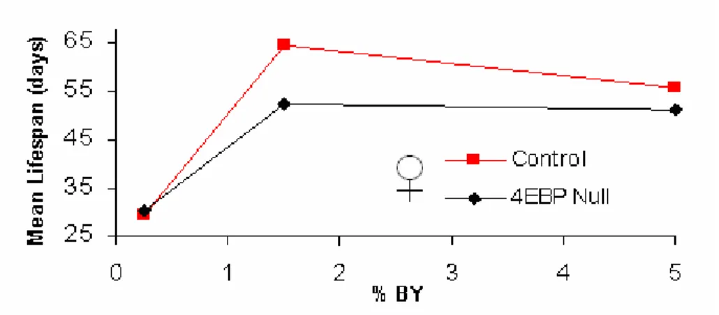
As many labs use different yeast sources in fly media, we also investigated the lifespan effect due to DR using Brewer’s Yeast (BY). Male and female control flies had a 24%
and 15% lifespan increase upon DR with BY, while neither male nor female d4EBP nulls showed a significant change in lifespan upon DR (Fig. 6a, 6b and Table S3). While an inverse correlation between reproduction and lifespan extension has been noted
(Partridge, Gems et al. 2005), recent experiments have shown that this link can be decoupled (Dillin, Crawford et al. 2002; Mair, Sgro et al. 2004). We found that both control and d4EBP null flies showed similar reductions in egg production upon YE restriction despite their differences in lifespan (Fig. 7). These observations suggest that d4EBP is necessary for the full lifespan extension upon DR, but not for reproductive changes.
a
b
Figure 6 d4EBP is necessary for the lifespan extension due to DR using Brewer’s
Yeast. Lifespan of male and female revertant (control) and d4EBP null flies on various YE concentrations. a, Male control and d4EBP null flies. b, Female control and d4EBP null flies.
Figure 7. 4EBP null flies show normal fecundity response to variations in YE.
d4EBP revertant (control) and d4EBP null flies both show increased egg laying in a dose dependent manner when YE is increased. 5 females and 3 males were put into individual
vials within 24 hrs of eclosion. The number of eggs laid were measured daily from day 4 to day 8 and averaged per fly per day ±SEM (n=5).
4.22 4EBP Overexpression
To ascertain whether elevated levels of 4EBP are sufficient to extend lifespan, a wild- type d4EBP (d4EBPwt), as well as two activated alleles of d4EBP, which bind more strongly to eIF4E (Miron, Verdu et al. 2001), were overexpressed with the ubiquitously expressed driver, armadillo-Gal4. The two activated alleles were previously classified as strong (d4EBPs) and weak (d4EBPw) based on their growth inhibition properties.
Overexpression of d4EBPwt caused no change in lifespan on rich food in males or females, while overexpression of the weak activated allele extended mean lifespan of females on rich food, but no significant lifespan extension was observed in male flies.
Induction of the strong allele extended both male and female lifespan on rich food (Fig.
8a, b and Table S4). In contrast, under DR (0.25% YE), there was no lifespan extension, beyond the effect of DR alone, in all of the 4EBP alleles tested (Fig. 8c, d and Table S4).
These observations are consistent with the hypothesis that lifespan extension during DR is mediated by an increase in d4EBP activity.
a
0%
20%
40%
60%
80%
100%
0 20 40 60 8
Time (days)
% Survival
0 4EBPwt/arm
4EBPw/arm 4EBPs/arm +/arm 4EBPwt/+
4EBPw/+
4EBPs/+
11%
p<0.0001
♂
5%YEb
0%
20%
40%
60%
80%
100%
0 20 40 60 8
Time (days)
% Survival
0 4EBPwt/arm
4EBPw/arm 4EBPs/arm +/arm 4EBPwt/+
4EBPw/+
4EBPs/+
22%
p<0.0001
14%
p<0.0005
♀
5%YEc
0%
20%
40%
60%
80%
100%
0 20 40 60 80
Time (days)
% Survival
4EBPwt/arm 4EBPw/arm 4EBPs/arm +/arm 4EBPwt/+
4EBPw/+
4EBPs/+
♂
0.25%YEd
0%
20%
40%
60%
80%
100%
0 20 40 60 80
Time (days)
% Survival
4EBPwt/arm 4EBPw/arm 4EBPs/arm +/arm 4EBPwt/+
4EBPw/+
4EBPs/+
♀
0.25%YEFigure 8 Overexpression of activated d4EBP extends lifespan in a nutrient-
dependent manner in Drosophila. Male flies overexpressing d4EBPLLs and female flies overexpressing d4EBPw or d4EBPs extend lifespan on high nutrition (5%YE) but not under DR (0.25% YE). a, Survival of male flies on high nutrition. b, Female flies on high nutrition c, Male flies under DR. d, Female flies on high nutrition. “+” is the Benzer Lab w1118 strain, into which each of these lines was outcrossed 6x. P values
were obtained by comparing the survival curves with GraphPad Prism Software using the longest lived control.
4.3 Metabolic Changes
To understand how 4EBP may be modulating the organism under DR, we investigated the composition of male flies. Upon DR there is a similar trend towards mass reduction in both the control and 4EBP null (Fig. 9a). The levels of total protein per mg of fly were also reduced in both lines upon DR, though 4EBP null flies had 20% more protein per mg of fly on DR compared to the control (Fig. 9b). Next, the storage metabolites,
triglycerides, and glycogen were measured. Upon DR there is an increase in triglycerides in both control and null flies, while the null flies have lower levels of triglycerides on both diets compared to the control (Fig. 9c). Glycogen levels were also found to increase upon DR in control and null flies, yet while 4EBP played no role in glycogen levels upon high nutrition, there was an almost 70% decrease in glycogen in flies missing 4EBP on DR (Fig. 9d).
a b
Mass Upon DR
0.6 0.65 0.7 0.75
Control 4EBP Null
mass/fly
4% YE 0.25% YE
*
Protein Content Upon DR
0 10 20 30 40 50
Control 4EBP Null
Protein/mg Fly
** ** **
c d
Triglyceride Content Upon DR
0 0.05 0.1 0.15 0.2 0.25
Control 4EBP Null
Triglyceride/Protein **
*** * ***
Glycogen Content Upon DR
0 0.03 0.06 0.09 0.12 0.15 0.18
Control 4EBP Null
Glycogen/protein
***
***
***
Figure 9 DR induces a shift towards increased storage metabolites that is altered in male flies lacking 4EBP. a, Control and 4EBP null flies have a trend towards decreased mass upon DR. b, DR decreases the protein content of the fly upon DR, though 4EBP null flies have more protein on DR compared to control flies. c, DR increases the triglyceride content of the control and 4EBP null flies. 4EBP null flies have decreased triglyceride contents on both food concentrations. d, On high nutrition control and 4EBP null flies have the same levels of glycogen. Upon DR controls have a 9 fold increase in glycogen levels, while 4EBP null flies increase only 3 fold.
4.4 Stress Resistance
It has previously been seen that 4EBP null flies are sensitive to starvation (Teleman, Chen et al. 2005; Tettweiler, Miron et al. 2005). As 4EBP null flies have altered composition that is diet-dependent, the starvation resistance of control and 4EBP null flies after 6 days on high nutrition or DR was investigated. While control flies have a 26% increase in starvation resistance upon DR, 4EBP null flies are sensitive to starvation and have no benefit from DR (Fig 10). This data implicates a shift in metabolism
towards storage metabolites upon DR, which 4EBP nulls are deficient in.
0%
20%
40%
60%
80%
100%
0 20 40 60 8
Time (Hrs)
% Surviva
0
l
0.25% Con 4% Con 0.25% Null 4% Null
Figure 10 Starvation resistance increases upon DR in a 4EBP dependent manner.
Starvation resistance of 6 day old male flies was measured on 1% agarose. Control flies show increased stress resistance in 0.25% YE, while 4EBP null flies are sensitive on all nutrient conditions and show no benefit from DR. Con 4% - 43.7 hrs (n=118), 0.25% - 55.1 hrs (n=106), 4EBP Null 4% - 25.2 hrs (n=103), 0.25% - 24.9 hrs (n=105).
Chapter 5 Genome Wide Translational Changes 5.1 Translation state array analysis
Given the role of 4EBP in regulating the translation initiation factor eIF4E, we
investigated the translational changes occurring under DR. Initiation, the binding of the ribosome to mRNA, is the rate-limiting step for the translation of most mRNAs
(Sonenberg, Hershey et al. 2000). Hence, the relative translation rate of an mRNA can be inferred from the number of ribosomes (polysomes) bound to it. To analyze the
translation profile of Drosophila, polysomes were fractionated according to size over sucrose gradients. Under DR, there was an overall reduction in the number of polysomes and ribosomal subunits (Fig. 11). Though overall translation was reduced upon DR,
translation state array analysis (TSAA) (Zong, Schummer et al. 1999; Arava, Wang et al.
2003) was performed to investigate if individual mRNAs might be comparatively up and downregulated upon DR. Sucrose gradients containing resolved polysomes were
fractionated to contain low (1 to 4) or high (5 or more) numbers of ribosomes bound per transcript. Each fraction was then hybridized to an Affymetrix microarray chip to determine the relative abundance in the high and low fractions.
Figure 11 Polysomal distribution of mRNAs of male flies on 4% YE and 0.25% YE Individual ribosomal subunits and the polysome peaks are noted, normalized to body weight of flies. RNA was prepared from the low (Lo) and high (Hi) translation fractions for microarray analysis performed in triplicate.
Using a false discovery rate (FDR) of less than 5%, 55 genes were translationally downregulated and 201 upregulated upon DR. We used Gene Ontology (GO)
classification (Ashburner, Ball et al. 2000) to identify biological themes overrepresented in the differentially translated genes. Categories enriched among the downregulated genes were carboxylic acid, cellular biosynthesis, and carbohydrate metabolism genes, while increased translation genes were enriched for mitochondrial ATP generation, oxidative phosphorylation, and protein folding genes (Table 1). Components of the mitochondria overrepresented were nuclear encoded Complex I and IV subunits of the electron transport chain and mitochondrial ribosomal proteins. Mitochondrial ribosomal proteins are necessary for the translation of mitochondrially encoded electron transport chain subunits.
Table 1 Biological Processes Translationally Changed Upon DR
5.2 5’UTR Analysis
eIF4E regulates the translation of mRNAs with extensive 5’UTR secondary structure (Koromilas, Lazaris-Karatzas et al. 1992). To assess the 5’UTR secondary structures of the differentially translated genes, theoretical folding free energies (ΔG) were calculated using DAMBE Software (Xia and Xie 2001). While exact folding energies would need to be experimentally calculated, DAMBE was used to estimate the folding energies for the individual mRNAs. The mean ΔG for the 5’UTRs of all mRNAs in the Drosophila genome is -47 kcal/mol. Under DR, translationally downregulated mRNAs have highly structured 5’UTRs with an average ΔG of -69 kcal/mol, whereas upregulated mRNAs have significantly less secondary structure, averaging -20 kcal/mol (Fig.12a, Table 2).
Since many genes from Complexes I and IV, and mitochondrial ribosomal proteins, were translationally upregulated, we examined the 5’UTR secondary structures of all subunits of these complexes and observed that most possess low secondary structure (Fig. 12b, Table 2). Upon further analysis, it was also found that translationally downregulated genes have, on average, longer 5’UTRs then the rest of the genome. Translationally upregulated and mitochondrial 5’UTRs were found to be shorter, with lower GC content then the rest of the genome (Table 2).
a
0%
10%
20%
30%
40%
50%
>-10 -10-30 -30-70 <-70 Folding Energy (kcal/mol)
%mRNAs
Whole Genom e Decreased Increased
b
0%
10%
20%
30%
40%
50%
60%
>-10 -10-30 -30-70 <-70
Folding Energy (kcal/mol)
%mRNAs
Whole Genom e Com plex I Com plex IV m RPs
Figure 12 5’UTR secondary structure analysis of differentially translated mRNAs upon DR and mitochondrial genes. a, Theoretical 5’UTR folding free energies, ΔG, were calculated for mRNAs which showed higher or lower translation ratios and the whole genome. The distribution of ΔG’s are shown for each class of translation regulation. b, 5’UTR folding free energies of genes in Complex I, Complex IV and ribosomal proteins of the mitochondria. P-values were calculated by generating sampling distributions for each experimental distribution, as described in the methods.
Table 2 Structural properties of the 5’UTRs of translationally regulated genes
N
ΔG(kcal/mol)
±SEM P-value
Length (bp)
±SEM P-value
%GC
±SEM P-value Whole Genome 9544 -46.7 ± 0.6 232 ± 3 42%±0.1%
Down 49 -69 ± 10 <0.01 342 ± 44 <0.01 44%±1% 0.122 Up 146 -20 ± 3 <0.0001 130 ± 21 <0.0001 37%±1% <0.0001 ComplexI 31 -21 ± 5 <0.01 129 ± 19 <0.01 39%±1% <0.01 ComplexIV 12 -7 ± 1 <0.05 74 ± 8 <0.01 36%±2% <0.01 mRP 64 -8 ± 1 <0.0001 75 ± 4 <0.0001 34%±1% <0.0001
5.3 Mitochondrial Measurements
These findings suggest coordinate translational upregulation of the mitochondrial electron transport chain. To validate and further investigate this finding, we examined protein levels or activity for mitochondrial Complexes I and IV under normal and DR conditions and in the presence and absence of d4EBP. Protein levels of CG12079, a Complex I subunit with low 5’UTR secondary structure, were measured by western blot.
Under DR, CG12079 protein levels increased 65% in control flies, while d4EBP nulls showed a smaller, non-significant increase upon DR and appeared to have reduced levels on both nutrient conditions (Fig. 13a). There were no significant changes in transcript levels of CG12079 upon manipulation of nutrients or d4EBP (Fig. 13b). The coordinate upregulation of Complex IV subunits was investigated by measuring cytochrome C oxidase (COX) activity. Upon DR, control flies showed a 60% increase in COX activity, while in d4EBP mutants COX activity was unchanged (Fig. 13c). Microarray analysis of total RNA from control flies upon DR shows no overall transcriptional changes in
Complex IV subunits (Fig. 13d). The respiration of flies upon DR was also investigated.
While control flies have a small but significant increase in CO2 output, 4EBP null flies show no significant difference in respiration upon DR and have reduced respiration on
low nutrition compared to control flies (Fig. 14). Similar to the lifespan experiments, where a small, but consistent DR response was seen in d4EBP nulls, both the Complex I subunit levels and COX activity showed a trend towards upregulation upon DR in d4EBP nulls. This may be because of 4EBP independent regulation of eIF4E, such as
phosphorylation (Arquier, Bourouis et al. 2005; Reiling, Doepfner et al. 2005), or alternatively there may be 4EBP/eIF4E independent pathways which also affect lifespan and mitochondrial function upon DR. Regardless, these data clearly show a post-
transcriptional upregulation of the mitochondria under DR that is partially d4EBP dependent and support our TSAA results that mitochondrial mRNAs are translationally upregulated upon DR.
a b
c d
Figure 13 DR induces the mitochondrial electron transport chain without affecting transcription a, Western blot of mitochondrial Complex I subunit, CG12079, in male revertant (control) and d4EBP null flies upon DR, with values normalized to tubulin (±SEM) (n=4). b, qRT-PCR of CG12079, in male control and d4EBP null flies upon DR, with values normalized to Actin5C (±SEM) (n=4). c, Cytochrome C oxidase (COX) activity measured on crude homogenates in male control and d4EBP null flies upon DR, normalized to protein content (±SEM) (n=7). d, Lack of transcriptional changes in genes of Complex IV upon DR. The ratio of total RNA on 0.25% YE vs 4% YE in control flies using Affymetrix microarrays. Complex IV gene list from the MitoDrome database (www2.ba.itb.cnr.it/MitoDrome) ( *p<0.05, ** p<0.01, *** p<0.001).
0.35 0.375 0.4 0.425
Control Null
CO2/mg fly
4% YE 0.25% YE
* **
Figure 14 Respiration increases upon DR in a 4EBP dependent manner. CO2
output of control and 4EBP null flies on 4% and 0.25% YE normalized to fly mass. (n=5) ( *p<0.05, ** p<0.01).
Chapter 6 5’UTR Analysis
6.1 Conserved Gene Ontology Categories
To further understand the biological role that secondary structure of 5’UTRs may play in Drosophila, we investigated the overrepresented biological processes of mRNAs which had strong predicted secondary structure (ΔG > -120 kcal/mol) and weak predicted 5’UTR secondary structures (ΔG > -5 kcal/mol). Previously it has been seen that in mammals signal transduction and growth genes have strong 5’UTR secondary structure (Kozak 1991). Similarly, upon investigation of the Drosophila genome, gene classes overrepresented for strong 5’UTR secondary structure include development, signal transduction, regulation of metabolism, growth, and transcription factors (Table 4). As the original mammalian 5’UTR analysis used a small subset of mRNAs, we analyzed the current annotation of 5’UTRs for the Human Genome (Table 4). There was strong
conservation in the overrepresentation of gene classes between Drosophila and Human including developmental, protein kinases, and regulation of metabolism in the human genome. Upon analysis of Drosophila weakly structured 5’UTRs, the immune response, electron transport, and proteolysis were all biological processes that were overrepresented (Table 5). In Human 5’UTRs, immune response, proteolysis, aerobic respiration, and the TCA cycle were all processes that were overrepresented for low structured 5’UTRs (Table 5).
Table 4
Table 5
As we have previously seen that mitochondrial genes were translationally upregulated upon DR in Drosophila and aerobic respiration was one of the classes overrepresented among human mRNAs, all the 5’UTRs for Complex I, IV, and the mitochondrial ribosomal proteins were further analyzed. Similar to Drosophila, we found that each of these complexes had a shift towards reduced 5’UTR secondary structure compared to the rest of the genome (Fig. 15).
Human 5'UTR Distribution
0%
10%
20%
30%
40%
50%
60%
> -20 -20 - -50 -50 - -100 -100 - -175 <-175 Folding Energy (Kcal/mol)
% mRNAs
All 5'UTRs Complex I Complex IV mRPs
Figure 15 5’UTR secondary structure analysis of human mitochondrial genes a, Theoretical 5’UTR folding free energies, ΔG, for genes in Complex I, Complex IV, and ribosomal proteins of the mitochondria.
6.2 In vivo Analysis of 5’UTR Function
The importance of the various 5’UTRs on translational control were investigated using luciferase reporter constructs. 5’UTRs were cloned upstream of the Firefly Luciferase (FLuc) gene under the control of the Act5C promoter (Fig. 16a). These constructs were then transfected into S2 cells with an internal control Renilla Luciferase (RLuc) vector as well as either an activated allele of 4EBP (4EBPLLAA) or a control, GFP construct, both under the Act5C promoter. Using various UTRs which had either strong, medium, or weak secondary structure (Table 6), we investigated their translational control upon expression of 4EBPLLAA using monocistronic constructs.
Table 6: 5’UTRs Analysed with reporter constructs
Gene Description ΔG Length %GC TSAA P Value
Tub αTub84B -32.6 204 45% 0.67 0.30
GPDH Glycerol 3 phosphate
dehydrogenase -89.9 428 43% 0.56 0.0061
CG1213 GlucoseTrans -149.9 661 41% 0.71 0.0093
mRPL30 mito Ribosomal Protein -6.1 90 33% 1.79 0.002
mRPL14 mito Ribosomal Protein -4.72 102 27% 1.67 0.062
CG3621 ComplexI Subunit -14.4 116 34% 1.66 0.0081
CG6463 ComplexI Subunit -15.4 89 42% 1.64 0.0059
6.21 Monocistronic Reporter
4EBPLLAA expression caused no significant expression change with the GPDH 5’UTR compared to the tubulin 5’UTR, which was used as a baseline control, while the glucose transporter, CG1213, had a comparative two fold decrease in reporter activity. 4EBPLLAA expression caused 2 to 4 fold increases when the mitochondrial 5’UTRs for two
mitochondrial ribosomal proteins and a Complex I subunit (CG3621) were present in the reporter construct (Fig 16b). The 5’UTR for CG6463, a Complex I subunit, showed no significant difference in reporter activity upon 4EBPLLAA expression, though there was a trend towards increased reporter activity (Fig. 16b). To further confirm that these changes in FLuc expression are due to translational control, the levels of FLuc mRNA were measured using qRT-PCR. Compared to tubulin, GPDH was translated greater than 20 fold more efficiently in control cells or upon 4EBPLLAA expression (Table 7). The two mRP 5’UTR constructs were inefficiently translated in control conditions compared to the tubulin 5’UTR construct (Table 7). While still inefficient compared to tubulin, 4EBPLLAA expression enhanced the translation of mRP 5’UTR reporters between 4-5 fold compared to control conditions (Table 7).
a
FLuc or RLuc
Act5C promoter EcoR
5’UTR
KpnI BamHI Cap
b
Monocistronic 5'UTR Reporter
0.0 1.0 2.0 3.0 4.0
Tub GPDH
CG1213
mRPL30 mRPL
14
CG6463
CG3621
4EBP/GFP
Figure 16 In vivo analysis of 5’UTR function using a monocistronic reporter. a, Schematic of construct used in analysis. b, The data are presented as the ratio of activity in the presence of 4EBPLLAA versus the presence of GFP, with all data normalized to the RLuc transfection control plasmid.
Table 4 Translational efficiency of various 5’UTRs.
FLuc Protein/FLuc mRNA Levels normalized to Tubulin
5’UTR
Plasmid GFP
Plasmid 4EBPLLAA
4EBPLLAA enhancement
Tub 1 1
GPDH 25 29 1.2
mRPL30 0.03 0.15 5.7
mRP14 0.03 0.13 4.2
6.22 Bicistronic Reporter
It is known that during times of high 4EBP activity there is a shift from cap-dependent translation to IRES translation. To investigate if mitochondrial 5’UTRs have IRES activity, they were inserted into a bicistronic construct (Fig. 17a). As the RLuc ORF is the first in the mRNA, it should be more sensitive to changes in cap-dependent
translation, while the FLuc should be dependent on internal ribosome entry. Therefore, the FLuc/RLuc ratio gives a measure of cap-independent vs cap-dependent translation initiation. Using this construct, Marr et al. have previously shown that the 5’UTR of the Drosophila insulin receptor (InR) has IRES activity when 4EBPLLAA is expressed (Marr, D'Alessio et al. 2007). In our hands, the positive control InR 5’UTR had a 2-fold
increase in cap-independent vs. cap-dependent translation (FLuc/RLuc) upon 4EBPLLAA expression, while the Tubulin and CG1213 5’UTRs showed no change in IRES activity upon 4EBPLLAA expression (Fig. 17b). The mitochondrial 5’UTRs showed increased IRES activity upon 4EBPLLAA expression (Fig. 17b). Interestingly, the 4EBP dependent increase in IRES activity was much higher in the mitochondrial 5’UTRs than the
previously published InR 5’UTR. While the trends for the mitochondrial 5’UTRs are similar between the monocistronic and bicistronic reporters, with mRPL14 and CG3621
5’UTRs conferring the greatest increase upon 4EBPLLAA expression, the bicistronic constructs had much larger increases in reporter activity (Fig. 16b,17b).
a
b
RLuc FLuc
5’UTR
NotI NcoI Act5C Promoter
Bicistronic 5'UTR Reporter
0 2 4 6 8 10 12
INR
P1 Tub
CG 1213
mRPL30
mRPL14 CG
6463 CG
3621
4EBP/GFP
Figure 17. Assessing IRES activity using a bicistronic construct . a Schematic of construct used in analysis. b The data are presented as the ratio of activity in the presence of 4EBPLLAA versus the presence of GFP, with all data FLuc activity normalized to the RLuc activity from the first cistron.
Concluding Remarks
Chapter 7 Summary of Results
We have shown that, in Drosophila, 4EBP is upregulated upon DR in a dFOXO- independent manner. While it has been shown that dFOXO is necessary for the
transcriptional induction of d4EBP under starvation (Teleman, Chen et al. 2005), we found that dFOXO is not necessary for the induction of the d4EBP protein in the DR conditions where YE is limited but sucrose is kept constant. We found that d4EBP is necessary for full lifespan extension upon DR and that upregulation of activated d4EBP is sufficient to extend the lifespan of the fly on a rich diet. This lifespan extension, from 4EBP overexpression, was correlated with the previously characterized growth inhibition properties of the various 4EBP insertions. Investigation of the metabolic changes upon DR found that there is a shift in metabolism towards metabolite storage that is partially dependent upon d4EBP. Given the role of 4EBP in inhibiting the translation initiation factor eIF4E, these data suggested that modulation of mRNA translation upon DR may alter the lifespan and metabolism of the fly. Investigation of genome-wide translation changes upon DR identified a correlation between mRNA translation changes and 5’UTR secondary structure. Control of mRNA translation by gross 5’UTR secondary structure may represent a novel means of regulating gene expression under nutrient limitation.
Such a regulatory mechanism would have the advantage of being faster than
transcriptional regulation, yet more energy-efficient than post-translational control.
Though global translation is decreased upon DR, mitochondrial genes are one class of genes which is comparatively, translationally upregulated. We show that Complex I and IV subunits of the electron transport chain and mitochondrial ribosomal proteins have weakly folded 5’UTR secondary structures.
Upon investigation of genome-wide 5’UTRs we found a high conservation in gene classes overrepresented for weak and strong 5’UTR secondary structure between
Drosophila and Humans. This included conservation of low 5’UTR secondary structure among mitochondrial mRNAs in Humans. Drosophila mitochondrial 5’UTRs were found to confer inefficient translation to reporter constructs during conditions of high cap-dependent translation, yet were translationally upregulated upon increased 4EBP activity. This translational upregulation conferred by mitochondrial 5’UTRs was found to be through increase IRES dependent translation.
Chapter 8 Discussion 8.1 IRES Translation
Upregulation of 4EBP in Drosophila has previously been shown to induce a shift in translational control from cap-dependent translation to cap-independent, internal ribosome entry site (IRES) translation for InR 5UTRs (Marr, D'Alessio et al. 2007).
While there have been many IRES genes found, conserved sequences between various UTRs have not been found. While it has been postulated that IRESs may posses secondary structures recognized by the ribosome or transacting factors, recently Xia observed a positive correlation between low 5’UTR secondary structure and increased IRES activity (Xia 2007). This correlation was found by reanalyzing data from yeast genes upregulated upon invasive growth which have IRESs (Gilbert, Zhou et al. 2007).
Gilbert et al. found that the IRES activity was dependent on eIF4G levels. The gene they focused on, YMR181c, had tracts of polyAs in its 5’UTRs, and its IRES activity was dependent on the PABP (Gilbert, Zhou et al. 2007). It is unclear if the IRES activity of the other genes were dependent on the PABP, but as eIF4G interacts with PABP, one could postulate that PABP may be able to bind the 5’UTR of unstructured genes, thereby