Ultrasound Imaging, Targeting and Triggering in
Introduction
The field of drug delivery has witnessed tremendous progress as smart vehicles are now capable of performing a multitude of complex functions within the body. Most modern drug delivery systems (DDS) are based on the integration of imaging modalities that work in conjunction with sensing, motor, or actuation components within the device.
General Principles of Ultrasound
- Ultrasound B-mode Imaging
- Contrast Imaging
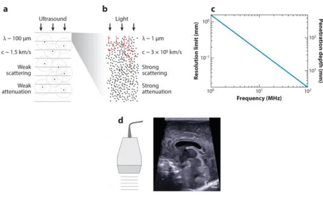
4). where ) is the transmitted amplitude, ) & is the incident amplitude, - is the depth, and + is the absorption coefficient, which varies linearly with frequency. As both resolution and attenuation increase with frequency, there is a trade-off between resolution and depth (Figure 1c).
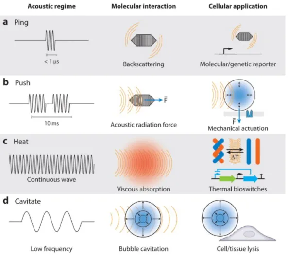
Ultrasound Actuation in vivo
- Heat Dissipation
- Limitations
- Cavitation
- Limitations
- Acoustic Radiation Force
- Limitations
Cavitation is one of the most common ways to trigger drug release and activation in the body. Finally, it is worth mentioning that ARF can be used in the biofabrication and patterning33 of delivery vehicles for controlled release applications.
Contrast Agents and Sonosensitizers
- Microbubbles
- Metal and Inorganic Particles
- Gas Vesicles
Genetically encoded GVs have also been used as sonosensitizers in cells29, but have yet to be incorporated into synthetic materials. Due to their size, biocompatibility, and structural strength, GVs have the potential to be used as ultrasound amplifiers for imaging and actuation of biomaterials and micromachines used for smart drug delivery.
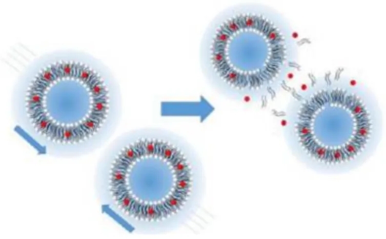
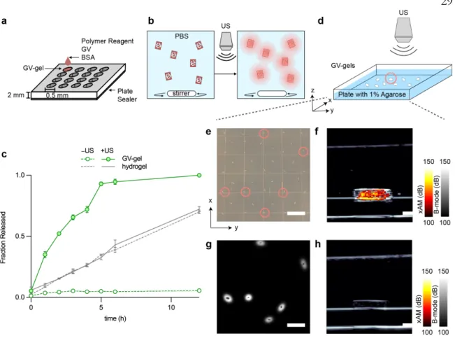
Delivery via acoustically switchable hydrogels (DASH) is enabled by adding cylindrical, air-filled protein nanostructures called gas vesicles (GVs) to hydrogels to block the diffusion of drug payloads from the gel (Fig. 1, a–b). The fraction released is defined as the fluorescence signal in the vessel, normalized to the fluorescence signal at the end of the experiment.
Ultrasound Controlled Drug Delivery by
Abstract
Devices that can be remotely controlled to precisely deliver biomedicine to disease sites are a major goal of biomedical research. Here we present a drug delivery platform comprising a simple protein-containing hydrogel that can be imaged and induced to release drugs at specific sites using conventional ultrasound imaging equipment. We show that the addition of air-filled cylindrical protein nanostructures called gas vesicles (GVs) to the hydrogel blocks the diffusion of the drug load out of the gel and allows the gel to be visualized by ultrasound.
An increase in ultrasound pressure causes the GVs to collapse when the gels reach a specific anatomical location, immediately converting these steric blockers into percolation channels, resulting in a 71.5-fold increase in drug diffusion and rapid release from the gel. We are evaluating this concept through in vitro measurements of ultrasound-modulated diffusion and drug release and targeted in vivo release in the lower gastrointestinal tract. Delivery by acoustically switchable hydrogels (DASH) enables local, image-guided drug delivery using a simple formulation and ubiquitously available ultrasound imaging equipment.
Introduction
In this study, we evaluate this concept with detailed measurements of ultrasound-modulated diffusion and drug release from DASH devices, and then demonstrate their use for protein release in the lower gastrointestinal tract following oral administration. Finally, we use this system to deliver etanercept to the duodenum of rats in a colitis model, resulting in reduced inflammation and improvement in the disease activity index. The DASH platform enables local, image-guided drug delivery using a simple formulation and ubiquitous ultrasound imaging equipment.
GV-containing hydrogels exhibit acoustically switchable
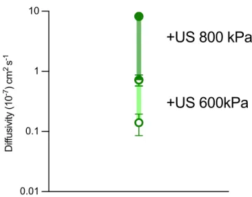
We found that the dynamic range of diffusivities between the "on" and "off" states of ultrasound was optimal at a GV and polymer concentration of 15%. We hypothesized that this acoustically switchable behavior can be further extended by forming GV clusters before gelation. To test this hypothesis, we clustered GVs using biotin and streptavidin, generating aggregates with hydrodynamic diameters of about 1 μm (Fig. 1h).
Incorporating these into the hydrogel reduced diffusion in the presence of intact GV clusters (at a 15% volume fraction) by a factor of four compared to unclustered GVs, while doubling its diffusivity after GV collapse (Fig. 1i) . These experiments demonstrate the ability of GVs to yield common hydrogel materials with acoustically switchable porosity that affects protein diffusivity. GV-gels of different compositions are polymerized inside glass capillary tubes and loaded with a BSA-AlexaFluor 647 reservoir.
DASH devices enable imaging and spatially targeted
The kinetics of cargo release is monitored by aliquoting samples from the vial every hour and determining the fluorescence intensity. GV-gels are dispersed on 1 wt. % agarose phantom, and an ultrasound probe is used to target individual gels for collapse.
DASH gels enable spatially selective ultrasound-triggered
3 In vivo spatially targeted imaging and payload delivery from GV gels a, Illustration of the spatially targeted in vivo imaging and delivery experiment. The composite BURST signal is calculated from the average intensity of manually drawn ROIs around gastrointestinal organs. Scale bars, 10 mm e, pre-collapse ultrasound images of the cecum with xAM (heat color map) and B-mode (grayscale).
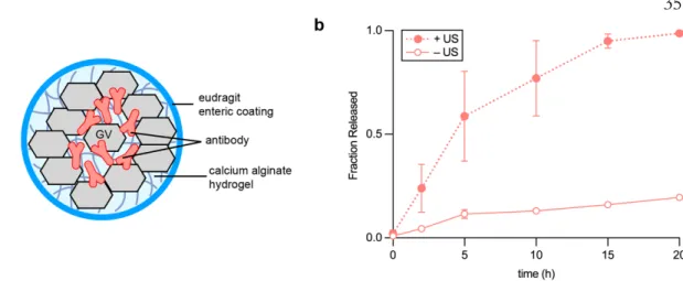
DASH release of etanercept enables the treatment of colitis
Since most gels reach the cecum 5 h after gavage, we triggered etanercept release by applying a high-pressure (550 kPa) ultrasound pulse to the abdominal region (Fig. 5a) of animals in the treatment group. All rats were sacrificed on day 12 for blinded histopathological analysis of the colon by an unaffiliated pathologist. The onset of severe colitis occurred on day 4 and can be described by a sudden decrease in the rate of weight gain compared to the healthy controls32 (Fig. 5b), an increase in the disease activity index (DAI) (Fig. 5c), colon tissue damage and colon shortening.
In DASH-etanercept-treated rats, we observed a statistically significant recovery of the weight gain rates (Fig. 5b) and a reversal of the DAI (Fig. 5c), colon shortening (Fig. 5e) and colon tissue damage trends (Fig. 5d ). Postmortem histological examination revealed that the treatment group had mild to no histological changes due to colitis at the end of treatment, with the regular structure of the lamina propria restored and comparable to that in the healthy rat controls. In contrast, rats given the DASH gels without ultrasound administration and rats in the untreated control group showed crypt abnormalities and inflammatory acute infiltration of the sub-epithelium and lamina propria (Fig. 5f).
Discussion
Notwithstanding the need for further research, the relative simplicity and versatility of the DASH approach and its compatibility with existing FDA-approved hydrogel materials, drugs, and ultrasound devices make this technology poised to enter exciting clinical applications.
Methods
Fluorescence intensities collected during the time series were then normalized to the final intensity at 72 h to determine the kinetics of GV-gel cargo release. BURST imaging was then performed on the entire rat abdomen 5 h after gavage to verify the arrival of the GV-gel bolus in the cecum and then trigger the release of fluorophores. A novel concept of smart NIR-light controlled drug release of black phosphorus nanostructures for cancer treatment.
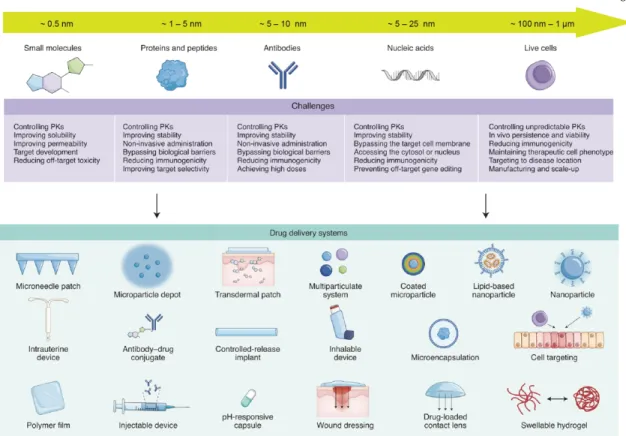
Each of the five generations of therapeutics (small molecules, proteins and peptides, antibodies, nucleic acids, and cell therapies (living cells)) has its own unique delivery challenges. Regardless of therapeutic class, drug delivery systems have adopted one or more drug modification or environmental modification strategies. Positioning is controlled by moving the transducer in the x-y plane using a micromanipulator and guided by real-time fluorescence imaging of the bacteria.
Supplementary Data
Ultrasound Controlled Smart GV-Hydrogel Systems
Abstract
Introduction
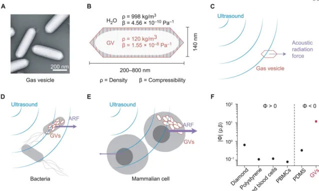
The wide range of collapse pressures accessible through the large GV opens new possibilities for the design and optimization of drug delivery systems using ultrasound. The field of drug delivery encompasses a wide range of therapies22, covering a broad length scale from about 0.5 nm in small molecules, proteins, peptides, antibodies and nucleic acids, to 1 µm in the case of cells (Fig. 3). Its wide range of accessible GV sizes and its hydrophilic exterior lend itself well to incorporation into a variety of existing hydrogel drug delivery systems and can be tailored specifically for different therapeutic payloads.
More experiments will need to be performed to optimize the dynamic range of the behavior depending on the selected drug load, and ultrasound imaging parameters will need to be adjusted specifically if newer, less documented GV types will be considered for implementation in synthetic materials. Nevertheless, the versatility of GVs will enable them to be easily incorporated into the many clinically approved hydrogel drug delivery systems. The microstructure of hydrogels plays a significant role in determining the mechanical and transport properties of biomaterials used in drug delivery implants and tissue scaffolds.
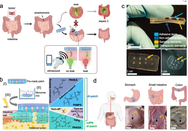
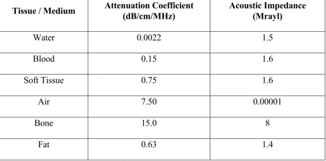
Materials to the left and right of the vertical dotted line exhibit positive and negative acoustic contrast in water, respectively. We designed one such hydrogel implant using GVs embedded in a polyacrylamide gel as one of two sensors to indicate leak originating from a hole in the abdomen (Fig. 9, b–d ).
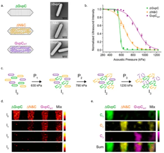
GV-gels enable acoustically multiplexed payload release
GV-gels can be tuned to deliver payloads across length scales
Despite the unique delivery challenges associated with the various types of therapeutics, hydrogels continue to play a prominent role in drug delivery platforms due to their biocompatibility and tunable mesh sizes from 10 nm to 1 µm. Bottom) Representative TEM image of gas vesicles purified from HEK293T cells transiently transfected with mARGs for 72 h.
GVs enable biomaterial patterning using acoustic radiation force
To test the ability of GVs to form patterns in 3D space, we created an acoustic hologram using a 3.5-MHz single-element transducer and a 3D-printed phase mask designed to form an "R" -shaped pressure profile (Fig. 8h) . As expected, the bacteria were immobilized in the gel in the desired spatial pattern (Fig. 8i). As an additional feature, the spatial distribution of GV-expressing cells could be imaged by ultrasound (Fig. 8j), providing a way to verify patterns in optically opaque media.
These results demonstrate the ability of GVs to enable acoustic trapping, patterning, and dynamic rearrangement of engineered bacteria and rapid biofabrication of living materials. The sound wave passes through a Mylar membrane, is reflected by the glass reflector and forms a standing wave near the reflector. A transducer and phase mask are adjusted so that the acoustic hologram is formed inside the sample chamber containing acoustic E.
GVs embedded in sealants enable post-surgical leak detection
Sensing elements are detectable (red arrows). ii), (d-ii) 2 hours after administration, sensing elements were no longer detectable, in line with the disappearance of the opaque dots (c-iii).