Vol 9 No 1 (2020): 2020 (1)
Second allogeneic stem cell transplantation in acute leukemia patients: single-centre experience
Mehmet Bakırtaş, Tuğçe Nur Yiğenoğlu, Semih Başcı, Bahar Uncu Ulu, Nurgül Özcan, Dicle İskender, Mehmet Sinan Dal, Merih Kızıl Çakar, Fevzi Altuntaş
87-96
Does platelet/ lymphocyte ratio a predictor of CD34+ peripheral blood stem cell yield in the healthy donors mobilized with GCSF?
Zafer Gokgoz 115-120
The potency of green grass jelly extract (Premna oblongifolia Merr) as antihyperlipidemia towards aorta histopathology representation of rat (Rattus norvegicus) induced with high fatty diet (HFD)
Eka Nora Vitaloka Aprilia Putri Winthoko, Anna Roosdiana, Dyah Ayu Oktaviani A Pratama, Jusak Nugraha, Marijam Purwanta, Muh Husni Rifa’I, Achmad Nur Rendy 97-102
The potential of traditional balinese spices against the growth of Salmonella sp in vitro I Nyoman Jirna, I Gede Sudarmanto, Surya Bayu Kurniawan, Gusti Ayu Made Ratih, Burhannuddin Rasyid
121-127
Second Allogeneic Stem Cell MEHMET BAKIRTAŞ Jurnal Teknologi Laboratorium
Vol.9, No.1, June 2020, pp. 87 – 96
ISSN 2580-0191(Online), ISSN 2338 – 5634(Print) DOI: 10.29238/teknolabjournal.v9i1.208
Journal homepage: https://www.teknolabjournal.com/index.php/Jtl/index
Original Research
Second allogeneic stem cell transplantation in acute leukemia patients: single- centre experience
Mehmet Bakırtaş1a, Tuğçe Nur Yiğenoğlu1b, Semih Başcı1c*,Bahar Uncu Ulu1d, Nurgül Özcan1e, Dicle İskender1f, Mehmet Sinan Dal1g, Merih Kızıl Çakar1h ,Fevzi Altuntaş1i
1 Department of Hematology and Bone Marrow Transplantation Center, Ankara Dr. Abdurrahman Yurtaslan Oncology Training and Research Hospital, University of Health Sciences, Ankara, Turkey
aE-mail address: [email protected]
bE-mail address: [email protected]
cE-mail address: [email protected]
dE-mail address: [email protected]
eE-mail address: [email protected]
fE-mail address: [email protected]
gE-mail address: [email protected]
hE-mail address: [email protected]
iE-mail address: [email protected]
HIGHLIGHTS
Relapsed acute leukaemia after the first allogeneic stem cell transplantation has a poor prognosis.
Second allogeneic transplantation may offer survival advantage for relapsed leukaemias.
ARTICLE INFO A B S T R A C T Article history
Received Date: March 01st, 2020 Revised Date: June 11st, 2020 Accepted Date: June 12nd, 2020
Keywords
Acute myeloid leukaemia Allogeneic stem cell transplant Relapse
Acute leukaemia patients who relapse after the first allogeneic stem cell transplantation (Allo-SCT) have a poor prognosis.
Participating in clinical trials is the best option for these patients. If patients cannot participate in clinical trials, as the treatment options are limited, the second allo-SCT constitutes the potential curative treatment option. The data of acute leukaemia patients who underwent second allo-SCT because of relapsed/refractory disease after the first allo-SCT at our centre between December 2009 and February 2019 were analyzed retrospectively. Three hundred nineteen acute leukaemia patients were performed allo- SCT at our centre. 20 of these 319 acute leukaemia patients relapsed after first allo-SCT and underwent second allo-SCT. 10 AML patients and 10 ALL patients were included in the study. After second allo-SCT overall survival (OS) was 26.1±10.8 weeks, and progression-free survival (PFS) was 19.9±8.6 weeks. If the patients cannot participate in clinical trials, second allo-SCT should be considered for patients with late (≥12 months) relapses after the first allo-SCT. If possible, haploidentical donors should be selected for second allo-SCT and patients should be in complete remission before the transplant.
This is an open-access article under the CC–BY-SA license.
Second Allogeneic Stem Cell MEHMET BAKIRTAŞ
1. INTRODUCTION
Acute leukaemias are haematological malignancies characterized by abnormal proliferation of blasts caused by hematopoietic myeloid or lymphoid precursors or both.
Monoclonal hematopoietic progenitor cells lose their skills to normally differentiate and proliferate.
The most frequently seen acute leukaemia type in adults is acute myeloid leukaemia (AML), and it has an incidence of 5-8 /100.000.1,2 On the other hand, acute lymphoblastic leukaemia (ALL) has an incidence of 1.28 / 100.000, and it is less commonly observed in adults compared to AML.3 Acute leukaemias become symptomatic in a short time due to their aggressive nature. In spite of intensive treatment methods, they have a poor prognosis. Better survivals have been achieved with improvements in intensive chemotherapies and supportive care. In addition to this, targeted therapies have been started to use in selected acute leukaemia patients. Despite these improvements and new agents in acute leukaemia treatment, the relapse rate is still high.
Allogeneic stem cell transplantation (Allo-SCT) is being used as a curative treatment method in the treatment of acute leukaemia. Some patients relapse after the first allo-SCT. The acute leukaemia patients who relapse after the first allo-SCT have poor prognoses.4 Participating in a clinical trial is the best option for these patients. If patients cannot participate in a clinical trial, as the treatment options for these patients are limited, second allo-SCT constitutes the potential curative treatment option.5 Some previous studies revealed that in acute leukaemia patients who relapsed after the first allo-SCT and underwent second allo-SCT, the survival was better than those of the patients who received only chemotherapy.6,7,8 However, data indicate that only a limited number of acute leukaemia patients who relapsed after the first allo-SCT could be taken to the second allo-SCT.6,9 Because these patients previously received multiple line therapies and generally had a bad performance.6,9 There is still a limited number of studies regarding the efficiency of second allo-SCT and the factors impacting the survival after the second allo-SCT.
The data in the literature generally come from retrospective studies conducted limited number of patients. In this study, we aimed to analyze the outcome of second allo-SCT in acute leukaemia patients who relapsed after the first allo-SCT and to find out the factors impacting the survival rates of second allo-SCT.
2. MATERIAL AND METHOD
The acute leukaemia patients who underwent the second allo-SCT due to relapsed/refractory disease after the first allo-SCT at Dr. Abdurrahman Yurtaslan Ankara Oncology Training and Research Hospital, Bone Marrow Transplantation Unit between December 2009 and February 2019 were included in the study. The local ethical committee approval received. Patients that underwent the second allo-SCT performed because of graft failure after the first allo-SCT was not included in the study. The data were analyzed retrospectively.
In acute leukaemia patients, complete response (CR) was described as having thrombocyte count ≥100.000/µL, neutrophil count ≥1000/µL in peripheric blood, blast ratio <5%
in the bone marrow and having normal maturation in all hematopoietic series of bone marrow in addition to transfusion independence and not having the extramedullary disease.10 After achieving CR, having blasts in peripheric blood or extramedullary area or having ≥20 blasts in bone marrow were defined as relapse.
*Corresponding Author:
Semih Başci
Department of Hematology and Bone Marrow Transplantation Center, Ankara Dr. Abdurrahman Yurtaslan Oncology Training and Research Hospital, University of Health Sciences, 06200 Yenimahalle, Ankara, Turkey
E-mail: [email protected] Phone: +90 5062929890
Second Allogeneic Stem Cell MEHMET BAKIRTAŞ
In both the first and second allo-SCT, peripheric blood-derived stem cells were used.
Human leukocytes antigen (HLA) typing (HLA, A, B, C, DR and DQ) was performed by high- resolution method. In second transplantations, full matched (10/10) and mismatched (9/10) siblings, relatives other than siblings, unrelated donors and haploidentical related donors were used.
The intensity of conditioning regimens was classified according to the published criteria of the Center of International Blood and Marrow Transplant Research (CIBMTR).11 The prophylaxis of graft-versus-host disease (GVHD) was carried out with cyclosporine. Acute GVHD was graded according to the severity index of the International Bone Marrow Transplant Registry (IBMTR).9 Chronic GVHD was graded according to the criteria of the National Institute of Health (NIH) 2015 consensus.12
The overall survival (OS) after allo-SCT was defined as the duration from transplant date to the date of death or to the date of the latest follow-up for the survivors. The progression-free survival (PFS) after allo-SCT was described as the duration from the date of the transplant to the first date when there was a progression or to the date of death or to the latest follow-up date for progression-free patients. The transplant-related mortality (TRM) was defined as the cumulative death within the first 100 days after allo-SCT without any evidence of disease progression. The engraftment definition for neutrophil was defined as the first day when the absolute neutrophil count (ANC) was >500/mm3 or 1000/mm3 for three consecutive days without any support, and for thrombocytes, it was defined as the first day when thrombocyte count was >20000/mm3 for three consecutive days without any support.13
The statistical analyses were performed with IBM SPSS Statistics v21 software. Chi-square test was used for descriptive statistics and comparisons among the groups for categorized data, and Mann Whitney U tests were applied for nonparametric figurative data. Survival rates were calculated by Kaplan-Meier survival analysis and Cox regression analysis. The impacts of the variables over OS and PFS were examined using a log-rank test. The cases where Type-1 error level was under 5% was accepted as statistically significant.
3. RESULTS AND DISCUSSION
Three hundred nineteen acute leukaemia patients were performed allo-SCT at our centre.
Among survivors, the median follow-up is 6.4 years. TRM is %12.4. Twenty of 319 acute leukaemia patients relapsed after first allo-SCT and underwent second allo-SCT at our centre.
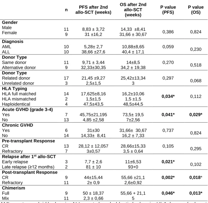
Ten AML patients and 10 ALL patients were included in the study. There were nine female and 11 male patients. The median age at the time of the second allo-SCT was 31 (range 18-55). The median duration between first allo-SCT and relapse was 25 weeks (range 8-219 weeks). When the patients who had relapsed within 12 months after the first allo-SCT were compared with the patients who had relapsed after 12 months, PFS after the second allo-SCT was found significantly shorter in early relapsed patients (p:0.021); however, there was no statistically significant difference regarding OS after second allo-SCT between early and late relapsed patients(p:0.102).
The median duration between the first and second allo-SCT was 50 weeks (range 14-236 weeks). While we did not find any statistically significant difference between post-transplant PFS and duration between the first and second allo-SCT (p:0.141), we found a statistically significant difference between post-transplant OS and the duration between the first and second allo-SCT (p:0.02*). Before the second allo-SCT, nine patients were full chimeric, 11 patients were mixed chimeric. In full chimeric patients, post-transplant PFS and OS were significantly longer than mix chimeric patients (p:0.013).
Second allo-SCT was performed from the donors’ of 10/10 compatible siblings in 11 patients, 10/10 compatible relatives other than siblings in 2 patients, 10/10 compatible unrelated in 1 patient, 9/10 compatible unrelated in 2 patients, and haploidentical donors in 4 patients.
Second Allogeneic Stem Cell MEHMET BAKIRTAŞ
In second allo-SCT; 13 full matched related donors(11 siblings, two relatives other than siblings), three unrelated donors (1 full matched, two mismatched), four haploidentical donors were used. In patients whose donors were related median post-transplant PFS was 21,45 ±9,27 weeks and post-transplant OS was 25,42±13,34 weeks. In patients with unrelated donors, median post-transplant PFS was only 2.5±1 weeks, and post-transplant OS was three weeks. In patients with haploidentical donors, post-transplant OS and PFS were longer than patients with matched/miss-matched donors.
While in 11 patients’ same donors were used in the second allo-SCT, different donors were used in 9 patients. We did not find any impacts of using alternative donors over PFS and OS.
Myeloablative conditioning was used in 11 patients, and reduced intensive conditioning (RIC) regimen was used in 9 patients. There was no significant difference between post-transplant OS and PFS and conditioning regimen (p:0.287; p:0.265, respectively). The patients had an average of 1 cycle chemotherapy (range 1-3) in the duration between relapse and the second allo-SCT.
While 13 patients were taken to the second allo-SCT with CR, seven patients were taken with active disease. There was no statistically significant difference between pre-transplant response and post-transplant PFS and OS (p:0.105 and p:0.295 respectively).
Median follow up duration was 34 weeks (range 20-257 weeks). 7 of 20 acute leukaemia patients (35%) performed second allo-SCT are still in remission. After second allo-SCT OS was 26.1±10.8 weeks and PFS was 19.9±8.6 weeks. The post-transplant PFS was 5.28±2.7 weeks, and OS were 14.88±8.65 weeks in AML patients while post-transplant PFS was 38.66±27.6 weeks, and OS was 40.4±17.1 weeks in ALL patients. There was no statistically significant difference between the patients with both diagnoses regarding the PFS and OS after second allo- SCT (p:0.059, p:0.230 respectively).
At the end of the follow-up time, nine patients died. While 3 of these deaths (15%) were related to relapse, 6 of them (30%) were related to TRM. TRM was found 20% among AML patients and 40% among ALL patients. In all patients, neutrophil engraftment occurred at a median of 14 days, and thrombocyte engraftment occurred at a median of 13 days. After the second allo-SCT, CR was observed in 9 patients (45%). Nine patients (45%) were full chimeric, 11 patients were mixed chimeric. Grade 3-4 acute GVHD was observed in 2 (10%) patients.
Chronic GVHD was observed in 6 (30%) patients. The relation between post-transplant PFS and OS and diagnosis, gender, donor type, alternative donors, the presence of acute or chronic GVHD, pre-transplant response, post-transplant response, chimerism is shown in the table (Table 1).
Acute leukaemia patients relapsing after allo-SCT generally have received multiple line chemotherapies including various agents previously, and they usually show resistance to the previously used chemotherapeutic agents.4 In such patients salvage chemotherapy, discontinuation of immunosuppression, donor lymphocyte infusion, second allo-SCT can be considered. If possible, patients should be encouraged to participate in clinical trials.
In the study conducted by Cerny et al. AML, ALL, myelodysplastic syndrome and chronic lymphocytic leukaemia patients that underwent the second allo-SCT, 1-year OS was found 44%, and the median post-transplant OS was found 8.9 months (range 0-27.6 months).14 In the study conducted by Aljasem et al., 3-year OS rate was found 35%.15 In another study, 1-year OS after second allo-SCT in AML patients was 20%.16 In an analysis of CIBMTR, 1-year OS in 369 patients that underwent second allo-SCT was 49%, and the median survival was found 12 months.17 In our study, on the other hand, the OS after the second allo-SCT was found 26.1±10.8 weeks and PFS was found 19.9±8.6 weeks, 1-year OS was 11%. In AML patients, the PFS after second allo- SCT was found 5.28±2.7 weeks, and the OS was 10.88±8.65. In ALL patients, the PFS after second allo-SCT was found 38.66±27.6 weeks and OS were found 40.4±17.1 weeks.
According to the literature, CR achievement decreases as the number of chemotherapy lines increase.18,19 Therefore, obtaining a CR in acute leukaemia patients relapsed after the first
Second Allogeneic Stem Cell MEHMET BAKIRTAŞ
allo-SCT is more difficult than the second allo-SCT. In addition to this, organ functions generally impair due to high-dose treatments.19 As a result, some of the patients are taken to second allo- SCT without having a CR. Although there are many studies conducted to increase the CR rate after relapse, the results of these studies are inadequate and more studies are required.5,18,20,21,22
In the study conducted by Chueh et al. pre-transplant response was found to be an independent factor impacting the success of second allo-SCT.23 In this study, 70.4% of the patients had CR before second allo-SCT. In our study, 65.2% of the patients had CR before second allo-SCT. We did not find any statistically significant difference between pre-transplant response and post- transplant PFS and OS (p:0.105 and p:0.295 respectively).
If the patients have related donors, they can use their relatives for the second allo-SCT;
however, using the same donor for the second allo-SCT is relatively more difficult if the previous donors are unrelated. In the study conducted by Chueh et al., there was no difference regarding the survival rates between the patients that underwent the second allo-SCT from an alternative or the same donors.23 In the study conducted by Aljasem et al., no relationship was observed between OS and alternative donor use.15 The previous studies could not show the advantages of selecting an alternative donor, either.9,19 Similarly, we did not find any statistically significant difference between the patients that underwent second allo-SCT from the same donors or alternative donors regarding post-transplant PFS and OS.
There are studies that show the positive effects of GVHD or HLA incompatibilities on the prevention of relapse. In some studies, while positive effects are observed on the rates of survival, in other benefits could not be indicated.24,25,26 In the study conducted by Chueh et al., no relationship was found between acute or chronic GVHD observed after second allo-SCT and survival.23 In our study, also, we did not find any statistically significant relationship between chronic GVHD and PFS and OS (p:0.737 and p:0.825 respectively); however, a statistically significant relationship was found between acute GVHD and PFS and OS (p:0.041 and p:0.029 respectively).
Some studies reported that the duration between the first allo-SCT and the second allo- SCT is an important prognostic factor, and it is related to better survival rates.27,28 In the study conducted by Chueh, there was no statistically significant survival difference between the patients with early relapses and late relapses.23 However in the study conducted by Aljasem et al., 3-year OS was 21% in early relapsed patients, and it was 46% in patients relapsed after 12 months (p:0.009) (20). In our study, we found shorter PFS in early relapsed patients (p:0.021), but there was no statistically significant difference regarding OS (p:0.102).
In the study conducted by Cerny et al. in second allo-SCT, median neutrophil engraftment was achieved in 15 days, and median thrombocyte engraftment was achieved in 21 days. The rate of TRM was found at 30%.14 In our study, neutrophil engraftment occurred at a median of 14 days, and thrombocyte engraftment occurred at a median of 13 days. Furthermore, the rate of TRM was similarly found 30%. The reason for early neutrophil and thrombocyte engraftment may be peripheric blood-derived stem cells.
In the majority of second allo-SCT in the literature reduced-intensity conditioning (RIC) regimens were used.29 In the study conducted by Aljasem et al., 3-year OS was found 42% in myeloablative regimens and 23% in non-myeloablative and RIC regimens; however, we did not find any statistically significant difference between myeloablative regimens and non- myeloablative and RIC regimens (p=0.08) (20). Similarly, in our study, no statistically significant relationship was found between OS and PFS and conditioning regimens (p:0.287; p:0.265).
4. CONCLUSION
Patients who were performed second allo-SCT had a poor prognosis. Development of new immune therapeutics with less toxicity and new strategies are required. Patients should be encouraged to participate in clinical trials. If the patients cannot participate in clinical trials, second
Second Allogeneic Stem Cell MEHMET BAKIRTAŞ
allo-transplant should be considered in patients who have late (≥12 months) relapses after the first allo-SCT, If possible, haploidentical donors should be selected for second allo-SCT and patients should be in CR before the transplant. More prospective studies are needed to design future treatment approaches.
DISCLOSURE STATEMENT
The authors reported no potential conflict of interest.
FUNDING INFORMATION
The authors declared that this case had received no financial support.
REFERENCES
1. Fey MF, Buske C, ESMO Guidelines Working Group. Acute myeloblastic leukaemias in adult patients: ESMO Clinical Practice Guidelines for diagnosis, treatment and follow-up.
Ann Oncol Off J Eur Soc Med Oncol. 2013;24 Suppl 6:vi138-43.
doi:10.1093/annonc/mdt320.
2. De Kouchkovsky I, Abdul-Hay M. ‘Acute myeloid leukemia: A comprehensive review and 2016 update.’ Blood Cancer J. 2016;6(7):e441. doi:10.1038/bcj.2016.50.
3. Hoelzer D, Bassan R, Dombret H, et al. Acute lymphoblastic leukaemia in adult patients:
ESMO clinical practice guidelines for diagnosis, treatment and follow-up. Ann Oncol.
2016;27:v69-v82. doi:10.1093/annonc/mdw025.
4. Kröger N. Approaches to relapse after allogeneic stem cell transplantation. Curr Opin Oncol. 2011;23(2):203-208. doi:10.1097/CCO.0b013e328342c6c8.
5. Collins RH, Shpilberg O, Drobyski WR, et al. Donor leukocyte infusions in 140 patients with relapsed malignancy after allogeneic bone marrow transplantation. J Clin Oncol.
1997;15(2):433-444. doi:10.1200/JCO.1997.15.2.433.
6. Thanarajasingam G, Kim HT, Cutler C, et al. Outcome and Prognostic Factors for Patients Who Relapse after Allogeneic Hematopoietic Stem Cell Transplantation. Biol Blood Marrow Transplant. 2013;19(12):1713-1718. doi:10.1016/j.bbmt.2013.09.011.
7. Porter DL, Alyea EP, Antin JH, et al. NCI First International Workshop on the Biology, Prevention, and Treatment of Relapse after Allogeneic Hematopoietic Stem Cell Transplantation: Report from the Committee on Treatment of Relapse after Allogeneic Hematopoietic Stem Cell Transplantation. Biol Blood Marrow Transplant.
2010;16(11):1467-1503. doi:10.1016/j.bbmt.2010.08.001.
8. Weisdorf D. The role of second transplants for leukemia. Best Pract Res Clin Haematol.
2016;29(4):359-364. doi:10.1016/j.beha.2016.10.011.
9. Eapen M, Giralt SA, Horowitz MM, et al. Second transplant for acute and chronic leukemia relapsing after first HLA-identical sibling transplant. Bone Marrow Transplant.
2004;34(8):721-727. doi:10.1038/sj.bmt.1704645.
10. Döhner H, Estey EHE, Amadori S, et al. Diagnosis and management of AML in adults_Recommendations from European Leukemia Net 2010. Blood. 2010;115(3):453- 474. doi:10.1182/blood-2009-07-235358.
11. Bacigalupo A, Ballen K, Rizzo D, et al. Defining the Intensity of Conditioning Regimens:
Working Definitions. Biol Blood Marrow Transplant. 2009;15(12):1628-1633.
doi:10.1016/j.bbmt.2009.07.004.
12. Jagasia MH, Greinix HT, Arora M, et al. National Institutes of Health Consensus Development Project on Criteria for Clinical Trials in Chronic Graft-versus-Host Disease: I.
The 2014 Diagnosis and Staging Working Group Report. Biol Blood Marrow Transplant.
2015;21(3):389-401.e1. doi:10.1016/j.bbmt.2014.12.001.
13. Rihn C, Cilley J, Naik P, Pedicano AVJ, Mehta J. Definition of myeloid engraftment after
Second Allogeneic Stem Cell MEHMET BAKIRTAŞ
allogeneic hematopoietic stem cell transplantation. Haematologica. 2004;89(6):763-764.
14. Cerny J, Ramanathan M, Raffel G, Shanahan L, Kilmer E, Nath R. Outcomes of Second Allogeneic Stem Cell Transplantation: Single Center Analysis. Biol Blood Marrow Transplant. 2017;23(3):S264. doi:10.1016/j.bbmt.2016.12.162.
15. Aljasem HA, Messner HA, Lipton JH, et al. Outcome following second allogeneic hematopoietic cell transplantation: A single-center experience. Eur J Haematol.
2018;100(3):308-314. doi:10.1111/ejh.13015.
16. Devillier R, Crocchiolo R, Etienne A, et al. Outcome of relapse after allogeneic stem cell transplant in patients with acute myeloid leukemia. Leuk Lymphoma. 2013;54(6):1228- 1234. doi:10.3109/10428194.2012.741230.
17. Bejanyan N, Weisdorf DJ, Logan BR, et al. Survival of Patients with Acute Myeloid Leukemia Relapsing after Allogeneic Hematopoietic Cell Transplantation: A Center for International Blood and Marrow Transplant Research Study. Biol Blood Marrow Transplant.
2015;21(3):454-459. doi:10.1016/j.bbmt.2014.11.007.
18. Radich JP, Sanders JE, Buckner CD, et al. Second allogeneic marrow transplantation for patients with recurrent leukemia after initial transplant with total-body irradiation-containing regimens. J Clin Oncol. 1993;11(2):304-313. doi:10.1200/JCO.1993.11.2.304.
19. Hartwig M, Ocheni S, Asenova S, et al. Second allogeneic stem cell transplantation in myeloid malignancies. Acta Haematol. 2009;122(4):185-192. doi:10.1159/000253025.
20. Ko RH, Ji L, Barnette P, et al. Outcome of patients treated for relapsed or refractory acute lymphoblastic leukemia: A therapeutic advances in childhood leukemia consortium study.
J Clin Oncol. 2010;28(4):648-654. doi:10.1200/JCO.2009.22.2950.
21. Nguyen S, Béziat V, Norol F, et al. Infusion of allogeneic natural killer cells in a patient with acute myeloid leukemia in relapse after haploidentical hematopoietic stem cell transplantation. Transfusion. 2011;51(8):1769-1778. doi:10.1111/j.1537- 2995.2010.03058.x.
22. Ueki T, Sumi M, Sato K, et al. Reduced-intensity cord blood transplantation without prior remission induction therapy induces durable remission in adult patients with relapsed acute leukemia after the first allogeneic transplantation. Eur J Haematol. 2011;86(3):268-271.
doi:10.1111/j.1600-0609.2010.01555.x.
23. Chueh HW, Lee SH, Sung KW, Yoo KH, Koo HH. Second allogeneic stem cell transplantation in hematologic malignancies: A single-center experience. J Pediatr Hematol Oncol. 2013;35(6):424-429. doi:10.1097/MPH.0b013e31829b7f58.
24. Morishima Y, Sasazuki T, Inoko H, et al. The clinical significance of human leukocyte antigen (HLA) allele compatibility in patients receiving a marrow transplant from serologically HLA-A, HLA-B, and HLA-DR matched unrelated donors. Blood.
2002;99(11):4200-4206. doi:10.1182/blood.V99.11.4200.
25. Le RQ, Melenhorst JJ, Battiwalla M, et al. Evolution of the donor T-cell repertoire in recipients in the second decade after allogeneic stem cell transplantation. Blood.
2011;117(19):5250-5256. doi:10.1182/blood-2011-01-329706.
26. Petersdorf EW, Anasetti C, Martin PJ, et al. Limits of HLA mismatching in unrelated hematopoietic cell transplantation. Blood. 2004;104(9):2976-2980. doi:10.1182/blood- 2004-04-1674.
27. Bosi A, Laszlo D, Labopin M, et al. Second allogeneic bone marrow transplantation in acute leukemia: Results of a survey by the European Cooperative Group for Blood and Marrow Transplantation. J Clin Oncol. 2001;19(16):3675-3684. doi:10.1200/JCO.2001.19.16.3675.
28. Kishi K, Takahashi S, Gondo H, et al. Second allogeneic bone marrow transplantation for post-transplant leukemia relapse: Results of a survey of 66 cases in 24 Japanese institutes.
Bone Marrow Transplant. 1997;19(5):461-466. doi:10.1038/sj.bmt.1700680.
29. Byrne BJ, Horwitz M, Long GD, et al. Outcomes of a second non-myeloablative allogeneic
Second Allogeneic Stem Cell MEHMET BAKIRTAŞ
stem cell transplantation following graft rejection. Bone Marrow Transplant. 2008;41(1):39- 43. doi:10.1038/sj.bmt.1705882.
Table 1. The variables that have an impact on post-transplant PFS and OS n PFS after 2nd
allo-SCT (weeks)
OS after 2nd allo-SCT
(weeks)
P value (PFS)
P value (OS) Gender
Male
Female 11
9
8,83 ± 3,72 31 ±16,2
14,33 ±8,41
31,66 ± 30,67 0,386 0,824 Diagnosis
AML ALL
10 10
5,28± 2,7 38,66 ±27,6
10,88±8,65 40,4 ± 17,1
0,059
0,230 Donor Type
Same donor Alternative donor
11 9
9,71 ± 3,44 32,33±30,35
14±8,5 34,2 ± 19,38
0,270
0,518 Donor Type
Related donor Unrelated donor
17 3
21,45 ±9,27 2,5±1,5
25,42±13,34 3
0,297
0,068 HLA Typing
HLA full matched HLA mismatched Haploidentical
14 2 4
17,625±8,16 1,5±1,5 47,5±43,5
16,2±10,06 1,5 ±1,5 48,5±44,5
0,034* 0,112
Acute GVHD (grade 3-4) Yes
No
7 13
45,75±21,195 4,85 ±2,58
73,5± 19,5 7±2,56
0,041* 0,029*
Chronic GVHD Yes
No
6 14
31±30 14,33± 8,41
31,66± 30,67 16,2 ± 7,33
0,737
0,824 Pre-transplant Response
CR Refractory
13 7
28,12 ± 12,057 3±0,57
28,66±15,33 3,5 ± 0,64
0,105
0,295 Relapse after 1st allo-SCT
Early relapse
Late relapse (≥12 months) 3 2
7,7 ± 2,6 81 ± 10
11±6,53 93+0
0,021*
0,102 Post-transplant Response
CR Refractory
9 11
44±15,44 2± 0,9
55,66 ±21,1 2,6±0,92
0,002* 0,018*
Chimerism Full
Mix
9 11
50 ± 18,37 2,3 ± 0,66
55,66 + 21,1 5
0,046* 0,013*
AML: acute myeloid leukemia, ALL: acute lymphoblastic leukemia, HLA: human leukocyte antigen, CR: complete response, GVHD: graft versus host disease, PFS: progression free survival, OS: overall survival
Second Allogeneic Stem Cell MEHMET BAKIRTAŞ
SHORT BIOGRAPHY
Mehmet Bakırtaş, MD
He was born in Köyceğiz, Turkey. He graduated from Black Sea Technical University, Medical Faculty in 2011. Between 2012 and 2016, he worked as a Research Assistant at Internal Medicine of Mediterranean University Faculty of Medicine. He worked in Van State Hospital in 2017 as an Internal Medicine specialist. He has been working in Ankara Oncology Training and Research Hospital, Hematology Service and Bone Marrow Transplant Unit as an Hematology fellow since 2017. He has many articles published in international SCI and national journals.
https://orcid.org/0000-0003-3216-482X
Tuğçe Nur Yiğenoğlu, MD
https://orcid.org/0000-0001-9962-8882
Semih Başcı, MD
https://orcid.org/0000-0003-4304-9245
Bahar Uncu Ulu, MD
https://orcid.org/0000-0002-6230-9519
Nurgül Özcan, MD
https://orcid.org/0000-0001-8819-5153
Second Allogeneic Stem Cell MEHMET BAKIRTAŞ
Dicle İskender, MD
https://orcid.org/0000-0002-6062-6422
Mehmet Sinan Dal, Assoc.Prof.
https://orcid.org/0000-0002-5994-2735
Merih Kızıl Çakar, Assoc.Prof.
https://orcid.org/0000-0003-0978-0923
Fevzi Altuntaş, Prof.
https://orcid.org/0000-0001-6872-3780
DOES PLATELET/LYMPHOCYTE RATIO A PREDICTOR OF CD34+ ZAFER GOKGOZ Jurnal Teknologi Laboratorium
Vol.9, No.1, June 2020, pp. 115 – 120
ISSN 2580-0191(Online), ISSN 2338 – 5634(Print) DOI: 10.29238/teknolabjournal.v9i1.209
Journal homepage: https://www.teknolabjournal.com/index.php/Jtl/index
Original Research
Does platelet/ lymphocyte ratio a predictor of CD34+ peripheral blood stem cell yield in the healthy donors mobilized with GCSF?
Zafer Gokgoz
Department of Hematology, Faculty of Medicine, Yuksek İhtisas University, Turkey E-mail address:[email protected]
HIGHLIGHTS
Consequently, a high platelet/lymphocyte ratio may be correlated with the number of collected CD34+ stem cells
ARTICLE INFO A B S T R A C T Article history
Received Date: March 04th, 2020 Revised Date: June 01st, 2020 Accepted Date: June 04th, 2020
Keywords Lymphocyte ratio CD34+
Stem Cells
This research aimed to determine whether there is a correlation between platelet/lymphocyte ratio the number of collected CD34+ stem cells. This retrospective study included 94 adult related stem cell donors who were healthy and volunteer by screening their files between the years 2016 and 2018. All donors were mobilized using 2.5 mcg/kg lenograstim or filgrastim and underwent peripheral stem cell apheresis. Complete blood counts were tested at baseline before G-CSF administration (pre–G-CSF), and before PBSC collection after mobilization with G-CSF administration. The patients were divided into two groups as aged below and over 50 years old. From these comparative data, only BMI value of the group aged below 50 years was statistically significantly lower than the other group, whereas no statistically significant difference was found between the groups in terms of other parameters. The numbers of the collected CD34+ stem cells of both age groups were similar, and no significant difference was found. The values of platelet/lymphocyte ratio, early measurement of CD34+ stem cells and the amount of the collected CD34+ stem cells of both groups at the first and second days of the procedure were found similar. This research show that a high platelet count and consequently, a high platelet/lymphocyte ratio may be correlated with the number of collected CD34+ stem cells but our hypotheses revealed insignificant outcomes.
*Corresponding Author:
Zafer Gokgoz
Department of Hematology, Faculty of Medicine, Yuksek İhtisas University, Turkey Karakaya Mahallesi, Baglum Bulvari No.1, 06291, Kecioren, Ankara, Turkey E-mail: [email protected]
Phone:+90 312 3297 425
This is an open-access article under the CC–BY-SA license.
DOES PLATELET/LYMPHOCYTE RATIO A PREDICTOR OF CD34+ ZAFER GOKGOZ
1. INTRODUCTION
Peripheral blood stem cell collection for allogeneic or autologous stem cell transplantation has become the standard treatment modality thanks to easy-applicability and rapid haematological reconstitution compared with stem cell collection from bone marrow harvest in many malignant and benign cases.1,2 Stem cell mobilization is performed with granulocyte-colony stimulating factor (GCSF) in healthy donors. The donor response to G-CSF influences the number of CD34+ cells and eventually transplantation outcomes. Low level of peripheral blood stem cell increases the risk for graft failure.3 On the other hand, transplantation of high CD34+ stem cell doses increases the risk for acute graft versus host disease (GVHD).4 Nevertheless, a high level of stem cell infusion in the patient is considered to be associated with faster immune reconstruction.5 As known, GCSF causes lymphocytosis, however, GCSF has several effects on T-cell subgroups such as especially mobilization of naive T lymphocytes, reduced adhesion molecules and chemokine receptors, reduced T-helper-1-related cytokines and increased regulatory T (T-reg) cells.6 It is also known that GCSF suppresses pro-inflammatory cytokines and increases secretion of IL-10. All these effects indicate the immunosuppressive nature of GCSF in healthy humans.7 On the other hand, it has been shown that platelet count in the peripheral circulation decreases due to G-CSF administration.8 The reduction in platelet count is directly proportional with GCSF dose.9 Baseline platelet counts prior to the onset of GCSF administration is strongly correlated with the amount of mobilized CD34+ stem cells.10,11
In the light of all these information from the literature, we have retrospectively evaluated whether there is a correlation between the platelet/lymphocyte ratio as a cheap and easy- applicable method and the number of collected CD34+ stem cells.
2. MATERIAL AND METHOD
This retrospective study included 94 adult related stem cell donors who were healthy and volunteer by screening their files in the Bone Marrow Transplant Unit of Medicana Ankara International Hospital between the years 2016 and 2018 (Ethics Committee Approval Dated 08.02.2019, Number:26). All donors were over 18 years old. A haematologist from the bone marrow transplant team approved the donors by evaluating their detailed anamnesis, physical examination, complete blood count, biochemistry and infection markers, blood grouping and Rh typing, and pregnancy testing in the females of childbearing age. The donors with a chronic disease and ongoing medication were excluded from the study. Body mass index (BMI) was calculated according to the formula BMI= weight (in kilograms)/height (in meters squared). All donors were mobilized using 2.5 mcg/kg lenograstim or filgrastim and underwent peripheral stem cell apheresis.
The onset of G-CSF administration was accepted as the 1st day, and the donors underwent mobilization procedure with Fresenius (Bad Homburg, Germany) device if the early measurement of CD34+stem cells resulted in 20x106 /L at the 5th day. The collection of minimum 2x10⁶ /kg body weight CD34+ stem cells was targeted. The donors underwent apheresis procedure for 1 or 2 days according to that target cell dose or transplant planning of the patient.
The blood volumes were processed for 2-4 times and coagulated with 40-90 mL/minute acid- citrate dextrose as the coagulant in the healthy donors (HD) determined to be performed the procedure at the second day. The healthy donors (HD) were administered 10% Calcium Gluconate. Complete blood counts were tested at baseline before G-CSF administration (pre–G- CSF), and before PBSC collection after mobilization with G-CSF administration.
The parameters were analyzed by SPSS for Windows Version 23.0. The continuous variables were expressed as median (minimum-maximum); numbers while percentages were used to express categorical variables. The differences between continuous variables of two groups were calculated by Mann-Whitney-U test. The repeated measures were compared by Wilcoxon test. Chi-square test was used in the analysis of categorical parameters. Spearman Rho
DOES PLATELET/LYMPHOCYTE RATIO A PREDICTOR OF CD34+ ZAFER GOKGOZ
correlation was used to calculate the correlation of two continuous variables. The p<0.05 value was accepted as the statistical significance level.
3. RESULTS AND DISCUSSION
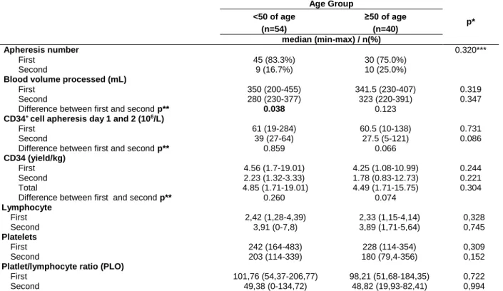
Totally 94 donors were included in the study. The study group consisted of 38 females and 56 males. The patients were divided into two groups as aged below and over 50 years old.
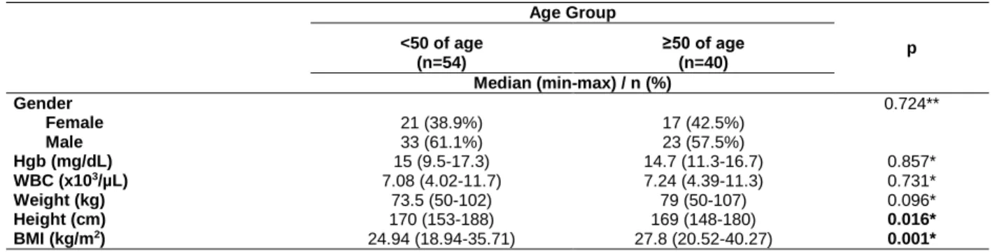
This distribution was based on the consideration that the platelet/lymphocyte ratio may have an impact on the amount of CD34+ stem cells depending on donor age. Median haemoglobin (Hg) value was 15 g/dl in the group aged below 50 years, whereas that value was 14.7 g/dl in the group aged ≥50 years. WBC counts of the groups were found 7.08x10³/mL and 7.24x10³/mL, respectively. Body mass index (BMI) value of the younger group was 24.9, whereas that value was 27.8 in the group aged ≥50 years (Table 1). From these comparative data, only BMI value of the group aged below 50 years was statistically significantly lower than the other group, whereas no statistically significant difference was found between the groups in terms of other parameters.
The numbers of the collected CD34+ stem cells of both age groups were similar, and no significant difference was found (Table 2). In addition, we have analyzed the platelet/lymphocyte counts in two groups as <100 and >100 (Table 3). The values of platelet/lymphocyte ratio, early measurement of CD34+ stem cells and the amount of the collected CD34+ stem cells of both groups at the first and second days of the procedure were found similar.
In the present study, we have analyzed the relationship between baseline platelet/lymphocyte ratio and the amount of CD34+ stem cells collected following mobilization.
The previous studies have suggested assumptions on predicting a number of the CD34+ stem cells based on various demographic and laboratory analyses and reported that the most effective factors on CD34+ stem cell apheresis yield are G-CSF dose administered in the donor as well as age, gender and the bodyweight of the donor,10 however, no study on platelet/lymphocyte ratio has been conducted yet as far as we know. It has been aimed to predict the amount of CD34+
stem cells that can be collected by performing an easy and low-cost method in this study.
The peripheral blood stem cell collection from healthy donors induced with GCSF is an easy technique with a low incidence of complication. The amount of the collected CD34+ stem cells has various effects on engraftment. The detection of these effects in the early pretransplantation period is important in the regulation and prediction of the posttransplantation period.
It is known that a high level of baseline platelet count before GCSF administration is proportional to the number of mobilized CD34+ stem cells.10,11,12 In the present study, we did not analyze the relationship of platelet count with baseline value before GCSF administration and number of the CD34+ stem cells before the procedure. In addition, some studies have suggested that the amount of the collected stem cells may decrease with advancing age, whereas some other studies assert the contrary to this conclusion.13 Various studies on donor age have presented different outcomes.14,15 De La Rubia et al. have reported that a higher number of CD34+ stem cells was collected in the younger donors. From this point of view, we distributed our donors into two groups as ones aged below and over 50 years old. We have determined that the platelet/lymphocyte ratio was not significantly correlated with the amount of CD34+ stem cells in both groups.
Our study has some limitations. As our study is retrospective, we could not obtain lymphocyte subgroups and also it would be better if we could search interleukins, tumour necrosis factors, and interferon-alpha because these mediators are effected by GCSF also, if we could not use one type of generic for all donors we had to use filgrastim or lenograstim both.
DOES PLATELET/LYMPHOCYTE RATIO A PREDICTOR OF CD34+ ZAFER GOKGOZ
4. CONCLUSION
As an overall conclusion, we have assumed that a high platelet count and consequently a high platelet/lymphocyte ratio may be correlated with the number of collected CD34+ stem cells or contrarily a platelet/lymphocyte ratio encountered below a certain threshold value due to extremely reduced platelet count after GCSF administration may also be proportional with the collected CD34+ stem cells in developing hypotheses of the study. However, both of our hypotheses revealed insignificant outcomes.
DISCLOSURE STATEMENT
The authors reported no potential conflict of interest.
FUNDING INFORMATION
The authors declared that this case had received no financial support.
REFERENCES
1. Gratwohl A, Baldomero H, Schmid O, Horisberger B, Bargetzi M, Urbano-Ispizua A.
Change in stem cell source for hematopoietic stem cell transplantation (HSCT) in Europe:
a report of the EBMT activity survey 2003. Bone Marrow Transplant. 2005;36(7):575-590.
doi:10.1038/sj.bmt.1705104.
2. Bensinger WI, Martin PJ, Storer B, et al. Transplantation of Bone Marrow as Compared with Peripheral-Blood Cells from HLA-Identical Relatives in Patients with Hematologic Cancers. N Engl J Med. 2001;344(3):175-181. doi:10.1056/NEJM200101183440303.
3. Storb R, Prentice RL, Thomas ED. Marrow Transplantation for Treatment of Aplastic Anemia. N Engl J Med. 1977;296(2):61-66. doi:10.1056/NEJM197701132960201.
4. Przepiorka D, Smith TL, Folloder J, et al. Risk factors for acute graft-versus-host disease after allogeneic blood stem cell transplantation. Blood. 1999;94(4):1465-1470.
http://www.ncbi.nlm.nih.gov/pubmed/10438735.
5. Savani BN, Rezvani K, Mielke S, et al. Factors associated with early molecular remission after T cell-depleted allogeneic stem cell transplantation for chronic myelogenous leukemia. Blood. 2006;107(4):1688-1695. doi:10.1182/blood-2005-05-1897.
6. Ringden O, Barrett AJ, Zhang M-J, et al. Decreased treatment failure in recipients of HLA- identical bone marrow or peripheral blood stem cell transplants with high CD34 cell doses.
Br J Haematol. 2003;121(6):874-885. doi:10.1046/j.1365-2141.2003.04364.x.
7. Melve GK, Ersvær E, Kittang AO, Bruserud Ø. The chemokine system in allogeneic stem- cell transplantation: a possible therapeutic target? Expert Rev Hematol. 2011;4(5):563- 576. doi:10.1586/ehm.11.54.
8. Falanga A, Marchetti M, Evangelista V, et al. Neutrophil activation and hemostatic changes in healthy donors receiving granulocyte colony-stimulating factor. Blood. 1999;93(8):2506- 2514. http://www.ncbi.nlm.nih.gov/pubmed/10194429.
9. Tassi C, Tazzari PL, Bonifazi F, et al. Short- and long-term haematological surveillance of healthy donors of allogeneic peripheral haematopoietic progenitors mobilized with G-CSF:
a single institution prospective study. Bone Marrow Transplant. 2005;36(4):289-294.
doi:10.1038/sj.bmt.1705066.
10. Vasu S, Leitman SF, Tisdale JF, et al. Donor demographic and laboratory predictors of allogeneic peripheral blood stem cell mobilization in an ethnically diverse population.
Blood. 2008;112(5):2092-2100. doi:10.1182/blood-2008-03-143677.
11. Zubair AC, Grant R, Wu W, et al. Platelet count is a sensitive predictor of autologous peripheral blood progenitor cell collection yield in previously treated plasma cell disease patients. Transfusion. 2008;48(6):1106-1114. doi:10.1111/j.1537-2995.2008.01651.x.
12. Suzuya H, Watanabe T, Nakagawa R, et al. Factors associated with granulocyte colony-
DOES PLATELET/LYMPHOCYTE RATIO A PREDICTOR OF CD34+ ZAFER GOKGOZ
stimulating factor-induced peripheral blood stem cell yield in healthy donors. Vox Sang.
2005;89(4):229-235. doi:10.1111/j.1423-0410.2005.00701.x.
13. Anderlini P, Przepiorka D, Seong C, et al. Factors affecting mobilization of CD34+ cells in normal donors treated with filgrastim. Transfusion. 1997;37(5):507-512.
doi:10.1046/j.1537-2995.1997.37597293882.x.
14. Favre G, Beksaç M, Bacigalupo A, et al. Differences between graft product and donor side effects following bone marrow or stem cell donation. Bone Marrow Transplant.
2003;32(9):873-880. doi:10.1038/sj.bmt.1704245.
15. De Lavallade H, Ladaique P, Lemarié C, et al. Older age does not influence allogeneic peripheral blood stem cell mobilization in a donor population of mostly white ethnic origin.
Blood. 2009;113(8):1868-1869. doi:10.1182/blood-2008-11-187773.
Table 1. The demographic characteristics of the donors
Age Group
<50 of age ≥50 of age p
(n=54) (n=40)
Median (min-max) / n (%)
Gender 0.724**
Female 21 (38.9%) 17 (42.5%)
Male 33 (61.1%) 23 (57.5%)
Hgb (mg/dL) 15 (9.5-17.3) 14.7 (11.3-16.7) 0.857*
WBC (x103/µL) 7.08 (4.02-11.7) 7.24 (4.39-11.3) 0.731*
Weight (kg) 73.5 (50-102) 79 (50-107) 0.096*
Height (cm) 170 (153-188) 169 (148-180) 0.016*
BMI (kg/m2) 24.94 (18.94-35.71) 27.8 (20.52-40.27) 0.001*
*Mann Whitney-U test. ** Chi-square test
Table 2. Efficacy and effect of HSC mobilization according to age group of the donors
Age Group
<50 of age ≥50 of age p*
(n=54) (n=40)
median (min-max) / n(%)
Apheresis number 0.320***
First 45 (83.3%) 30 (75.0%)
Second 9 (16.7%) 10 (25.0%)
Blood volume processed (mL)
First 350 (200-455) 341.5 (230-407) 0.319
Second 280 (230-377) 323 (220-391) 0.347
Difference between first and second p** 0.038 0.123
CD34+ cell apheresis day 1 and 2 (106/L)
First 61 (19-284) 60.5 (10-138) 0.731
Second 39 (27-64) 27.5 (5-121) 0.086
Difference between first and second p** 0.859 0.066
CD34 (yield/kg)
First 4.56 (1.7-19.01) 4.25 (1.08-10.99) 0.244
Second 2.23 (1.32-3.33) 1.78 (0.83-12.73) 0.221
Total 4.85 (1.71-19.01) 4.49 (1.71-15.75) 0.304
Difference between first and second p** 0.260 0.074
Lymphocyte
First 2,42 (1,28-4,39) 2,33 (1,15-4,14) 0,328
Second 3,91 (0-7,8) 3,89 (1,71-5,64) 0,745
Platelets
First 242 (164-483) 228 (114-354) 0,309
Second 203 (114-339) 180 (79,4-356) 0,152
Platlet/lymphocyte ratio (PLO)
First 101,76 (54,37-206,77) 98,21 (51,68-184,35) 0,722
Second 49,38 (0-134,72) 48,82 (19,93-82,41) 0,994
*Mann Whitney-U test. ** Wilcoxon test. *** Chi-square test
DOES PLATELET/LYMPHOCYTE RATIO A PREDICTOR OF CD34+ ZAFER GOKGOZ Table 3. Platelet lymphocyte ratio comparisons
PLO_1 p PLO_2 p
<100 ≥100 <100 ≥100
CD34 count (apheresis day 1)
61 (10-215) 60.5 (19-284) 0.967 61 (10-284) 37 (37-37) 0.246
CD34 count (apheresis day 2)
24 (5-55) 38 (16-121) 0.272 38 (5-121) 28 (28-28) 0.584
CD34_kg_1 4.41 (1.08-18.3) 4.32 (1.64-19.01) 0.487 4.4 (1.08-19.01) 2.75 (2.75-2.75) 0.261 CD34_kg_2 1.88 (0.83-3.33) 2.12 (1.32-12.73) 0.353 2 (0.83-12.73) 2.54 (2.54-2.54) 0.465 CD34_kg_T 4.67 (1.98-18.3) 4.65 (1.71-19.01) 0.800 4.66 (1.71-19.01) 5.29 (5.29-5.29) 0.699
The Potency of Green Grass Jelly Extract EKA NORA PITALOKA Vol.9, No.1, June 2020, pp. 97 – 102
ISSN 2580-0191 (Online), ISSN 2338 – 5634 (Print) DOI: 10.29238/teknolabjournal.v9i1.174
Journal homepage: https://www.teknolabjournal.com/index.php/Jtl/index
Original Research
The potency of green grass jelly extract (Premna oblongifolia Merr) as antihyperlipidemia towards aorta histopathology representation of rat (Rattus norvegicus) induced with high fatty diet (HFD)
Eka Nora Vitaloka Aprilia P.W1a* , Anna Roosdiana2b, Dyah Ayu Oktaviani A.Pratama3c , Jusak Nugraha4d, Marijam Purwanta5e, Muh Husni Rifa’I3f, Achmad Nur Rendy3g
1 Post-Graduates School of Immunology, Universitas Airlangga, Indonesia
2 Department of Chemistry, Faculty of Mathematic and Science, Universitas Brawijaya, Indonesia
3 Department of Veterinery, Faculty of Veteriner, Universitas Brawijaya, Indonesia
4 Department of Clinical Pathology, Faculty of Medicine, Universitas Airlangga, Indonesia
5 Department of Pharmacy, Faculty of Pharmacy, Universitas Airlangga, Indonesia
a Email address: [email protected]
b Email address: [email protected]
c Email address: [email protected]
d Email address: [email protected]
e Email address: [email protected]
f Email address: [email protected]
g Email address: [email protected]
HIGHLIGHTS
Green grass jelly extract (Premna oblongifolia Merr) could prevent fatty acid cell infiltration and prevent macrophag infiltration in rat (Rattus norvegicus) hyperlipidemia model
ARTICLE INFO A B S T R A C T
Article history
Received date: June 25th, 2019 Revised date: May 05th, 2020 Accepted date: April 14th, 2020
Keywords Hyperlipidemia High Fatty Diet (HFD) green grass jelly Aorta Histopathology
Green grass jelly (Premna oblongifolia merr) is a plant containing fiber and chlorophyll which can lower cholesterol and triglyceride levels. This study have an aim to investigate the potency of green grass jelly extract (Premna oblongifolia Merr) to prevent hyperlipidemia. The animal mode used for this study is male Rattus norvegicus, Wistar strain, the age of 8 weeks, and weight of 200 g which is divided into 5 groups of treatment namely group Kn (negative control), Kp (positive control), Kp1, Kp2, and Kp3 induced with HFD and green grass jelly extract at a dose of 5.27 g/ kg BW/ daily, 8.43 g/ kg BW/ daily, 9.37 g/ kg BW/ daily. The data of infiltrasi fatty cells and makrophag on aorta histopatholgy was analyzed by description. This research showed that treatment of green grass jelly extract (Premna oblongifolia Merr) to animal of hyperlipidemia model reduced infiltration fatty cells and makrophag. The conclusion of this study was the green grass jelly extract was able to prevent increase of fatty cells and makropha infiltration of rat (Rattus noervegicus) induced with HFD on dose 9,37 g/
kg/ BW/ daily.
This is an open-access article under the CC–BY-SA license.
*Corresponding Author:
Eka Nora Vitaloka Aprilia Putri W
Sekolah Pascasarjana, Universitas Airlangga
Jalan Airlangga No 4-6, Kertajaya, Surabaya, Jawa Timur, Indonesia, 60282 Email: [email protected]
The Potency of Green Grass Jelly Extract EKA NORA PITALOKA
Hyperlipidemia is a condition of increase blood lipid level consist cholesterol, triglyceride, and LDL level (low-density lipoprotein) but decreases blood lipid-like HDL level (high-density lipoprotein).
HDL level normal on blood plasma Rattus ≥35 mg/dL, the normal LDL level is 7-27.2mg/dl, and the normal cholesterol level is 10-54mg/dl.1
Hyperlipidemia could happen on pets (cat and dog). Chronic hyperlipidemia could cause endothelial dysfunction. The endothelial dysfunction caused by blood vessels increased, which make LDL easy to include at the blood vessel. Stress oxidation cause LDL easier to change to be LDL-ox, and than the macrophage scavenger receptor will catch it and change to be a foam cell. The accumulation of LDL on blood vessel walls could be cell foam.2
The high fatty diet was the food which compounds fat, and it can make the stress oxidation inside of the body.2 Stress oxidative caused by free radical which binding with another compound to make a stable compound and could destroy another macromolecule, like the cell membrane of lipid, DNA and protein.3 It could increase blood lipid, which is cholesterol and triglyceride level, which called hyperlipidemia.4 Free radical can be caused LDL increased in the blood and also inflammation in the wall of the blood vessel. It’s an infiltration of fatty cells and macrophage at tunica adventitia.
The inflammation stimulated cell immunocompetent which is lymphocyte, neutrophil, monocyte and macrophage.5
The therapy was already used to patient hyperlipidemia, usually consist of SSRI (selective serotonin reactive inhibitor). But, a therapy used synthetic medicine has a negative effect and cause systemic distribution, vital organs like liver and ren. Green grass jelly (Premna oblongifolia Merr) is safe and easy therapy as preventive to hyperlipidemia. It is a plant consists of soluble fiber like pectin.6 Because it, green grass jelly, called soluble fiber, which could decrease total cholesterol levels and serum LDL. Pectin is soluble fiber at the digestion tract, and it’s used to bind bile acid. It’s a final result of cholesterol and will be the end of the metabolism process as feces. It’s expected could be to decrease the cholesterol level in the body.5 This study has a purpose in knowing the fatty cell infiltration and the macrophage infiltration on aorta histopathology representation of rat hyperlipidemia model after giving preventive therapy by green grass jelly extract.
2. MATERIALS AND METHOD
Preparation Experimental number 421-KEP-UB Animal
Kn group is rat without giving any treatment (negative control/Kn), Kp group is ratt with hyperlipidemia condition (positive control /Kp), treatment group 1 (P1) is ratt with green grass jelly extract 5.27 gram/kg BW/day as preventive treatment and diet HFD (high Fatty Diet), treatment group 2 (P2) is ratt with green grass jelly extract 8.43 gram/kg BW/day as preventive treatment and diet HFD (high Fatty Diet), treatment group 3 (P3) is ratt with green grass jelly extract 9.37 gram/kg BW/day as preventive treatment and diet HFD. First, we have to adaptation the animal trial with laboratory area for seven days by giving them standard food to all rat. The ingredients of standard food are carbohydrate, protein, fat, mineral, vitamin, and water.
Preparation of Green Grass Jelly Extract
The sample we use is green grass jelly (Premna oblongifolia merr) which harvest at April 2018 in the morning in Batu, Malang. The green grass jelly we get, we cleaned it and drained it at the oven with temperature 30˚C. The green grass jelly extract then weighed with three dose different which is 1.35 g then dissolved with 8mL water (aquades), 2.7 g then dissolved with 10 mL water and 5.4 g then dissolved with 18 ml water and then just let stand every extract at bowl one by one, then homogenous it or stir it, then pour it to filter and press it, then collect the filtrate. Take the filtrate use spuit 3 mL until getting the green grass jelly with contains soluble in a small volume.
Preparation Animal Model Hyperlipidemia
Rat (Rattus norvegicus) can use be an animal model of obesity is male rat strain Wistar, 8 weeks, 200-gram body weight, with one-week acclimatization in the laboratory.7 The researcher was chosen ratt (Rattus norvegicus) strain Wistar by easy to get, easy to care and have a fast metabolic.
That was a benefit in experimental research which involved with metabolism.8 Rat 6-8 weeks still not effected by growth hormone and sexual hormone.9
The male Wistar Rat, 20 rats, which is age 8 weeks, was adaptation one week at the laboratory. High fatty diet was compound by 1 gram quail yolk egg: 2 gram margarin: 2 fat cow.10 A week after food adaptation, we give green grass jelly extract as preventive as the group treatment in
The Potency of Green Grass Jelly Extract EKA NORA PITALOKA
Then we did the next step of treatment like give a high-fat diet an hour after green grass jelly extract treatment as long two weeks.
After that, the rat didn’t give food and water (fasting) a day for did a checkup total cholesterol level. After that, to make sure the animal model already on the hyperlipidemia phase, then we need to check the cholesterol level in animal model hyperlipidemia. The result of the cholesterol level will be higher than in normal conditions.
The Green Grass Jelly Extract Treatment
The green grass jelly extract treatment starts giving after adaptation, giving by oral used sonde method to gastric rat as long 21 days. The green grass jelly extract (Premna oblongifolia Merr) was giving to rat with 3 concentration different which isKn group (negative control) normal rat, Kp group (positive control)rat with hyperlipidemia not give any treatment, P1 treatment (treatment 1)each rat with average weight 200 gram with 5.27 g/kg BW dose (2.5 mL), groupP2 (treatment 2) the rat with average weight 200 gram giving 8.43 g/kg BW (2.5mL) dose and group P3 (treatment 3) the rat with average weight 200 gram giving 9.37 g/kg BW (2.5mL) dose once a day.
The Analysis Histopathology Aorta Representation use H and E staining
The analysis