_______________________________________________________________________________________________________________________________
Open Access Maced J Med Sci. 2019 Jul 30; 7(14):2221-2225. 2221 ID Design Press, Skopje, Republic of Macedonia
Open Access Macedonian Journal of Medical Sciences. 2019 Jul 30; 7(14):2221-2225.
https://doi.org/10.3889/oamjms.2019.621 eISSN: 1857-9655
Basic Science
The Vascular Endothelium in Patients with Dengue Haemorrhagic Fever
Mutiara1, Stephen C.L. Koh2*, Adang Bachtiar3, Herman Hariman2
1Murni Teguh Memorial Hospital and Faculty of Medicine, University of North Sumatera, Medan, Indonesia; 2Department of Clinical Pathology, Faculty of Medicine, University of North Sumatera, Haj Adam Malik Hospital, Medan, Indonesia;
3Department of Public Health Program, Cipto Mangunkusuma Hospital, University of Indonesia, Jakarta, Indonesia
Citation: Mutiara, Koh SCL, Bachtiar A, Hariman H.The Vascular Endothelium in Patients with Dengue Haemorrhagic Fever. Open Access Maced J Med Sci.
2019 Jul 30; 7(14):2221-2225.
https://doi.org/10.3889/oamjms.2019.621 Keywords: DHF; Endothelium status
*Correspondence: Stephen C.L. Koh. Division of Tropical Medicine, Murni Teguh Memorial Hospital, Medan, Indonesia. E-mail: [email protected] Received: 09-Jun-2019; Revised: 15-Jul-2019;
Accepted: 16-Jul-2019; Online first: 12-Jul-2019 Copyright: © 2019 Mutiara, Stephen C.L. Koh, Adang Bachtiar, Herman Hariman. This is an open-access article distributed under the terms of the Creative Commons Attribution-NonCommercial 4.0 International License (CC BY-NC 4.0)
Funding: This research did not receive any financial support
Competing Interests: The authors have declared that no competing interests exist
Abstract
BACKGROUND: Dengue fever is the most serious consequence of mosquito-borne infection worldwide. The pathophysiology of DHF in human is complex, which involve endothelial cell activation and impaired endothelial barrier leading to plasma leakage triggering the activation of the haemostatic system. The increased vascular permeability may lead to hypovolemia, hypotension and shock, which is life-threatening.
AIM: The objective of the study was to determine the effects of dengue haemorrhagic fever on the vascular endothelium.
METHODS: Fifty patients (males 34, females 16), were recruited, Grade 1 (n = 41), Grade 2 (n = 6), Grade 3 (n = 2) and Grade 4 (n = 1) DHF. Blood sampling was performed at the febrile, defervescence and convalescent phases for the determination of haemoglobin, haematocrit, platelets, prothrombin fragment F1 + 2, Von Willebrand Factor (VWF), vascular endothelial growth factor (VEGF) and D-dimer levels. Fifteen normal subjects were recruited to serve as normal controls.
RESULTS: The patients aged between 4 and 54 years old. Grades 1 & 2 DHF showed no significant differences in the parameters studied. However, thrombocytopenia, elevated F1 + 2, VWF, VEGF and D-dimer levels were evident in febrile, defervescence and convalescent phases suggesting endothelial activation and plasma leakage.
Pleural effusion was observed only in severe DHF. The three patients with Grades 3 and 4 DHF had similar study results. No mortality was recorded in the study.
CONCLUSION: In dengue haemorrhagic fever, the vascular endothelium is activated, causing plasma leakage triggering the activation of the haemostatic system creating a hypercoagulable and enhanced fibrinolytic state evident by marked fibrinolysis.
Introduction
Dengue fever is the most serious consequence of mosquito-borne infection worldwide.
There are more than 2.5 billion persons at risk of infection and occur mainly in the sub-tropical regions of Asia, Africa, and America [1] and the attacks have shifted mainly to adults [2]. The actual numbers of dengue cases are underreported or misclassified [3].
One study estimated that 3.9 billion people in 128 countries are at risk of infection with dengue viruses [4]. In Indonesia, the overall incidence increased significantly from 0.05 / 100,000 in 1968 to 35-40 / 100,000 in 2013 [5]. Clinical manifestations of DF include mild or marked febrile syndromes of abrupt onset with headache, pain behind the eyes muscle and bone pain, nausea, vomiting and rash. There is no specific treatment for dengue fever, but
maintaining patients’ body fluid volume is critical.
Dengue as defined by WHO [6] as dengue with and without warning signs of plasma leakage and defined into four grades (Grades 1 to 4).
The pathophysiology of DHF in human is complex and the clinical symptoms due mainly to immune response, which also involve endothelial cell activation leading to plasma leakage and triggering the activation of the haemostatic system. The endothelium plays an important regulatory role in the circulation as a physical barrier and involved in the control of thrombosis and thrombolysis, vascular tone and growth of blood vessels [7]. It plays a critical role in a variety of human disorders. Endothelial injury is associated with elevated Von Willebrand Factor (VWF) and vascular endothelial growth factor (VEGF) a known potent regulator of vascular permeability and angiogenesis is released by platelets [8], [9]. The
_______________________________________________________________________________________________________________________________
_______________________________________________________________________________________________________________________________
platelets are the main transporter of VEGF [10].
Endothelial activation may be responsible for plasma leakage and shock [11]. D-dimer, the lysis product of cross-linked fibrin indicates hyperfibrinolysis in response to clotting activation and fibrin formation [12]
It is also a marker for hypercoagulability and has been used to determine thrombosis in myeloproliferative disease [13], [14]. Thrombocytopenia is commonly observed in both mild and severe dengue syndrome and associated with clinical outcome [6], [15], [16], [17]. This may be due to bone marrow suppression, destruction and lengthening of the platelet life cycle [18], [19]. The level of platelet count correlates with severity of DHF, and high haematocrit with marked thrombocytopenia support the diagnosis of dengue shock syndrome (DSS) [2]. It has been considered as an important factor responsible for bleeding events in DHF [20]. Platelet activation is significantly increased in dengue-patients, especially with thrombocytopenia, which exhibited signs of apoptosis pathway activation [21]. Increased activation of coagulation (prothrombin fragment 1 + 2) was reported in a critical phase of severe dengue infection associated with plasma leakage and thrombocytopenia [2]. In the Brazilian study, it was reported that elevated D-dimer and thrombocytopenia with reduced thrombin generation and excessive fibrinolysis are associated with bleeding complications [23].
The objective of the study was to determine the effects of dengue haemorrhagic fever on the vascular endothelium.
Material and Method
The study received ethical approval from the Health Research Ethical Committee No 418 / TGL / KEPK FK USU-HAM / 2018, Faculty of Medicine, University of North Sumatera, Indonesia. The study was conducted at the Murni Teguh Memorial Hospital, Medan Indonesia.
Subjects
The patients admitted to the hospital were mainly from grade 1 DHF with some grade 2 and a few severe DHF. Fifty patients (males 34, females 16) admitted to the hospital with fever were recruited and diagnosed according to WHO protocol (6) to have Grade 1 (n = 41), Grade 2 (n = 6), Grade 3 (n = 2) and Grade 4 (n = 1) DHF. The Inclusion criteria: patients who met WHO criteria for dengue fever and willing to take part in the study and had one or more dengue serology positive for either IgM/IgG antibodies or NS1antigen, Exclusion criteria: patients with other infections and systemic diseases and not willing to take part in the study.
Normal Controls
Fifteen normal subjects (males n = 14, female n = 1) who are normotensive, had not taken any medication recently and no history of health issues was recruited to serve as normal controls for the DHF study. Their mean age was 22.9 ± 1.1 years and ranged between 18 years and 33 years old.
Blood Sampling and Laboratory Investigation
From a clean venepuncture 3 mL EDTA blood was used for routine determination of haemoglobin (Hb), haematocrit (Hct) and platelets performed in the Siemens high volume haematological analyser (ADVIA 2120 / 1), and plasma for serological tests for IgG / IgM antibodies and NS1 antigen (SD Bioscience, Ingbert, Germany). 10 mL of citrated blood was spun in the refrigerated centrifuge at 2500g for10 minutes and the plasma aliquoted and stored at -80°C.
Citrated-plasma was used for Elisa analysis of prothrombin fragment F1 + 2 (F1+2), Von Willebrand Factor (VWF), vascular endothelial growth factor (VEGF) (USCN Life Sciences, Wuhan, China) and D- dimer (Vidas D-dimer Exclusion II, Biomerieux SA France).
Statistical Analysis
The Statistical Package for Social Sciences (SPSS 22 IBM Corp) was used to perform statistical analysis. The independent t-test for differences between groups at different DHF phases was performed together with one-way Analysis of Variance (ANOVA). A P value of < 0.05 was considered statistically significant.
Results
Characteristics of patients with dengue haemorrhagic fever
Petechiae or rash, headaches/bone and pain behind eyes are seen in all patients; Epistaxis is seen in grades 2 and 3 patients while the grade 4 patient was unconscious at admission, had bled into the brain at defervescence phase as evident from CT-scan.
Pleural effusion was only observed in grades 3 and 4 DHF. The liver enlargement was seen in grades 2, 3 and 4 and 14.6% (6 / 41) in grade 1. The patients were discharged in an afebrile state. The clinical characteristics of DHF patients are shown in Table 1.
Comparison of parameters studied in DHF (Grade 1) between cohorts at age seventeen years and below and above seventeen years.
Mutiara et al.The Vascular Endothelium in Patients with Dengue Haemorrhagic Fever _______________________________________________________________________________________________________________________________
_______________________________________________________________________________________________________________________________
Open Access Maced J Med Sci. 2019 Jul 30; 7(14):2221-2225. 2223 Table 1: Characteristics of patients with dengue haemorrhagic
fever
Grade 1 Grade 2 Grade 3 Grade 4
N 41 6 2 1
Age mean (SD) years 20.6 (11.8) 30.8 (8.7) 38, 43 54
Sex males/females 28/13 4/2 2/0 1/0
Petechiae/rash 41 6 2 1
Pain:
headache/bones/behind eyes
41 6 2 1
Nausea 19 6 2 1
Cough 15 4 1 0
Bleeding: epitaxis 0 6 2 1*
Pleural effusion 0 0 2 1
Liver enlargement 6 6 2 1
*Bleeding to the brain (CT scan).
There were twenty cohorts (males n = 13, females n = 7) at seventeen years and below and twenty-one cohorts (males n = 14, females n = 7) above 17 years old. Except for the significance in age (P ≤ 0.001) and lower mean trend for platelets in the above 17 years cohorts which did not reach statistical significance (P = 0.05), there were no statistical differences in the other parameters studied (not shown). They were therefore combined (Grade 1) for further statistical analysis.
Dengue haemorrhagic fever: Comparison of parameters studied between Grades 1 and 2 at febrile, defervescence and convalescence phases and comparison to febrile phase.
The combined Grade 1 DHF cohorts are significantly younger than the Grade 2 cohorts (P = 0.03). There were no significant differences in the other parameters studied between the two groups of cohorts at different phases of DHF.
Platelets had higher mean numbers at a convalescent phase in both grades 1 and 2 DHF, but they did not reach statistical differences even when compared to the febrile phase. Thrombocytopenia with elevated F1 + 2, VWF, VEGF and D-dimer was observed. Moreover, there was also no significant differences in the parameters studied when defervescence and convalescence phases were compared to febrile phase, except for D-dimer (Grade 2 DHF) which showed a significant decrease (P = 0.01) at convalescence compared with febrile phase even though it remained elevated (Table 2).
Analysis of Variance (ANOVA), One-way ANOVA analysis for Hb, Hct, platelets, F1 + 2, VWF.
VEGF in either Grades 1 or 2 between different DHF phases showed no statistical differences except for D- dimer (Grade 2 DHF) showed a significant decrease ((P = 0.04) at convalescence (not shown). When combined {Grades 1 & 2}, ANOVA analysis showed no significant differences in the parameters studied.
Comparison between normal controls against combined DHF (Grades 1 & 2) at different phases for F1 + 2, VWF, VEGF and D-dimer.
Grades 1 & 2 DHF were combined to analyse against normal controls. There were significant differences (P ≤ 0.001) at all phases of DHF for elevated F1 + 2, VWF, VEGF and D-dimer levels compared with normal controls (Table 3).
Table 2: Dengue haemorrhage fever: Comparison of parameters studied (mean ± SD) between Grades 1 & 2 at febrile, defervescence and convalescence phases and compared to febrile phase
Grade 1 Gr1 – P vs Febrile phase
Grade 2 Gr2- P vs Febrile phase
P Gr1 vs Gr2) Febrile
N (male/female) 41 (28/13) 6 (4/2)
Age years 20.6 (11.8) 30.8 (8.7) 0.03
Haemoglobin g//L
13.6 (1.8) 14.3 (2.1) 0.46
Haematocrit % 40.6 (5.6) 42.8 (6.1 0.45
Platelets x 109/L 94.8 (70.9 70.5 (60.4) 0.40
F1 + 2 pg/mL 293.1 (171.5) 296.0 (157.0) 0.97
VWF ng/mL 109.7 (29.6) 120.0 (29.1) 0.45
VEGF pg/mL 270.4 (248.6 253.5 (78.1) 0.74
D-dimer ng/mL 1770.4 (789.3) 1988.6 (472.1) 0.42
Defervescence Haemoglobin g//L
13.6 (1.7) 0.93 14.2 (2.0) 0.91 0.49
Haematocrit % 41.2 (6.6) 0.64 41.4 (8.3) 0.75 0.96
Platelets x 109/L 78.4 (51.3) 0.23 66.5 (413) 0.90 0.54
F1 + 2 pg/mL 350.5 (197.4) 0.93 378.9 (129.7) 0.34 0.66
VWF ng/mL 114.0 (24.9) 0.48 112.3 (26.1) 0.37 0.89
VEGF pg/mL 384.7(430.2) 0.27 312.2 (108.5) 0.31 0.65
D-dimer ng/mL 1829.0 (1499.4) 0.85 1525.2 (617.7) 0.22 0.45
Convalescence Haemoglobin g//L
13.2 (1.7) 0.35 13.6 (1.8) 0.65 0.63
Haematocrit % 40.1 (5.3) 0.68 40.4 (5.6) 0.50 0.91
Platelets x 109/L 101.8 (58.8) 0.63 97.0 (55.3) 0.45 0.85
F1 + 2 pg/mL 313.8 (264,8) 0.67 336.8 (176.2) 0.68 0.79
VWF ng/mL 113.7 (24.8) 0.51 114.3 (13. 0) 0.68 0.93
VEGF pg/mL 349.0 (433.8 0.32 467.5 (534.0) 0.37 0.62
D-dimer ng/mL 1528.1 (1422.8) 0.47 1085.2 (480.7) 0.01 0.16
This suggests that there is endothelial activation, plasma leakage triggering the activation of coagulation, creating a hypercoagulable and fibrinolytic state in DHF.
Table 3. Comparison between normal controls and combined DHF (Grades 1 & 2) at different phases for F1+2, VWF, VEGF and D-dimer (mean ± SD)
Normal-Control DHF-Febrile DHF- Defervescence
DHF- Convalescence
N 15 47 47 47
Prothrombin Fragment F1+2 pg/mL
ND 293.4 (166.1) 354.1 (189.2) 316.8 (263.4)
P < 0.001 < 0.001 < 0.001
VWF ng/mL 1.9 (31.4)* 111.0 (29.6) 113.8 (24.8) 113.8 (23.6)
P < 0.001 < 0.001 < 0.001
VEGF pg/mL 71.7 (27.9) 268.3 (233.4) 340.5 (323.3) 364.2 (442.9
P < 0.001 < 0.001 < 0.001
D-dimer ng/mL < 500 1800.7 (751.9) 1785.6 (1405.1) 1463.8 (1334.8)
P < 0.001 < 0.001 < 0.001
ND = not detectable (F1 + 2 sensitivity < 28.1 pg/mL); * ND (n = 13), VWF sensitivity <
0.94 ng/mL.
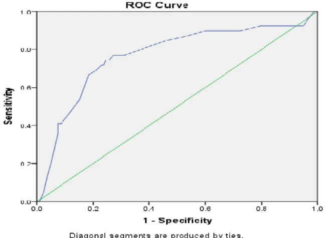
The combined Grades 1 & 2 DHF for VWF, VEGF, F1 + 2 and D-dimer at febrile, defervescence and convalescence phases with ANOVA analysis and normal controls are shown in Figure 1. The results from the three severe DHF patients recruited had elevated VWF, F1 + 2, VEGF and D-dimer with thrombocytopenia similar with grades 1&2 DHF but had lower haemoglobin levels. However, the patient with severe DHF (grade 4) was unconscious when admitted and found to have cerebral bleeding (CT scan) at defervescence phase with pleural effusion, hypervolemic shock. Thrombocytopenia with platelets at 43 x 109/L and elevated D-dimer of 1620 ng/mL at admission were given electrolyte and crystalloid infusions. The platelet rose to 88 x 109/L, and D-dimer level fell to 809 ng/mL at convalescence. His condition improved and discharged after two weeks in the hospital. The other two patients with grade 3 DHF also had pleural effusion and enlarged livers.
_______________________________________________________________________________________________________________________________
_______________________________________________________________________________________________________________________________
Figure 1: Mean levels of VWF, VEGF, F1 + 2 and D-dimer levels and ANOVA analysis in combined DHF Grades 1 & 2 at febrile, defervescence and convalescence phases
Thrombocytopenia was seen in one patient (platelets 87x 109/L) at febrile phase and fell to 11 x 109/L at defervescence phase but rose to 141 x 109/L at convalescence. The D-dimer levels of 5296.2 ng/mL at the febrile phase fell to 620.4 ng/mL at the convalescence phase. The other patient had normal platelets at admission (173 x 109/L), but severe thrombocytopenia was seen in defervescence and convalescence phases (4 x 10⁹/L and 37 x 109/l) respectively. The D-dimer was 2307.8 ng/mL at admission and 2060 ng/ml at convalescence. They were given electrolyte infusion and other medications and discharged one week later in an afebrile state.
Discussion
Dengue fever is the most serious consequence of mosquito-borne infection worldwide.
The pathophysiology of DHF in human is complex as its clinical symptoms are mainly due to an immune response involving the production of cytokine/chemokines as well as endothelial activation, T-lymphocytes, monocytes and platelets. Endothelial damage may also be caused by the virus itself.
Thrombocytopenia is responsible for bleeding events in DHF [20], [23] but many factors can contribute to the onset of thrombocytopenia from a reactive immune response against platelets and decreased platelet production [11], [24], platelet activation and apoptosis [21], Dengue virus could bind directly to prothrombin inhibiting the conversion to thrombin [24]
causing decreased coagulation activation, reduced thrombin generation and may be associated with bleeding complications in Brazilians with DHF [23]
The relationship between dengue and activation of coagulation is controversial [27]. However, activation of coagulation in critical DHF phase was reported in Indonesian patients (22), which was contrary to the
Brazilian study [23]. Bleeding manifestations and plasma leakage are complications seen in dengue and bleeding manifestation in adults may occur in the absence of plasma leakage [28].
In our study, petechiae or rash was observed in DHF besides the symptoms of pain in the bones, behind the eyes and headaches. Bleeding episodes like epitaxial were seen in grades 2 and 3 DHF while bleeding to the brain occurred in our grade 4 patient.
Pleural effusion was seen only in severe DHF with liver enlargement present in Grades 2, 3 and 4 and about 14.6% (6/41) in Grade 1 DHF.
Thrombocytopenia was observed in all phases of DHF even though in the convalescence phase, the mean platelet numbers were higher than in febrile and defervescence phases they did not reach statistical significance between grades 1 & 2 DHF. Normal haemoglobin and no haemo-concentration were observed, but elevated activation of coagulation (F1 + 2), VWF, VEGF and D-dimer suggest endothelial activation, plasma leakage and activation of coagulation in DHF. Activation of coagulation was reported earlier in critical DHF [22] in Indonesian patients, but contrary to this, Orsi and co-workers [23]
reported reduced thrombin generation and enhanced fibrinolysis contributing to the bleeding episodes in Brazilian patients. Reduced thrombin generation could result from the dengue virus binding directly to prothrombin inhibiting the conversion to thrombin [26].
Activation of coagulation and elevated D-dimer levels also indicates hypercoagulability and enhanced fibrinolysis. Endothelial activation evident by elevated VWF and VEGF suggests plasma leakage triggering the activation of the coagulation system, creating hypercoagulation and enhanced fibrin-lysis state.
Elevated D-dimer was seen in DHF even at convalescence. Normal haemoglobin and no haemoconcentration was observed in DHF grades 1 and 2. No mortality was recorded. Demographic differences and genetic make-up may contribute to these differences. Identifying the mechanisms affecting DHF would improve diagnosis and management therapy limiting morbidity and mortality.
In conclusion, in dengue haemorrhagic fever, the vascular endothelium is activated, causing plasma leakage triggering the activation of the haemostatic system creating a hypercoagulable and enhanced fibrinolytic state evident by marked fibrin-lysis.
Acknowledgements
The authors wish to express their sincere gratitude to the staff of the Research Laboratories at the Medical Faculty, Universitas Sumatera Utara and Murni Teguh Memorial Hospital for their expert technical assistance.
Mutiara et al.The Vascular Endothelium in Patients with Dengue Haemorrhagic Fever _______________________________________________________________________________________________________________________________
_______________________________________________________________________________________________________________________________
Open Access Maced J Med Sci. 2019 Jul 30; 7(14):2221-2225. 2225
References
1. World Health Organisation (WHO). Dengue and dengue haemorrhagic fever Factsheet No. 117, Geneva, Switzerland WHO, 2008.
2. World Health Organisation (WHO). Comprehensive guidelines for prevention and control of dengue and dengue haemorrhagic fever. Revised and expanded version WHO, 2011.
3 World Health Organisation Media Centre. Dengue and severe dengue. WHO Factsheet updated, 2017.
4 Brady OJ, Gething PW, Bhatt S, Messina JP, Brownstein JS, Hoen AG, Moyes CL, Farlow AW, Scott TW, Hay SI. Refining the global spatial limits of dengue virus transmission by evidence- based consensus. PLoS neglected tropical diseases. 2012;
6(8):e1760. https://doi.org/10.1371/journal.pntd.0001760 PMid:22880140 PMCid:PMC3413714
5 Karayanti MK, Ulterwaal CSPM, Kusriantuti R et al. The changing incidence of dengue haemorrhagic fever in Indonesia: a 45-year registry-based analysis. BMC Infectious Diseases 2014; 14:412.
https://doi.org/10.1186/1471-2334-14-412 PMid:25064368 PMCid:PMC4122763
6 World Health Organisation (WHO). Dengue: guidelines for diagnosis, treatment, prevention and control New ed., Geneva Switzerland, World Health Organisation, 2009
7 Verhamme P, Hoylaerts MF. The pivotal role of the endothelium in haemostasis and thrombosis. Acta Clinica Belgica. 2006;
61(5):213-9. https://doi.org/10.1179/acb.2006.036 PMid:17240734 8 Connolly DT. Vascular permeability factor: a unique regulator of blood vessel function. J Cell Biochem. 1991; 47:219-23.
https://doi.org/10.1002/jcb.240470306 PMid:1791186 9 Mohle R, Green D, Moore MAS, Nachman RL, Rafil S.
Constitutive production of thrombin-induced release of vascular endothelial growth factor by human megakaryocytes and platelet.
Proc Natl Acad Sci. 1997; 94:663-8.
https://doi.org/10.1073/pnas.94.2.663 PMid:9012841 PMCid:PMC19570
10 Verhuel HM, Hoekman K, Lulx-de Bakker S et al. Platelet transporter of vascular growth factor. Clin Cs Res. 19997; 3(12 Pt1):9815-90.
11 De Castro RA, De Castro JA, Barez MY, Frias MV, Dixit J, Genereux M. Thrombocytopenia associated with dengue hemorrhagic fever responds to intravenous administration of anti- D(Rh(O)-1) immune globulin. Am J Trop Med Hyg. 2007; 76:737- 42. https://doi.org/10.4269/ajtmh.2007.76.737 PMid:17426181 12 Falanga AM, Marchett M, Vignoli A. Coagulation and cancer:
Biological and clinical aspects. J Thromb Haemostas. 2013;
11:223-33. https://doi.org/10.1111/jth.12075 PMid:23279708 13 Kleinegris MC, Ten Cate H, Ten Cate HAJ. D-dimer as a marker for cardiovascular and arterial thrombotic events in patients with peripheral arterial disease. A systemic review. Thromb Haemostas.
2013; 110 (2):23. https://doi.org/10.1160/TH13-01-0032 PMid:23784703
14 Gomez K, Tudderham EGD, McVoy JH. Normal Haemostasis.
6th ed. Post Graduate Haematology Wiley Blackwell, 2011:747- 771. https://doi.org/10.1002/9781444323160.ch39
15 Mourao MP, Lacerda MV, Macedo VO, Santos JB.
Thrombocytopenia in patients with dengue virus infection in the Brazilian Amazon. Platelets. 2007; 18:605-1.
https://doi.org/10.1080/09537100701426604 PMid:18041652 16 Schexneider KL, Reedy EA. Thrombocytopenia in dengue fever.
Curr Hematol Rep 2005:145-8.
17 Honda S, Saito M, Dimano EM et al. Increased of phagocytosis of platelets from patients with secondary dengue virus infection by human macrophages. Am J Trop Med Hyg. 2009; 80:841-5.
https://doi.org/10.4269/ajtmh.2009.80.841
18 Nimmanitya S. Dengue hemorrhagic fever: disorders of hemostasis. Proceeding International Congress of Hematology.
Asia-Pacific Division 1999.
19 Srichaikul T, Nammannitya S. Hematology in dengue and dengue hemorrhagic fever. Baillieres Best Pract Res Clin Hematol.
2000; 13:261-76. https://doi.org/10.1053/beha.2000.0073 PMid:10942625
20 Diaz-Quijano FA, Villa-Centeno LA, Marinez-Vega RA.
Predictors of spontaneous bleeding in patients with acute febrile syndrome from a dengue endemic area. J Clin Virol. 2010; 49:11-5.
https://doi.org/10.1016/j.jcv.2010.06.011 PMid:20663710
21 Hottz ED, Oliviera MF, Nunes CG et al. Dengue induces platelet activation, mitochondrial dysfunction and cell death through mechanisms that involve DC-SIGN and caspases. J Thromb Haemostas. 2013; 11:951-62. https://doi.org/10.1111/jth.12178 PMid:23433144 PMCid:PMC3971842
22 Pudjianto S, Setiabudy RD, Nainggolan L, Setiabudy R.
Prothrombin fragment 1.2 (F1.2) in relation with plasma leakage and thrombocytopenia in dengue infection. Health Sci J Indonesia.
2016; 7:37-43. https://doi.org/10.22435/hsji.v7i1.4913.37-43 23 Orsi FA, Angerami RN, Mazetto BM et al. Reduced thrombin formation and excessive fibrinolysis are associated with bleeding complications in patients with dengue fever: a case-control study comparing dengue fever patients with and without bleeding. BMC Infect Dis. 2013; 13:250-6. https://doi.org/10.1186/1471-2334-13- 350 PMid:23890510 PMCid:PMC3733705
24 Lin CF, Lei HY, Liu CC et al. Generation of IgM anti-platelet autoantibody in dengue patients. J Med Virol. 2001; 63 (2):143-9.
https://doi.org/10.1002/1096-9071(20000201)63:2<143::AID- JMV1009>3.0.CO;2-L
25 Saito M, Oishi K, Inoue S et al. Association of increased platelet-associated immunoglobulins with thrombocytopenia and the severity of disease in secondary dengue virus infections. Clin Exp Immunol. 2004; 138 (2):299-303.
https://doi.org/10.1111/j.1365-2249.2004.02626.x PMid:15498040 PMCid:PMC1809201
26 Lin SW, Chuang YC, Lin YS, Lei HY, Liu HS, Yeh TM. Dengue virus non-structured protein NS1 binds to prothrombin/thrombin and inhibits prothrombin activation. J Infect. 2011; 64 (3):325-334.
https://doi.org/10.1016/j.jinf.2011.11.023 PMid:22138554 27 Mairuhu AT, Mac Gillavry MR, Setiati TE et al. Is clinical outcome of dengue-virus infections influenced by coagulation and fibrinolysis? A critical review of the evidence. Lancet Infect Dis.
2003; 3 (1):33-41. https://doi.org/10.1016/S1473-3099(03)00487-0 28 Wichmann O, Hongsinwon S, Bowonwatanuwong C,
Chotivanich K, Sukthana K, Pukrittayakmee S. Risk factors and clinical features associated with severe dengue infection in adults and children during the 2001 epidemic in Chonburi, Thailand. Trop Med Int Health. 2004; 9(9):1022-9. https://doi.org/10.1111/j.1365- 3156.2004.01295.x PMid:15361117
_______________________________________________________________________________________________________________________________
ID Design Press, Skopje, Republic of Macedonia
Open Access Macedonian Journal of Medical Sciences. 2019 Jul 30; 7(14):2226-2231.
https://doi.org/10.3889/oamjms.2019.651 eISSN: 1857-9655
Basic Science
Effect of Nanoherbal Andaliman (Zanthoxylum acanthopodium) and Extra Virgin Olive Oil Combination on Preeclamptic Rats Liver Histology
Putri Cahaya Situmorang1, Syafruddin Ilyas2*, Salomo Hutahaean1, Rosidah Rosidah2
1Department of Biology, Faculty of Mathematics and Natural Sciences, Universitas Sumatera Utara, Medan 20155, Indonesia; 2Faculty of Pharmacy, Universitas Sumatera Utara, Medan 20155, Indonesia
Citation: Situmorang PC, Ilyas S, Hutahaean S, Rosidah R. Effect of Nanoherbal Andaliman (Zanthoxylum Acanthopodium) and Extra Virgin Olive Oil Combination on Preeclamptic Rats Liver Histology. Open Access Maced J Med Sci. 2019 Jul 30; 7(14):2226-2231.
https://doi.org/10.3889/oamjms.2019.651
Keywords: Zanthoxylum acanthopodium; EVOO;
Preeclampsia; Liver
*Correspondence: Syafruddin Ilyas. Department of Biology, Faculty of Mathematics and Natural Sciences, Universitas Sumatera Utara, Medan 20155, Indonesia. E- mail: [email protected]
Received: 05-May-2019; Revised: 01-Jul-2019;
Accepted: 12-Jul-2019; Online first: 14-Jul-2019 Copyright: © 2019 Putri Cahaya Situmorang, Syafruddin Ilyas, Salomo Hutahaean, Rosidah Rosidah. This is an open-access article distributed under the terms of the Creative Commons Attribution-NonCommercial 4.0 International License (CC BY-NC 4.0)
Funding: This research was financially supported by the Directorate of research and community service, Directorate general of research and development, Ministry of research, Technology, and Higher education, Indonesia - Grant of PMDSU 2019 (Master's Education towards Doctorate 2019)
Competing Interests: The authors have declared that no competing interests exist
Abstract
BACKGROUND: Andaliman (Zanthoxylum acanthopodium) is a spice traditional Northen Sumatera, Indonesia and these fruits contain alkaloids, steroids and terpenoids. Extra Virgin Olive Oil (EVOO) contains antioxidants.
Combination of this plant have activities to reduce preeclampsia.
AIM: To know the safety of the combination of nano herbal andaliman and Extra virgin olive oil (EVOO) on preeclampsia patients’ liver.
METHODS: Pregnant rats were made to have preeclampsia with 3 ml of NaCl 6% injections. This research consists of 5 groups: K- (negative control): normal pregnant rats, K+: preeclampsia rats; P1: PE rats were given nano herbal andaliman 1 ml EVOO / day / 20 gBW from the 13th to the 19th day of pregnancy, P2: PE rats were given nano herbal andaliman 100 mg/day / 200 gBW from the 13th to the 19th day of pregnancy, P3: PE was given the combination of 1 ml EVOO/day / 200 gBW and andaliman nano herbal 100 mg/day/200 gBW on the 13th day of pregnancy to the 19th day given orally. Then on the 20th day of pregnancy, the subjects were dissected.
RESULTS: There were significant differences (p < 0.05) on the value of SGOT, SGPT, and the average damage of the hepatocyte cells except parenchymatous degeneration after being given the nano herbal andaliman and EVOO. The compared mean of normal hepatocytes cell, hydropic degeneration and necrosis value between all groups were p < 0.05 and p < 0.01 compared to (K-). The non-significant difference was found in the mean of parenchymatous degeneration between the groups (p = 0.058).
CONCLUSION: The combination of nano herbal andaliman (Zanthoxylum acanthopodium) and EVOO affected the level of necrosis in hepatocyte cells on preeclampsia rats.
Introduction
Preeclampsia (PE) is a multi-system disorder that is a major cause of maternal morbidity and mortality worldwide. Recent data show that the contribution of preeclampsia is estimated to be about 5 times that of morbidity & maternal-newborn mortality [1].PE is a life-threatening disease for mother and fetus in Indonesia. According to the Indonesian Demographic and Health Survey in 2007, PE contributed up to 24% of maternal deaths in Indonesia and made it the second leading cause of maternal mortality in Indonesia [2]. The international non- governmental organisation forum on Indonesian Development stated that Indonesia is a country in
Southeast Asia with the highest maternal mortality rate of 359/100.000 births [3].
Andaliman (Zanthoxylum acanthopodium.) is a spice that is used for traditional Batak cuisine, Norther Sumatera, Indonesia [4]. This plant has been used as a contraceptive for generations as an anti- fertility. Andaliman extract contains chemicals in the form of alkaloids, steroids and terpenoids, which have antioxidant activity and antimicrobial, repellent and kill insects [5], [6], [7]. The content of this plant is thought to have activities to reduce PE because andaliman fruit has also been reported to have anti-inflammatory activity and antioxidant activity [7].Extra Virgin Olive Oil (EVOO) from Olive fruit contains antioxidants, namely Vitamin E, hydroxityrosol and tyrosol [8].
Nanotechnology is a system that has several
Situmorang et al.Nanoherbal Andaliman (Zanthoxylum Acanthopodium) and Extra Virgin Olive Oil Combination on Preeclamptic Rats Liver Histology _______________________________________________________________________________________________________________________________
_______________________________________________________________________________________________________________________________
Open Access Maced J Med Sci. 2019 Jul 30; 7(14):2226-2231. 2227
advantages, namely being able to modify the characteristics of the surface, small size, high loading capacity so that it can be given in high concentrations [9]. Constraints that often occur in herbal medicines are difficult active substances to penetrate the lipid membrane of body cells because they have a large molecular size and low solubility in water that causes poor absorption and bioavailability [10]. Changes in drug molecules into nanometer scales provide a significant change in physicochemical properties and can improve the efficacy of these drug molecules [11].
The liver is the largest organ in the body that plays a role in detoxifying poisons in the blood, breaking down or changing the nature of toxic substances so that it can be released through urine.
The impact of the combination of these two herbs needs to be seen in the liver in terms of their safety effects in preeclamptic patients.
Material and methods
Nanoherbal Andaliman (Zanthoxylum acanthopodium)
Andaliman fruit used comes from the Dairi District of Northen Sumatera. Andaliman is washed thoroughly; then the wind is dried for 3 days at room temperature, then blended until smooth and then sized to nano size using High energy milling (HEM).
Simplicia as a destructive medium is inserted into the jar container and then inserted a ball with a larger diameter size and continued by inserting a small ball and the sample is placed lastly. The total volume of the ball and the sample inserted do not exceed 2 / 3 of the tube volume. The sample was tightly closed and then placed on a tube inside the HEM device, then HEM was turned on for 2 hours [12].
Phytochemical screening
Alkaloids: 1 g of nano herbal andaliman was put in a test tube then added 18 ml of distilled water and 2 ml of 2 N hydrochloric acids then heated for 2 minutes. The trial was conducted with Meyer reagent.
Positive alkaloids if sediment or turbidity occurs.
Glycosides: 3 g of nano herbal andaliman extracted in 30 ml mixture of 7 parts ethanol 96% and 3 parts of distilled water, then added concentrated sulfuric acid and refluxed in 10 minutes. After chilling 20 ml of filtrate added 10 ml of distilled water and 10 ml of lead (II) acetate 0.4 M were shaken and left for 5 minutes. The filtrate that has been filtered in the juice with 20 ml mixture of chloroform and isopropanol (3:
2) to be tested against sugar compounds and non- sugar compounds [13].
Flavonoids: 1 g of nano herbal andaliman
mixed in 20 ml of methanol then reflux 10 minutes.
After cooling, 10 ml of kerosene ether is added and then shaken and let stand until separation, the methanol layer is taken and then evaporated at 40°C, the remainder is dissolved in ethyl acetate and filtered by filtrate to be tested with 0.5 g zinc powder and 0.1 g powder magnesium [13].
Saponin: 1 g of nano herbal andaliman and 20 ml of hot water have been shaken for 20 seconds.
The saponin is positive if there is foam in not less than 10 minutes as high as 1-10 cm after adding 2 drops of hydrochloric acid 2 N foam was not lost.
Steroids/Terpenoids: 2 g nano herbal andaliman were macerated with 40 ml ether for 2 hours. The filtrate was filtered and evaporated. The remaining 4 drops of Lierbermann-Burchard reagent, if there is a red/purple colour changing to blue or blue- green means there is steroid.
Tanin: 0.5 g of nano herbal andaliman mixed in 50 ml of distilled water, then filtered, and then added 1 drop of 1% iron (III) chloride solution. If bluish-green was formed, show tannin compounds.
Antioxidant test with 1.1-diphenyl-2- picrylhydrazyl (DPPH) method
Nanoherbal andaliman was dissolved with methanol so that it becomes 250 μg/mL then homogenised for 10 minutes for 4 times then centrifuged to take the clear solution on top. Two mL of DPPH solution was added with 0.5 mL of nano herbal andaliman solution with 3 repetitions for each extract solution 6,25; 12.5; 25 and 50μg mL, then the absorbance was measured against methanol at a wavelength of 517 nm using a UV-Visible spectrophotometer.
Animal
This study used 25 pregnant Rattus norvergicus. Rats were mated at the Biology Laboratory animal house, University of Sumatera Utara. Pregnant rats are made into preeclampsia model by injecting 3 ml of 6% NaCl/day/200 gBW at 6 to 12 days gestation subcutaneously. Preeclampsia rats were evaluated by blood pressure more than 125/80 mmHg, MDA levels and proteunaria values more than 3 g/L [14]. This study consisted of 5 groups: K- (negative control): Normal pregnant rats; K+ (positive control): Preeclampsia (PE) pregnant rats, P1: PE rats given 1 ml EVOO/day/200 gBW in pregnancies 13 to 19 pregnancy day orally, P2: PE rats were given nanoherbal andaliman 100 mg/day/200 gBW at 13 to 19 days gestation orally, P3: PE rats were given a combination of 1 ml EVOO/day/200 gBW and nanoherbal andaliman 100 mg/days/200 gBW at 13 to 19 days of gestation orally.
Pregnant rats were dissected on the 20th day of pregnancy, for blood and liver to be taken, and then
_______________________________________________________________________________________________________________________________
_______________________________________________________________________________________________________________________________
liver preparations were made with paraffin blocks and Hematoxylin Eosin (HE) staining.
Examination of Serum Glutamic Oxaloacetic Transaminase (SGOT) and Glutamic Serum Pyruvate Transaminase (SGPT)
SGOT: Blood was centrifuged for ± 15 minutes at a speed of 5000 rpm. The blood serum then pipetted 200 µL aqua dest into the test tube, added 2000 µL of SGOT reagent 1 and then incubated for 5 minutes at 37°C. 500 µL of reagent 2 SGOT was added after homogeneous absorbance was measured at 365 nm wavelength with a spectrophotometer.
SGPT: Blood is centrifuged for ± 15 minutes at a speed of 5000 rpm. Blood serum piped 200 µL aquadest into the test tube then added 2000 µL of reagent 1 SGPT, then homogenised. After incubation for 5 minutes at 37°C. We have added 500 µL of reagent 2 SGPT then absorbance was measured at a wavelength of 365 nm with a spectrophotometer
Analysis of Data
The data were calculated the average score of liver histopathology changes from five fields of view with the Manja Roenigk Histopathology Scoring model. Then data were analysed by Anova test and non-parametric data by Kruskal Wallis test in SPSS 22 program.
Results
Phytochemical screening and DPPH test Based on the research that has been done, andaliman fruit in nanosize has the content of alkaloids, flavonoids, glycosides, steroids and terpenoids. The content of these compounds was similar to the content of andaliman extract in previous studies. DPPH test results on nano herbal andaliman with 3 repetitions for each extract solution 6,25; 12.5;
25 and 50μg / mL is IC50 48.5 µg / mL. Nanoherbal andaliman have very strong antioxidants.
Bodyweight and liver weight
Based on statistical data, ANOVA test on pregnant rat body weight showed that there was no significant difference in each treatment with p > 0.05 (p = 0.060). However, there were significant differences in liver weight (p < 0.05). The highest average weight is in K+ and the lowest group in the K- and P1 groups. Based on these data, nano herbal
andaliman, EVOO, and a combination of both can affect the weight of pregnant rat hearts. That means the liver here acts in the detoxification of foreign substances that first entered the body of the rat due to the content of secondary metabolites in the nano herbal andaliman and EVOO. The metabolic process in the liver is the process that affects its weight.
SGOT and SGPT
Based on statistical data on SGOT values in pregnant rats, there were significant differences (p <
0.05) in each treatment (Figure 1). However, the highest average value was found in the K+ group and the lowest in the K- and P3 groups. Nanoherbal andaliman can increase SGOT values higher in pregnant rats than EVOO and a combination of both.
Figure 1: Data are expressed with Kruskal Wallis test; Mann- Whitney test was applied to compare SGOT value between all groups; **p < 0.01 compared to control (K-); *p < 0.05 compared to K+; K-: Pregnant normal; K+: PE rats; P1: PE rats after given EVOO; P2: PE rats after given nano herbal andaliman; P3: PE rats after given EVOO and nano herbal andaliman
The SGPT value also has a significant difference with a value of < 0.05. The highest average SGPT value was found in P2, and the lowest was found in the control group (K-). This means that there is a high level of damage in the PE liver treatment there is P2 so that the value of SGOT and SGPT is higher than other treatments. Based on this data nanoherbal andaliman and EVOO can increase the value of SGOT and SGPT in pregnant rats (Figure 2).
Figure 2: Data are expressed Kruskal Wallis test; Mann-Whitney test was applied to compare SGPT value between all groups; **p <
0.01 compared to control; *p < 0.05 compared to K+; K-: Pregnant normal; K+: PE rats; P1: PE rats after given EVOO; P2: PE rats after given nano herbal andaliman; P3: PE rats after given EVOO and nano herbal andaliman
Situmorang et al.Nanoherbal Andaliman (Zanthoxylum Acanthopodium) and Extra Virgin Olive Oil Combination on Preeclamptic Rats Liver Histology _______________________________________________________________________________________________________________________________
_______________________________________________________________________________________________________________________________
Open Access Maced J Med Sci. 2019 Jul 30; 7(14):2226-2231. 2229
Histology of rat's liver
Based on statistical data on normal hepatocyte cell values, parenchymatous degeneration, hydrophilic degeneration and necrosis in pregnant rats using the cruciferous Wallis test there were no significant differences (p > 0.05). But based on the highest average normal hepatocyte cells were found in K- and P1 while the lowest was in K+ and P1 (Table 1). However, there was no significant difference in parenchymatic degeneration (p > 0.05).
The highest parenchymatous degeneration at K+ and lowest at K-. The highest hydropic degeneration was also found in P1 and P2 while the lowest was K-. The data proved significantly with a value of p < 0.05. The highest necrosis is at K+ and lowest at K- and P3.
Based on the statistics of nano herbal andaliman, EVOO and the combination of both causes liver damage with a degree of damage in the form of parenchymatous degeneration, hydrophilic degeneration and necrosis.
Table 1: Average of normal hepatocytes cells and liver damages on preeclamptic rats
Treatments Normal Parenchymatous Degeneration
Hydropic
Degeneration Necrosis
K- 13 ± 1.46 5.52 ± 1.76 5.52 ± 3.20 9.76 ± 3.67
K+ 5.36 ± 1. 49** 8.32 ± 2.69 11 ± 4.72** 27.2 ± 10.00**
P1 12.2 ± 1.31* 5.52 ± 1.94 11.5 ± 2.43 10.88 ± 3.56*
P2 8.56 ± 1.33** 6.72 ± 2.15 11.5 ± 3.43** 16.96 ± 4.21**
P3 11.2 ± 1.48* 7.2 ± 2.45 7.8 ± 3.00* 10.24 ± 4.18*
Kruskal Wallis test and Post-hock test were applied to compare mean of normal hepatocytes cell, hydropic degeneration and necrosis value between all groups; *p < 0.05 compared to control (K-); **p < 0.01 compared to K-; Non-significant difference was found in the mean of parenchymatous degeneration between the groups (p = 0.058).
Based on the average pattern of hepatocyte cell damage in pregnant rats, the greatest damage was found in the K+ group and also P2, while the lowest damage was found in K- and P3 (Figure 3).
Based on this data, pregnant women who often want to consume andaliman are better combined with EVOO to prevent liver cell damage.
Figure 3: Data are expressed Kruskal Wallis test; Mann-Whitney test was applied to compare trophoblast cells value between all groups; **p < 0.01 compared to control; *p < 0.05 compared to K+;
K-: Pregnant normal; K+: PE rats; P1: PE rats after given EVOO;
P2: PE rats after given nano herbal andaliman; P3: PE rats after given EVOO and nano herbal andaliman
Based on histological observations, it was seen that K+ (PE) had the greatest damage compared to other treatments. Liver hepatocyte damage was also seen after being given the herbal andaliman EVOO and a combination of both but not as much as K+ (PE) (Figure 4). This means that the compounds contained in nano herbal andaliman cause liver necrosis. Fat degeneration (parenchymatous degeneration) is also seen in histology such as fat accumulation in the cell cytoplasm where fat in the cytoplasm pushes the cell nucleus to the side, due to interference with hepatocytes so that lipoproteins are not formed. In group P1 there are more parenchymatous degeneration characterised by varied fats and vacuoles but cannot be seen hydrophilic degeneration is also evident in K+, P1 and P2 where cell forms are like cell swelling (Figure 4).
This degeneration is more severe damage; there are vacuoles containing water and cytoplasm that do not contain fat and glycogen.
Figure 4: Histology of rats liver; A) K- (Pregnant normal); B) K+(PE rats); C) P1 (PE rats after given EVOO); D) P2 (PE rats after given nano herbal andaliman); E) P3 (PE rats after given EVOO and nano herbal andaliman); A) Normal; B) Parenchymatous degeneration;
C) Hydropic degeneration; D) Necrosis (H & E) 40x
Discussion
PE can cause interference with the liver. Liver dysfunction can occur due to vasoconstriction and oedema that shows damage to the liver, muscles, kidneys, pancreas and red blood cells [15]. Liver damage is always associated with necrosis and administration of exogenous antioxidants, may be beneficial in protecting the liver [16]. Natural antioxidants are known to have beneficial effects on hepatitis or liver disorders caused by antitubercular agents [17]. Andaliman possesses unique flavour properties and bioactive compounds. The chemical compound in this herbal was Monoterpenes (46.54%), hydrocarbon monoterpenes (19.75%). The primary volatile compounds in andaliman (relative peak area >
10%) are geranyl acetate (32.04%) and limonene (15.80%) [18]. Andaliman in nanosize also contains alkaloids, flavonoids, glycosides, steroids and terpenoids as well as extracts [5], [6], [7]. Andaliman
_______________________________________________________________________________________________________________________________
_______________________________________________________________________________________________________________________________
fruits and leaves contain terpenoids, alkaloids, flavonoids, and other phenolics, which can function as antioxidants [19]. The ethyl acetate extract of andaliman fruit has antioxidant activity and with an IC50 value of 66.91 ppm and isolating EA.X.6.1 has antioxidant activity and with an IC505.55 ppm value [20]. Andaliman in nanosize has IC50 value of 48.5 µg/mL so that antioxidant properties are very strong compared to extract. Antioxidants are components that can inhibit free radicals and it is estimated that in scala nano can reduce oxidative stress in PE.
According to Tensisca et al., [21] andaliman fruit extract with ethanol and hexane has different antioxidant activity which is highest in water systems in emulsion and oily systems though having moderate activity. The content of important compounds is relatively stable during heating, but is heating up to 175°C; it can reduce up to 17%. Andaliman in the form of extract also has different contents and activities when exposed to heat, fluorescent light and ultraviolet [22]. Olive oil is a vegetable oil obtained from olive plants (Olea europaea) in the packaging of Extra Virgin Olive Oil (EVOO) in low doses can control serum levels of Hsp70, so the process of apoptosis does not occur excessively especially in preeclampsia [23]. The combination of these two plants can reduce hepatocyte cell damage because of the incorporation of antioxidants and vitamin E.
The value of SGPT and SGOT in PE can decrease with the combination of these two herbs (Figure 1 and 2). SGPT and SGOT will come out of liver cells if the liver cells are damaged so that it will cause an increase in SGPT and SGOT levels in blood serum [24], [25]. Increased SGOT treatment is also caused by stress. Hepatocytes are the type of cell that forms most of the liver. These cells are located between sinusoids, which are full of blood, and bile ducts. The liver is often the target organ of toxic substances because most poisons enter the body through the digestive system, then after being absorbed, carried by the portal vein to the liver — the highest parenchymatous degeneration at K+ and lowest at K-. Parenchymatous degeneration is the lightest level of category of degeneration. Cells that become parenchymal degeneration are found in granules in the cytoplasm, due to the deposits that cause the cytoplasm to become cloudy and followed by swelling in cells [26]. The highest hydropic degeneration is also in K+ (Table 1). Hydropic degeneration is a more severe level of damage.
These changes are generally a result of metabolic disorders, such as hypoxia or chemical poisoning.
This degeneration is also reversible even though it is possible to be irreversible if the cause of the injury persists [26]. Cells that have been injured can cause damage to the plasma membrane and changes in the nucleus. Pregnant women that often consume andaliman should also consume it with EVOO to prevent damage to the liver cells. EVOO contains vitamin E (tocopherol) that is anti-apoptosis [23].
Olives that are converted to Extra virgin olive oil
(EVOO) have analgesic, anti-inflammatory and anticancer properties [27]. So, using andaliman nano herbal is better along with EVOO to reduce necrosis in the liver.
Between P1, P2 and P3, it appears that more damage is found in P2 (Giving nano herbal andaliman only) means that the compounds contained in andaliman nanoherbal cause liver necrosis. Liver disease in PE have a high risk of pregnancy disorder, although no reports of maternal death but the birth of premature infants [28]. Liver disorders in PE dieaseas may increase liver enzymes, autoimmune, hyperemesis gravidarum, acute fatty liver, and intrahepatic cholestasis [29]. This is in accordance with Emita's study[30]. where there was a change in the color and texture of the liver surface, as well as an increase in hepatocyte damage. Liver disorders in PE can increase liver enzymes, low platelets (HELLP), acute fatty liver, hyperemesis gravidarum, intrahepatic cholestasis, and autoimmune liver [29]. Regulations of damaged liver in the metabolic system can cause gestational hypertension during the first pregnancy and can cause PE, hemolysis, increased liver enzymes, and low platelet syndrome (HELLP) [31].
EVOO contains exogenous antioxidants has anti- inflammatory, anticancer and analgesic properties and neuroprotective activities that can fight oxidative damage to the brain [32]. Thus, andaliman in combination with EVOO is better and safer than just andaliman.
In conclusion, the combination of nanoherbal andaliman (Zanthoxylum acanthopodium) and Extra Virgin Olive Oil (EVOO) can reduce parenchymatous degeneration, hydrophic degeneration and hepatocyte cell necrosis in preeclampsia rats (p < 0.05). Further testing with immunohistochemistry is recommended.
Acknowledgements
Authors are grateful to the Directorate of research and community service, Directorate general of research and development, Ministry of research, Technology, and Higher education have funded our research in Grant of PMDSU 2019 (Master's Education towards Doctorate 2019).
References
1. Bilano VL, Ota E, Ganchimeg T, Mori R, Souza JP. Risk Factors of Pre-eclampsia/Eclampsia and Its Adverse Outcomes in Low- and Middle-Income Countries: A WHO Secondary Analysis. 2014;
21:9(3):e91198. https://doi.org/10.1371/journal.pone.0091198 PMid:24657964 PMCid:PMC3962376
2. SKDI. Number of Maternal Mortality. In: RI D, editor. Jakarta:
Directorate of Maternal Health, 2007.
Situmorang et al.Nanoherbal Andaliman (Zanthoxylum Acanthopodium) and Extra Virgin Olive Oil Combination on Preeclamptic Rats Liver Histology _______________________________________________________________________________________________________________________________
_______________________________________________________________________________________________________________________________
Open Access Maced J Med Sci. 2019 Jul 30; 7(14):2226-2231. 2231 3. Ministri of healthy (Kemenkes RI). Situasi Kesehatan Ibu. Pusat
Data dan Informasi Kementerian Kesehatan Republik Indonesia.
Jakarta: Kemenetrian Kesehatan Republik Indonesia, 2014 4. Siregar BL. Deskripsi dan Perkecambahan Andaliman (Zanthoxylum acanthopodium DC.) di Sumatera Utara. Hayati.
2003; 10:1:38-40.
5. Wijaya CH, Hadiprodjo IT, Apriyantono A. Komponen volatil dan karakterisasi komponen kunci aroma buah andaliman
(Zanthoxylum acanthopodium DC.). J. Teknologi Industri Pangan.
2001; 12:117-125.
6. Parhusip A, Yasni S, Elisabeth Y. Kajian metode ekstraksi andaliman (Zanthoxylum acanthopodium DC.) terhadap mikroba patogen dan perusak pangan. J Ilmu Teknologi Pangan. 2003;
1:112-123.
7. Suryanto E, Sastrohamidjojo H, Raharjo S, Tranggono. Singlet oxygen quenching effect of andaliman. (Zanthoxylum
acanthopodium DC.). Indonesian Food and Nutrition Progress.
2004; 11:48-55.
8. Satria D, Jansen S, Ginda H, Ilyas S, Anjelisa P. Antioxidant and Antiproliferative Activities of an Ethylacetate Fraction of Picria Fel- Terrae Lour. Herbs. Asian Pac J Cancer Prev. 2017; 18(2):399- 403.
9. Dewandari KT, Yasni S, Yuliani S. Ekstraksi dan Karakterisasi Nanopartikel Ekstrak Sirih Merah (Piper Crocatum). Jurnal Pascapanen. 2013; 10(2):58-65.
https://doi.org/10.21082/jpasca.v10n2.2013.58-65
10. Saraf S, Ajazuddin A. Applications of Novel Drug Delivery System for Herbal Formulations. Fitoterapia. 2010; 81(7):680-689.
https://doi.org/10.1016/j.fitote.2010.05.001 PMid:20471457 11. Müller D, Schiffer M. Preeclampsia from a renal point of view:
Insides into disease models, biomarkers and therapy. World J Nephrol. 2014; 3(4):169-181. https://doi.org/10.5527/wjn.v3.i4.169 PMid:25374810 PMCid:PMC4220349
12. Situmorang PC, Ilyas S. Description of Testes Histology of Mus musculus after giving nano hebal Rhodomyrtus tomentosa (Haramonting). Asian J Pharm Clin Res. 2018; 11:11.
https://doi.org/10.22159/ajpcr.2018.v11i11.29042
13. Departemen Kesehatan RI. Farmakope Indonesia Edisi IV.
Jakarta: Departemen Kesehatan RI, 1995
14. Situmorang PC, Ilyas S, Hutahaean S. Study of Combination of Nanoherbal Andaliman (Zanthoxylum acanthopodium) and Extra Virgin Olive Oil (EVOO) Effects in the Expression of MDA, HSP-70 and Placental Histology of Preeclamptic Rats. Pharm. Sci. 2019;
https://doi.org/10.1088/1755-1315/305/1/012081
15. Billington M, Stevenson M. Critical care in childbearing for midwives. Blackwell Publishing; 2007.
16. Pramodh, K, Deval RG, Lakshmayya, Ramchandra SS.
Antioxidant and hepatoprotective activity of tubers of Momordica tuberosa cogn. Againts CCl4 i duced liver in rats. Indian J Exp Biol.
2008; 46:510-513.
17. Limsuwan S, Trip EN, Kouwen TR, Piersma S, Hiranrat A, Mahabusarakam W, et al. Rhodomyrtone: A new candidate as natural antibacterial drug from Rhodomyrtus tomentosa.
Phytomedicine. 2009; 16(6):645-651.
https://doi.org/10.1016/j.phymed.2009.01.010 PMid:19303274 18. Wijaya CH, Napitupulu FI, Karnady V, Indariani S. A review of the bioactivity and flavor properties of the exotic spice "andaliman"
(Zanthoxylum acanthopodium DC.). Food Reviews International.
2018; 1-19. https://doi.org/10.1080/87559129.2018.1438470 19. Rana VS and Blazquez MA. Terpenoid Constituents of
Zanthoxylum Acanthopodium DC. Leaves. J Essen Oil Res. 2008;
20:515−516. https://doi.org/10.1080/10412905.2008.9700075 20. Winarti W, Simanjuntak P, Syahidin MF. Identifikasi Senyawa Kimia Aktif antioksidan dari ekstrak etil asetat buah andaliman (Zanthoxylum acanthopodium DC). TM Conference Series 01.
2018; 162-166. https://doi.org/10.32734/tm.v1i3.283
21. Tensiska C, Hanny W, Nuri. Antioxidative Activity of andaliman fruit extract (Z. acanthopodium DC.) on several food system and its antioxidative stability on temperature and pH Influence. Journal of Food Technology and Industry. 2003; 14(1):29-39.