Case description: A 44-year-old male came to Wahidin Sudirohusodo Hospital on March 13, 2018 with chest pain typical duration >30 minutes onset > 48 hours, with coronary risk factor diabetes mellitus. Physical examination within normal limits, with electrocardiography showing ST elevation in leads II,III,aVF,V7‐V9, laboratory findings show increased troponin I, and HbA1c. Echocardiography shows reduced EF (43%), with hypokinetics in the inferior segment. The patient diagnosed with inferoposterior STEMI onset >24 hours and threatens conservatively with fondaparinux, DAPT, ACE inhibitor, statins and insulin. Background: Ischemic stroke is uncommon but one of the most feared complications of acute myocardial infarction. Case Illustration and Discussion: A 16-year-old male patient was admitted to the emergency department with chest pain 12 hours prior to admission, along with sweating.
Brugada Syndrome Unmasked by the Full Stomach Test in an Asymptomatic Patient with Family History of Sudden Death: A Case Report
Delusional in a Female Patient with Sacubitril/Valsartan Therapy
Cardiogenic Shock in Right Ventricular Myocardial Infarction: A Challenging Management in Rural Public Hospital
Managing Acute Pulmonary Edema in Congestive Heart Failure with Multiple Comorbidities: A Case Report
Patient With Acute Cardiogenic Pulmonary Edema Patients (ACPE): When Do We Need Non‐
T Wave Inversion Mimic Acute Coronary Syndrome In Intracranial Hemorrhage Patient: A Case Report
A case: Optimal management of pregnancy with rheumatic heart disease Oktaliani, R.,1 Supriadi, E.,1,2 Herlambang1,3 Oktaliani, R.,1 Supriadi, E.,1,2 Herlambang1,3. Conclusion: It has been reported that a 27-year-old pregnant woman is in the 39th week of pregnancy and diagnosed with heart failure and causes rheumatic heart disease. The most prominent principle of treatment of pregnancy with heart disease is early detection and reduction of excessive cardiac load from the first trimester to the puerperium.
Hyperkalemia in Latent Autoimmune Diabetes in Adult: A Case Report in a Rural Area in Indonesia
Chest pain and Bradyarrhythmia manifestation in Dengue Fever : A Rare Case Report Franky Santoso 1 , Markus Tjahjono 2
Left Ventricle Aneurysm Mimicking Acute Anterior ST Elevation Myocardial Infarction: How Important of Serial Electrocardiography
Left ventricular aneurysm mimicking acute anterior ST elevation myocardial infarction: how important serial electrocardiography. High thrombus burden in late presentation of inferior ST elevation myocardial infarction managed by primary percutaneous coronary intervention: is there still room for thrombus.
High Thrombus Burden in Late Presentation of Inferior ST‐Elevation Myocardial Infarction Managed by Primary Percutanous Coronary Intervention: Is There Still Place for Thrombus
Achieving Complete Peripheral Revascularization through Complex Percutaneous Peripheral Intervention Accessed Antegradely via Newly Grafted Femoral Artery Bypass Segment in a
End-stage renal disease with aneurysmal and critical coronary artery stenosis: Myocardial revascularization strategy. In this case, coronary revascularization was performed for an ESRD patient with aneurysmal and critical coronary artery stenosis. Diagnostic coronary angiography (DCA) was performed on the patient and the result was double vessel disease + left main disease with aneurysmal artery proximal to left anterior descending (LAD) coronary artery and critical stenosis at the distal of aneurysmal coronary artery.
Newly Diagnosed Rheumatic Valvular Heart Disease Admitted For Treatment of Heart Failure and Atrial Fibrilation: A Case Report
Background: Malignant anomaly of the right coronary artery is a rare anomaly that originates from the left main coronary artery and then passes between the aorta and the pulmonary trunk. CCTA performed The right coronary artery was visualized as abnormal, arising from the left main coronary artery, which followed between the aorta and the pulmonary trunk. Finally, we describe a patient with a malignant anomaly of the right coronary artery presenting with heart failure and polycythemia.
Ventricular Arrhythmias Originating from Papillary Muscles in The Right Ventricle: A Rare Case Report Study
Nonsignificant disease presenting as myocardial infarction in the absence of obstructive coronary artery disease (MINOCA) in men: a case report.
Non‐significant Disease Presenting as Myocardial Infarction in the Absent of Obstructive Coronary Artery Disease ( MINOCA) in Men: a Case Report
Apical Hypertrophic Cardiomyopathy Mimicking Wellen Type B A Challenging Diagnostic and Clinical Presentation
Background: Brugada syndrome (BrS) is a type of arrhythmic disorder characterized by the abnormal finding of several ECG patterns, such as incomplete right bundle branch block, ST junction elevation (J point) in the anterior precordial leads, and normal QT interval without proven structural heart disease. A 12-lead ECG was performed, which showed rounded ST elevation in V2-V6 with incomplete right bundle branch block, a specific diagnostic criterion for Brugada syndrome. Vital signs became stable and a laboratory test was performed with a chest X-ray while the patient was transferred to the intensive care unit.
The Role of Synchronized Electrical Cardioversion vs Pharmacological Cardioversion in Monophormic Ventricular Tachycardia post CABG : A Case Report in Emergency Room
VSD can be associated with several complications such as heart failure and aortic regurgitation (AR). Case Illustration and Discussion: We present a 12-year-old boy brought to the emergency room with palpitations. Case illustration: A 52-year-old woman came to the emergency room 3 hours before admission with complaints of palpitations.
Patient with Severe Rheumatic Mitral Stenosis: When We Should Call The Cardiothoracic Surgeon?
Rivaroxaban, a new oral anticoagulant, is an option for the treatment of GVT and has been evaluated in several studies. On the 3rd day of taking the drug, the patient's left leg was swollen and there was pain on dorsiflexion (Homans sign). Rivaroxaban is an option in postpartum and lactating women for DVT without an increased risk of bleeding for the patient and her baby.
A Case Report: Atrial Fibrillation with Aberrancy in Young Man with Symptomatic Wolff Parkinson White (WPW) Syndrome
Furthermore, T3 reduces action potential duration, the refractory period of atrial myocardium and atrial/ventricular nodal.3 The most common arrhythmia in TS is sinus tachycardia and atrial fibrillation, rarely supraventricular tachycardia.4. Unfortunately, the patient discharged at his own request and we lost track of the progress of his treatment. Cardiac enzyme and electrolyte test were within normal limits and the patient was given Bisoprolol.
Ischemic Stroke due to Atrial Fibrillation : A Challenge for the Diagnosis and Treatment in Rural Areas
Conclusion: New myocardial infarction at another site after thrombolytics may occur in some cases due to the increase of catecholamines and the inflammatory response that causes myocardial infarction elsewhere. With the patient's clinical condition and distance to the nearest hospital, transfer was impossible. Administration of vasodilators to "open" the microcirculation is avoided due to low arterial pressure during the PCI procedure.
Ventricular Septal Rupture after Acute Myocardial Infarction : A Case Report
Patients with clinical ALI should be referred to the emergency center with a vascular team for diagnosis and treatment. Antiplatelet aggregation inhibitors are used in patients with LEAD to prevent limb-related and general CV events. DAPT can be considered in patients with multiple coronary artery disease, diabetic patients with incomplete revascularization.
Discussion: Patients with clinical ALI should be referred to emergency center with vascular team for diagnosis and management. Eisenmenger's syndrome in a 12-year-old girl suffering from Ebstein's anomaly and atrial septal defect with a spike helmet ECG pattern: A case report. 3General Practitioner at Wiradadi Husada General Hospital, Banyumas, Indonesia; 4Cardiologist at Sunan Kudus Islamic Hospital, Kudus, Indonesia.
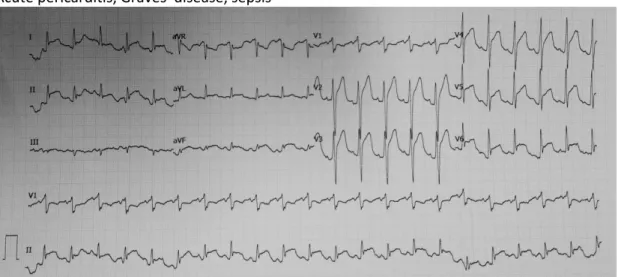
In this way we try to highlight how the detection of critically ill patient using the new potential ECG marker called spiked helmet pattern. The spiked helmet pattern is described as ST-segment elevation with upshift beginning before the QRS complex. Some reports said it was linked to a patient in critical condition, but the mechanism remains unknown.
Conclusion: Undiagnosed EA patients with ASD may lead to ES and arrhythmias with high mortality.
THE SPECTRUM OF ACUTE CORONARY SYNDROME IN 61 YEARS OLD MAN A CASE REPORT
Warapsari 1 , A. Tonang 2
A Case Report of Teenager with Heart Failure and Atrial Fibrillation Associated with Hyperthyroidism
The patient was diagnosed with thyroid heart disease and is receiving furosemide, spironolactone, candesartan, digoxin, propylthiouracil, bisoprolol and methylprednisolone. The patient has a history of similar palpitations a year ago and acute coronary syndrome 10 years ago. The patient was then treated with concor and clopidogrel in the intensive care unit and discharged after the third hospital day in stable condition.
2 Department of Cardiology and Vascular Medicine, Faculty of Medicine Universitas Indonesia - Harapan Kita National Cardiovascular Center, Jakarta, Indonesia. The patient and her husband should know that pregnancy may increase the risks to the mother and the fetus. The patient was taken to RSUD Sumedang, diagnosed as severe aortic regurgitation, receiving heart failure medications and Ceftriaxone.
Initially, the patient appeared stable, with anemic conjunctiva, cardiomegaly with aortic regurgitation murmur, Janeway lesions, and no signs of chronic severe AR. Patient was given ampicillin and gentamycin scheduled for urgent DVR and vegetation evacuation surgery in two weeks, but during hospitalization the patient went into shock with acute renal failure. Vasopressor and inotropic drugs were given, hemodialysis was advised, but the patient eventually refused further therapy.
The patient received 80-mg aspila once daily during treatment and required further investigations, such as MRI or TCD examination, to identify a defect in the posterior circulation.

Improvement Ejection Fraction After 11 Days Treatment in Pediatric Dilated Cardiomyopathy : Case Report
After further investigation by echocardiography, the patient was found to have a giant unruptured SVA with LVOT obstruction. The patient was scheduled for a cardiac MRI scan, but is still cost-limited and the patient was not covered by health insurance. On the third day, the patient suddenly developed a fever of 39 degrees Celsius, difficulty breathing and oxygen desaturation (81 percent).
The patient responded excellently to therapy, showed a negative test result from the second RT-PCR study and was discharged on the 16th day after admission. Identification of thrombus by echocardiography, management strategies using anticoagulants and patient follow-up should be applied to avoid poor outcomes. The patient was treated with 5000 IU/ml intravenous bolus heparin, continued with titration of 450 IU/hr, intravenous furosemide 2x40 mg, ramipril 1x5 mg, spironolactone 1x25 mg and bisoprolol 1x1.25 mg.
The patient was discharged after his condition improved and continued with ambulatory care given warfarin 1x3 mg. Conclusion: Clinicians should always be aware of aortic dissection even though the patient presents with typical chest pain. Unfortunately, the patient decided to go home before we could perform further examinations to evaluate the patient.
After receiving full anti-ischemic and anti-inflammatory treatment, the patient was symptom-free during hospitalization.

DIAGNOSIS AND MANAGEMENT OF UPPER EXTREMITY DEEP‐VEIN THROMBOSIS IN PATIENT WITH ADENOCARCINOMA LUNG MUTATION EXON 19 EGFR (+)
The appropriate preparation of the patient with asymptomatic CAVB for non-cardiac surgery is controversial. Conclusion: The appropriate preparation of the patient with asymptomatic CAVB for non-cardiac surgery is controversial. Normal cardiac enzyme was found and the patient was diagnosed with unstable angina pectoris high lateral Killip I.
Most myxomas are found in the left atrium, only 20% are found in the right atrium. The incidence of cardiac myxoma peaks between the ages of 40 and 60, with a female-to-male ratio of approximately 3:1. The patient was originally scheduled for PCI, but could not because of a suspected COVID-19 infection. Most fibroids are found in infants younger than 1 year of age.1 The incidence of fibroids is reported to be approximately 25.
On the third day of treatment, the patient was unconscious and the shortness of breath worsened. In the middle of the thrombolytic process, the patient suddenly developed extreme chest pain accompanied by hemoptysis. The patient then referred to Harapan Kita Hospital and was scheduled for the Bental method, total gestation replacement, Elephant Trunk and Thoracic Endovascular Aortic Repair (TEVAR) procedure. The diagnostic approach is fundamental in AD as it could mimic ACS.
Brugada syndrome is associated with inherited mutations in SCN5A.7 SCN5A mutations impair cardiac sodium channel gating more strongly at higher temperatures, resulting in prolongation of the PR and QRS intervals.7 Another factor that could trigger arrhythmias was inflammation.7,8 A study of the association between CRP levels and BrS symptoms suggests that inflammation might play a role in the pathophysiology of BrS arrhythmias.8 All cytokines are elevated in malaria patients compared to controls.9 Another report showed that BrS can be revealed by fever, which caused by malaria.3 . Pericardial fluid was analyzed: Ziehl-Neelsen stain negative, Rivalta positive, total cells 550 ul with 99% MN and 1% PMN, fluid total protein 6.5 d/dL, serum total protein 6.9 d/dL , LDH fluid 2265 U/L, LDH serum 691 U/L, glucose fluid 89 mg/dL. The patient was transferred to the Intensive Care Unit, treated with prednisone (20 mg/day) and referred to a pulmonologist. Background: Systemic sclerosis (SSS) is a rare chronic autoimmune disorder characterized by fibrosis of the skin and visceral organs.1 The most common cause of death in SSC is cardiopulmonary dysfunction, including pulmonary arterial hypertension with a 3-year survival rate of 52%. 2.

R D Putra
Background: We usually hypothesize the ischemic-related artery (IRA) in acute myocardial infarction (AMI) based on the appearance of ST elevation in corresponding electrographic leads. In this case, however, an acute complete occlusion in the right middle coronary artery (RCA) presented with ST elevation in the precordial lead V1-V3. Recording electrocardiogram (ECG) showed sinus rhythm with ST elevation in precordial leads V1-V3, with no significant ST segment changes in inferior leads.
But the anterior wall is infarcted due to occlusion of RCA collateral flow to LAD. It made ST elevation in lead V1-V3 without ST-segment changes in inferior lead even though the IRA was RCA. Conclusion: There are many underlying mechanisms that cause occlusion of RCA with anterior ST elevation.
But in this rare case, occlusion due to stenosis and thrombus was found in the middle LAD. Introduction: Heart valve disease is often underdiagnosed due to asymptomatic manifestation in the early stages of the disease. Right ventricular double outflow in the newborn: a type of critical congenital heart disease that is often misdiagnosed.
The main treatment of pre-excited AF in Indonesia is electrical cardioversion, regardless of the patient's hemodynamic status due to the lack of intravenous ibutilide, procainamide, propafenone, and flecainide.

