Carolus Linnaeus (1753) classified the plants into 24 classes using only one character: “the number and morphological arrangement of the stamens. The duplication and reduction of the number of chromosomes associated with the alternation of generations was later recognized by Strassburger (1894), who determined that in a life cycle there is not only an alternation between two different morphological forms, but that there is also an alternation between two different morphological forms. at the level of ploidy – the gametophyte with a single set of chromosomes and the sporophyte with a double set of chromosomes. They swim in the water film and reach up to the neck of the arehegonium.
This nomenclature is considered more scientific as it is also in accordance with the recommendations of the "International Code of Botanical Nomenclature" (ie the correct ending of the class word is – opsida and the subclass is – idae).
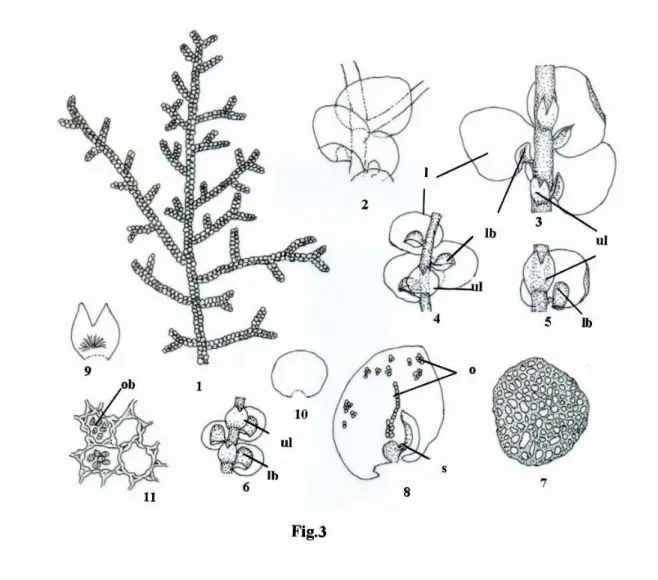
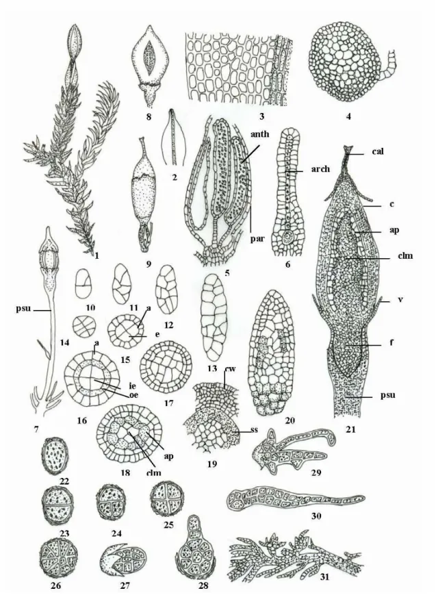
Jungermanniales Limpr. (in Cohn 1876)
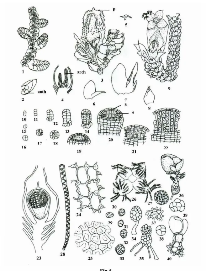
The cells of the leaves are thick-walled, and the thickenings are more pronounced in the corners, called trigones (Fig. 3: 11). The zygote first divides transversely, forming the outer epibasal cell and the inner hypobasal cell (Fig. 4: 11). Apical cells (upper layer) divide periclinally and differentiate into external amphithecium and internal endothecium (Fig. 4: 14-18).
The inner four endothelium divides only vertically and the cells thus produced are longer in the middle part of the capsule and shorter towards the periphery (Fig.4: 19, 20).
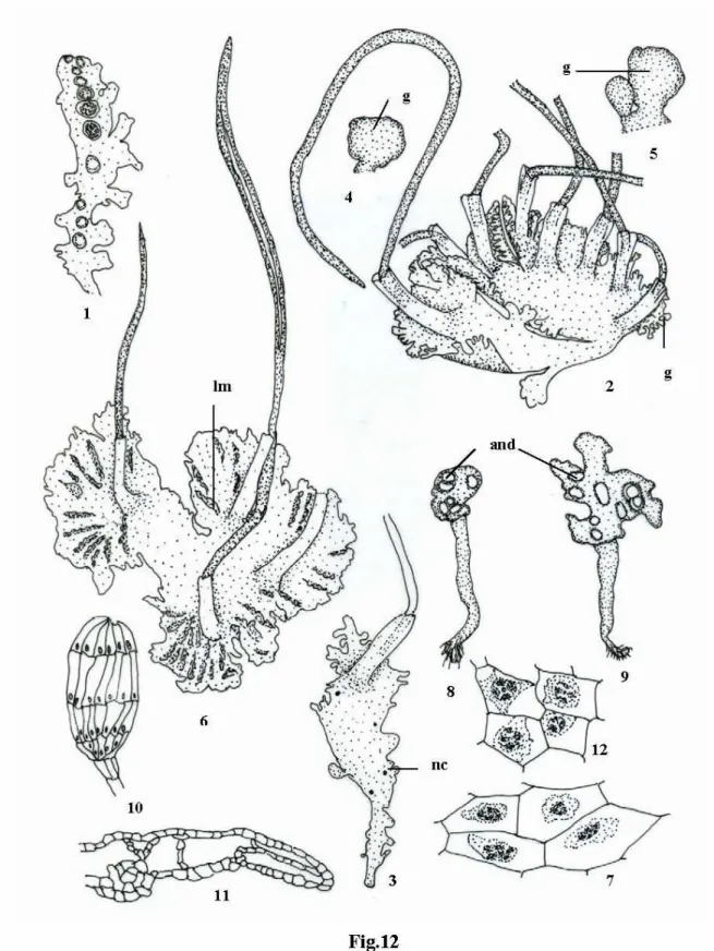
Metzgeriales Schuster ex Schjakov
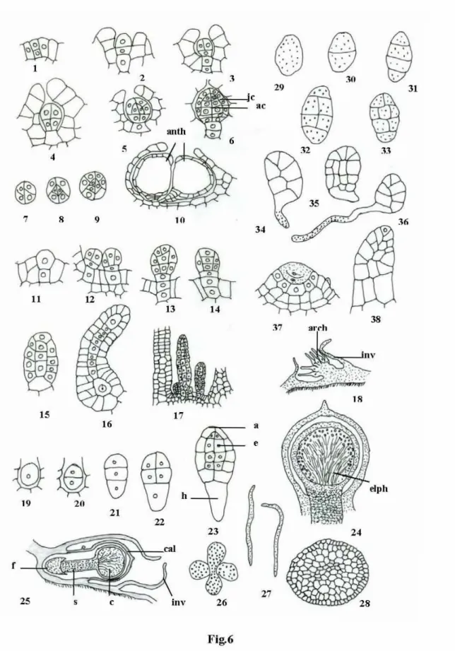
The antheridial chambers are somewhat raised from the thallus surface and have an apical opening (Fig. 6: 10). The cells in the outer layer of the capsule wall are polygonal with nodular thickening in the corners and intermediate nodular thickening (Fig. 5: 12). The cells in the inner layer of the capsule wall are somewhat narrow, elongated with semicircular bands of thickening (Fig. 5: 13).
During the process (germination), the spore divides transversely, forming first a 2-celled and then a 4-celled filamentous spore formation (Fig. 6: 29-31).
Monocleales Schuster
Sphaerocarpales Cavers
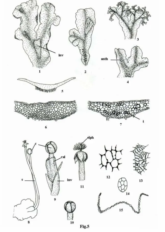
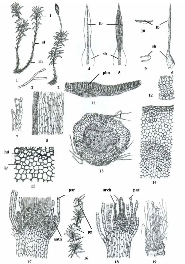
Marchantiales Limpr
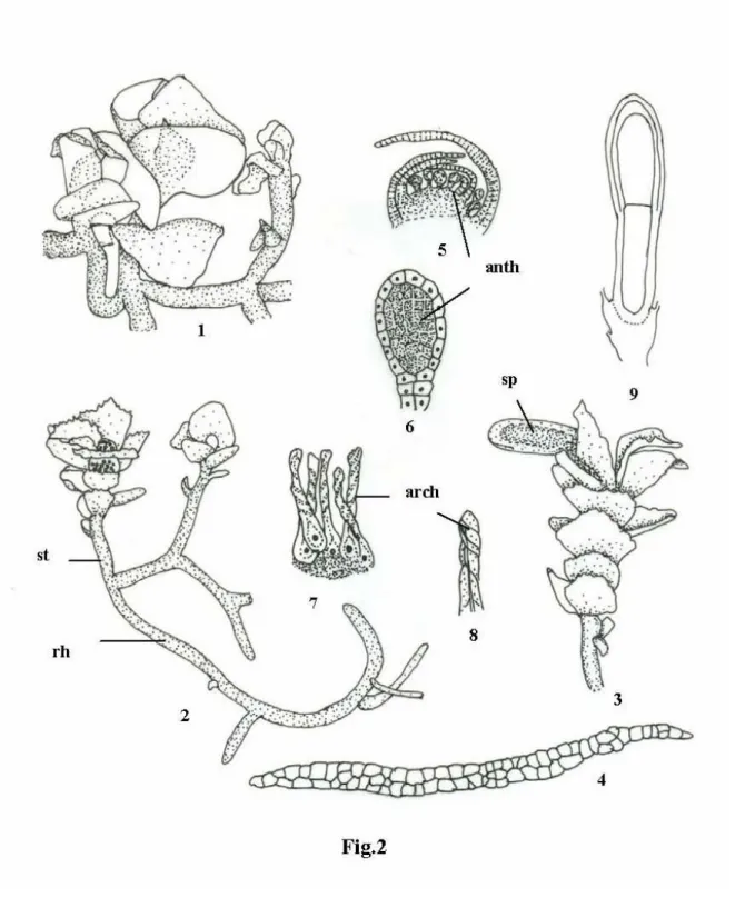
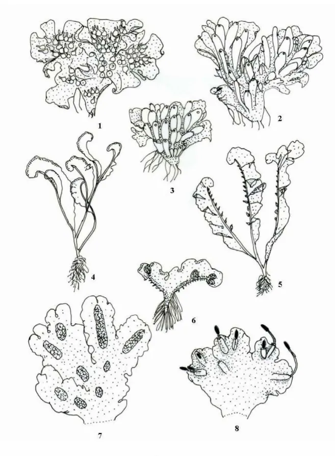
The thallus differentiates internally into the upper, narrow assimilative (photosynthetic) zone and the lower, wide storage zone (Fig. 8: 14). These jeweled cups develop near the growing point and have a number of gemmae attached to the base of the cups accompanied by numerous small, clavate, mucilage papillae (Fig. 8: 26). Archegoniophores are also terminal and always present at the tips of the thallus (Fig. 9: 13).
In the initial stage, these archegonia are in a dorsal position, but as they grow, after fertilization, the lobes curve down pushing the archegonia towards the lower side of the disc (Fig. 9: 14-17). The archegonial initial makes it appear as a projection (papillate growth) on the lobe of the archegonial disc (Fig. 9: 18). The cells of the capsule wall have ribbon-like thickening bands (Fig. 10: 26).
The elaters are long, narrow, pointed at both ends with 2-3 spiral thick bands (Fig. 10: 24). Now the cells of the embryo divide into several levels that form a more or less spherical multicellular structure (Fig. 10: 13). These cells elongate, separate from each other and are differentiated into (i) broader fertile cells and (ii) narrow elongated sterile cells (Fig. 10: 16).
After this, parent cell wall disintegrates and spore mother cells are released, which divide meiotically to form spore tetrads (Fig. 10: 22). Due to loss of moisture in the cells of the capsule wall, the capsule divides irregularly into 6-8 valves (Fig. 11: 1-4).
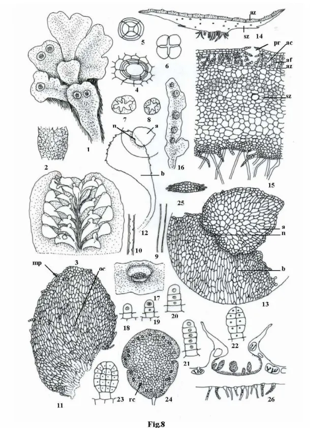
Anthocerotopsida
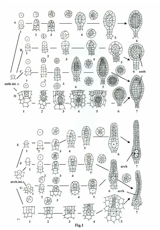
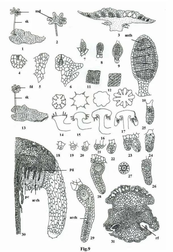
Epidermal cells of the dorsal side of the thallus usually have 1, rarely 2 chloroplasts with central small, distinct pyrenoid bodies (Fig. 13: 2,3). Within the androecial chamber, antheridia are present in variable numbers at different stages of development (Fig. 13: 16). The wall initial cell divides by periclinal and then anticlinal divisions, forming a two-layered wall of the androecium (Fig. 13: 16).
The free end of the basal cell divides by an oblique wall forming a triangular lateral segment or knob (Fig. 13: 22). The (ii) inner cell is the primary ventral cell, which also divides transversely forming ventral duct cell and egg (Fig. 13: 30). The three peripheral cells first divide vertically into 6 peripheral cells (Fig. 13: 32), which then divide transversely forming the envelope of the archegonium, but they are indistinguishable from thallus cells.
This slime mass remains in continuation with the mound of slime present on the surface of the thallus at the top of the archegonium (Fig. 13: 36). In the apical part of the capsule, the archesporium arches over the columella and becomes massive with several layers of cells (Fig. 14: 8-11). It is present from the base to the top of the capsule and consists of 16 rows of cells arranged in a full square (Fig. 14: 20).
The capsular region of the embryo divides periclinally to form the outer amphithecium and inner endothecium (Fig. 14:4). The lower ruptured part of the involucre forms a collar around the base of the sporophyte and aids in fluid retention (Fig. 14:21).
Bryopsida
During its existence, the genus Takakia remained a controversial genus regarding its systematic position, whether it is an interesting liverwort or an interesting moss, since at that time vegetative plants were known only with archegonia (Grolle, 1963). However, following the discovery of the antheridium and sporophyte (Smith, 1990; Smith and Davison, 1993; Schuster, 1997; Higuchi and Zhang, 1998), the genus was shown to be a moss because of its moss-like antheridia and sporophyte. Smith and Davison (1993) proposed a new systematic place for the taxon in Andreaeopsida together with Andreaea and Andreaeobryum.
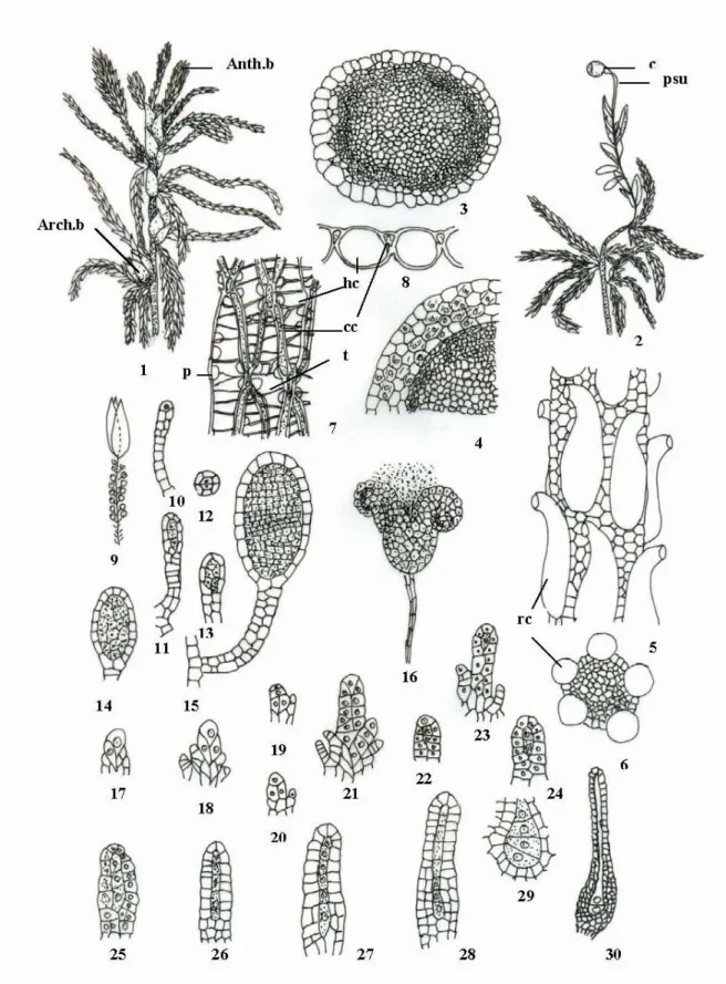
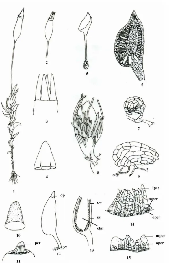
Sphagnales
The growth of the axis takes place by means of an apical cell with three cutting planes. Vegetatively, the plants reproduce by death and decay of older part of plants, which leads to separation of lateral branches, which develop into new plant. They have long stalk with 2 – 4 rows of cells and spherical antheridial body with single layer jacket and number of androcytes (Fig. 15: 15).
At maturity, the antheridium breaks up into a number of lobes, which turn around and release the androcytes – a characteristic similar to the liverwort: Porella (Fig. 15:16). The archegonial branch has a short bud and resembles large, bright green, conspicuous perichaetial leaves with 1-3 archegonia (Fig. 15: 1,3). Of these, one is primary archegonium that develops from the apical cell and the other two are secondary archegonia, which are not developed from the apical cell but from the segments produced by the apical cell (Fig. 15:21).
The capsule has multi-layered capsule wall with rudimentary stomata, dome-shaped arehesporium, which arches over the central columella (Fig. The dehiscence of the capsule is somewhat special through 'Gun shot Mechanism' or 'Air Gun Mechanism', where the operculum is thrown off with a jerk (Fig. It germinates and forms exosporic thalloid protonema, which can further develop into secondary protonema and buds that give rise to a new gametophyte (Fig.
A hangren (antheridial bracts removed), 10-15. anth. b – antheridial branch, arch. b – archegonial branch, psu – pseudopodium, c – capsule, cc – chlorophyll cells, hc – hyaline cells, p – pore, t – band of thickening, rc – retort cell).
Andreaeales
A mature sporophyte with unfertilized archegonia, 10-20. anth – antheridia, arch – archegonia, par – paraphysis, psu – pseudopodium, clm – columella, c – capsule, f – foot, cal – calyptra, ap – archesporium, v – vaginula, a – amphithecium, e – endothecium, oe - outer endothecium, ie - inner endothecium, ss - spore sac, cw - capsule wall).
Takakiales
Polytrichales
The antheridial mother cell is divided by two oblique walls that intersect to form an apical cell with two cutting surfaces (Fig. 20: 1-3). The larger cell then divides vertically, resulting in the formation of 3 cells in a row (Figure 20: 10). They divide into several planes and form a 2-3 layer thick wall of the archegone venter (Fig. 20: 25).
The apical cell can cut several segments or be directly separated by a transverse wall forming the outer cell and the central cell (Fig. 20: 19). With these changes, the central cell comes to the base of the archegonium and divides transversely into a ventral channel cell and an egg (Fig. 20: 20). The capsule is more or less elongated with three main regions: (i) lower region - apophysis (ii) middle region - theca and (iii) apical region - opercular (Fig. 21: 7).
At the mouth of the capsule there is a thin, membranous, disc-like cover, which is called the epiphragm (Fig. 21: 3). The teeth are fixed composed of number of cells with central straight cells bordered by 'U' shaped (curved) cells on both sides (Fig. 21: 8). The columella is an extension of the apophysis on the underside and with the epiphragm on the upper side (Fig. 22: 5,6).
Each of the amphithecial cells divides radially (anticlinally), resulting in 16 amphithecial cells enclosing four endothecial cells (Fig. 20: 31). Out of seven layers of amphithecium, the outermost three layers form the capsule wall, the next two layers form outer trabeculae, and the inner two layers form the outer wall of the spore sac (Fig. 20: 34).
Tetraphidales
In Pogonatum the body of the capsule is smooth, without ridges and is more or less circular in diameter, while in Polytrichum the body of the capsule is angled with 4-6 distinct ridges and is somewhat square in diameter (Fig. 22:2- 9). The male plant is represented by a single, unistratose perigonial leaf / male bract enclosing a single short-stalked antheridium and spherical antheridial body (Fig. 23: 7,9). Female plant is also highly reduced to have 3-4 small, unistratose perichaetial leaves with 1-2 archegonia (Fig. 23: 8).
Between the wall of the capsule and the spore sac are trabeculae and air spaces (Fig. 23: 6,13). The plants are small, erect and differentiated into axes and spirally arranged leaves (Fig. 24: . 1-4). They are simple, ovoid-elongated, wide at the base and pointed at the top, with smooth edges and a distinct middle rib (Fig. 24: 6,7).
It has an outer single-layered epidermis, followed by 3-5 layers of cortex with thick-walled cells in mature plants enclosing central conducting region with narrow elongated cells (Fig.24: 8,9). The marginal cells are somewhat projected near the apex giving an appearance of tooth-shaped margin (Fig. 24: 6). The archegonia are also present in groups at the apex on archegonial shoot associated with uniform paraphyses (Fig.24: 11).
A mature archegonium is petiolate and flask-shaped with a narrow, elongated neck and a swollen archegonial venter (Fig. 24:12). Annulus is located just above the rim and has 5-6 cell layers with elongated upper cells, which aid in the separation of the operculum (Fig. 24:15).
A revised classification of the Anthocerotopsida and a checklist of the Hornworts of North America, North of Mexico.