Employment of Synthetic Ion Transporters to Disrupt Ionic Homeostasis in LUVs and
Cells
A dissertation submitted to the Indian Institute of Technology Guwahati
as partial fulfilment for the Degree of Doctor of Philosophy in Chemistry
By
Ms. Oindrila Biswas
Department of Chemistry
Indian Institute of Technology Guwahati Guwahati 781039, Assam, India
To
My Loving Mother and Sister
6th October, 2022 I hereby declare that the thesis entitled “Employment of Synthetic Ion Transporters to Disrupt Ionic Homeostasis in LUVs and Cells” is the outcome of the research findings executed by me under the guidance of Prof. Debasis Manna, Department of Chemistry, Indian Institute of Technology Guwahati. The thesis has been submitted by me in the Department of Chemistry, Indian Institute of Technology Guwahati, for granting the degree of Doctor of Philosophy. In case of collaborations where the reported work has been done in association with other researchers, due credit has been given. I further declare that this work has not been submitted anywhere else for any degree, diploma, associateship or membership etc. of any Institute or University to the best of my knowledge.
(Ms. Oindrila Biswas)
To whom it may concern
6th October, 2022 This is to certify that the thesis titled “Employment of Synthetic Ion Transporters to Disrupt Ionic Homeostasis in LUVs and Cells” is being submitted by Ms. Oindrila Biswas (Roll No. 176122033) for the award of PhD degree in Chemistry, Indian Institute of Technology Guwahati, is genuinely her own research executed by her.
The data and findings were carried out by her under strictly following my laboratory guidelines. This thesis or any part of the thesis has not been submitted elsewhere for award of any degree/diploma before.
(Prof. Debasis Manna)
Indian Institute of Technology Guwahati
Prof. Debasis Manna Department of Chemistry Phone: +91-0361-258-2325 Fax: +91-0361-258-2349 E-mail: [email protected]
Contents
Acknowledgements I
Abstract III
List of Abbreviation V
Synopsis Report XI
Chapter 1
Synthetic Anion Transporters and their Potential Biological Applications
1.1. Introduction
1.2. Different modes of transport of the ion transporters 1.3. Therapeutic applications of ion transport
1.4. Ion transport and cell death 1.5. Artificial anionophores
1.5.1. Diarylisophthalamide-based anionophores 1.5.2. Diamidocarbazole-based anionophores
1.5.3. ortho-phenylenediamine-based anionophores 1.5.4. trans-decalin-based anionophores
1.5.5. Anthracene 1,8-bisureas -based anionophores 1.5.6. Valinomycin-like carriers
1.5.7. Tren-based urea and thiourea derivatives 1.5.8. Calixarene-based ion transporters
1.5.9. Bispidine-based transporters 1.5.10. Cyclic peptide-based transporters 1.5.11. Bis-amido imidazole derivatives 1.5.12. Sulfonamide-based ion transporters 1.5.13. Triazine-based ion transporters 1.5.14. Phosphazene-based ion transporters
1.5.15. Tetraurea macrocycle-based ion transporter 1.5.16. Fatty acid-based ion transporters
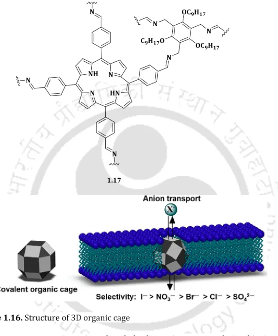
1.5.17. Organic cage-based ion transporter 1.5.18. Metal-organic synthetic ion transporters 1.5.19. Triazole quartet-based anion channel 1.5.20. Diarylisophthalamide-based ion channels 1.5.21. Non-toxic anionophores
1 2 3 5 8 8 9 10 10 11 11 12 13 14 14 15 16 16 17 17 18 19 21 22 23 24
1.7. Objective of research work 1.8. References
26 27 Chapter 2
Chloride Ion Transport by PITENINs Across the Phospholipid Bilayers of Vesicles and Cells
2.1. Background and objective of present work 2.2. Results and discussions
2.2.1. Design and synthesis of the acylthiourea derivatives 2.2.2. Anion binding studies of the acylthiourea derivatives 2.2.3. Chloride ion transport activities of the acylthiourea
derivatives
2.2.4. Relative cation and anion selectivity studies of the acylthiourea derivatives
2.2.5. Mechanism of chloride ion transport activities of the acylthiourea derivatives
2.2.6. Pleckstrin Homology Domain Binding Ability of the PITENINs
2.2.7. Activities of the PITENINs under Cellular Environment 2.3. Summary
2.4. Experimental section
2.4.1. Synthesis of compounds 2.4.1.1. General information
2.4.1.2. Synthetic Procedure of the Compounds 2.4.2. Solution phase anion binding studies
2.4.2.1. 1H-NMR Titrations 2.4.2.2. UV-Vis Titrations
2.4.3. The density functional theory (DFT) calculations using B3LYP/6-31+G(d) and B3LYP/6-31++G (d,p) methods
2.4.4. Ion Transport Activity Studies
2.4.4.1. Preparation of EYPC-LUVsHPTS
2.4.4.2. Ion transport activity across EYPC/CHOL-LUVHPTS 2.4.4.3. Quantitative measurement of transport activity from HPTS assay
2.4.4.4. Ion transport activity studies using ion-selective electrode-based assay
2.4.4.5. Ion Selectivity Studies
2.4.4.6. Chloride Efflux Studies at Different pH Using ISE
33 35 35 36 37 38 41 44 44 49 49 49 49 50 54 54 57 57 59 59 60 60 61 64 65
2.4.4.8. Evidence for mobile carrier mechanism
2.4.4.9. Test for the leaching-out of the compounds from the membrane bilayer environment
2.4.4.10. Evidence of vesicle stability in the presence of the compounds
2.4.4.11. Surface Plasmon Resonance (SPR) Binding Analysis
2.4.4.12. Biological Activity Studies
2.4.4.13. Transportation of Chloride Ion across Giant Unilamellar Vesicles (GUVs)
2.4.5. HPLC analysis of the compounds and stability test for the potent compounds
2.5. 1H NMR and 13C NMR of the compounds 2.6. References
71 72 73 73 74 78 80 85 91
Chapter 3
Photoinduced Generation of the Active Chloride/Phosphate Anionophore from its Inactive Proanionophore
3.1. Background and objective of present work 3.2. Results and discussions
3.2.1. Design and synthesis of the guanidine macrocycles
3.2.2. Anion binding studies of the guanidine-based macrocycle 3.2.3. Ion transport activities of the guanidine-based macrocyclic
derivatives
3.2.4. Mechanism of chloride ion transport activities of the guanidine-based macrocyclic derivatives
3.2.5. Chloride-mediated phosphate transport by the guanidine macrocyclic anionophore across GUVs
3.2.6. Cl-/NaH2PO4 antiport transportation of guanidine-based macrocyclic cpmpound across the vesicles using 31P NMR experiment
3.2.7. Molecular dynamics (MD) simulation studies of guanidine- based macrocyclic compound with a model membrane
3.2.8. Regeneration of the active guanidine-based macrocyclic anionophore from proanionophore
3.2.9. Biological activity studies of guanidine-based macrocyclic compound
3.3. Summary
95 96 96 96 97 98 101 102
103 104 106 106
3.4.1.1. General information
3.4.1.2. Synthetic Procedure of the guanidine-based macrocyclic Compounds
3.4.2. Binding studies of guanidine-based macrocyclic compounds
3.4.2.1. Spectrophotometric titration studies 3.4.3. Lucigenin fluorescence quenching measurements of
guanidine-based macrocyclic compounds in the presence of different anions
3.4.4. Ion transport activity studies of guanidine-based macrocyclic compounds
3.4.4.1. Ion transport activity studies using the fluorescence-based assay
3.4.4.2. Measurement of half-maximal effective
concentrations (EC50) of the compounds for different anions 3.4.4.3. Lucigenin-based selectivity studies
3.4.4.4. Ion transport activity studies using the ion- selective electrode-based assay
3.4.4.5. U-tube assay
3.4.4.6. Evidence of mechanistic pathway of transport of the anionophore
3.4.4.7. Ion transport activity studies using HPTS-based fluorescence assay
3.4.4.8. Evaluation of transport of phosphate using Tb(III) fluorescent chemosensor
3.4.4.9. Transport of phosphate ion through GUVs (Giant Unilamellar Vesicles)
3.4.4.10. Direct evidence for phosphate transport by 31P NMR measurements
3.4.4.11. Molecular dynamics (MD) simulation studies 3.4.4.12. Regeneration of the active anionophore from proanionophore
3.4.4.13. Biological activity studies of guanidine-based macrocyclic compounds
3.5. 1H and 13C NMR spectra of synthesized guanidine-based macrocyclic compounds
3.6. References
107 107 109 109 112
113 113 113 115 116 117 118 119 120 121 123 124 125 126 128 132
Chapter 4
Targeted Delivery of Pro-anionophore: Photo-induced transport of Cl- ion Across the Lipid Bilayer
4.1. Background and objective of present work 4.2. Results and discussions
4.2.1. Design and synthesis of the synthetic ionophore derivatives 4.2.2. Anion binding studies of the synthetic ionophore
derivatives
4.2.3. Chloride ion transport activities of the synthetic ionophore derivatives
4.2.4. Relative cation and anion selectivity studies of the synthetic ionophore derivatives
4.2.5. Mechanism of chloride ion transport activities of the synthetic ionophore derivatives
4.2.6. Regeneration of the active anionophore from proanionophore
4.2.7. Activities of the synthetic ionophore under Cellular Environment
4.3. Summary
4.4. Experimental Procedures 4.4.1. General information
4.4.2. Synthesis and characterization of compounds 4.4.3. UV-Vis Titrations
4.4.4. Ion transport activity studies
4.4.4.1. Ion transport activity studies using the fluorescence-based assay
4.4.4.2. HPTS-based selectivity studies
4.4.4.3. Ion transport activity studies using the ion- selective electrode-based assay
4.4.4.4. Evidence for mobile carrier mechanism 4.4.4.5. Regeneration studies using proanionophore 4.4.5. Biological Activity Studies
4.4.5.1. MTT-based cytotoxicity assay
4.4.5.2. MTT-based cytotoxicity assay using photo-trigger 4.4.5.3. Chloride mediated cell death
4.5. 1H and 13C NMR spectra of synthesized compounds 4.6. References
135 136 136 138 138 139 141 141 143 146 146 146 146 150 150 150 151 152 152 153 155 155 155 155 156 162
6. Future Prospects 165
Annexure I 167
Annexure II 173
Annexure III 175
Publication 177
I
Acknowledgements
This thesis is the outcome of the constant sacrifices made by my family, endless encouragement provided by supervisor, colleagues and friends.
First and foremost, I would like to thank the Almighty for providing me the constant zeal and strength to complete the task.
Next, I would like to thank my supervisor Prof. Debasis Manna, through whom I have been introduced to this fascinating world. By setting an example of punctuality, persistant-hardwork and continuous quest for novelty, he has influenced me throughout my research career. He has always extended his helpful hand towards me whenever I hit a hurdle in the road. He has enriched my experience of working in a scientific laboratory by his trustworthy recommendations and ceaseless suggestions. I really consider myself truly privileged to have been able to be part of his group. He has been my guide and mentor throughout my research work in his laboratory which has left a lasting impression in my personality.
I want to extend my heartfelt gratitude towards my collaborators Dr. Sachin Kumar (Dept. of BSBE, IIT Guwahati, India) and Dr. Niladri Patra IIT (ISM) Dhanbad for true guidance throughout the period of my research work. I want to specially mention my doctoral committee members Prof. Kalyanasis Sahu, Dr. Dipankar Srimani and Dr. Rajkumar P. Thummer for their constant scientific inputs and rigorous evaluation of my research work. Their encouragement has provided me with immense confidence about my work. I want to offer my sincere thanks to all he faculty members and office staff members of the Department of Chemistry, IIT Guwahati for their consistent assistance in all ways possible. I am immensely indebted to Central Instruments Facility (IIT Guwahati) and the Department of Chemistry for allowing me free access to all the instruments required or experimental purpose.
I would also like to aprreciate all my past and present lab members including Dr.
Saurav Paul, Dr. Sreeparna Das, Dr. Subhankar Panda, Dr. Ashalata Roy, Dr. Abhishek Saha, Mr. Dipjoyti Talukdar, Dr. Nirmalya Pradhan, Dr. Nasim Akhtar, Mr. Subhasis Dey, Mr. Sribash Das, Ms. Anjali Patel, Ms. Priyanka Mazumder, Mr. Gunanka Hazarika, Mr. Biswa Mohan Prusty, Ms. Soumya Srimayee, Ms. Nikumoni Das and Mr. Mohit Kumar for maintaining a positive and thriving atmosphere in the lab.
Lastly but not the least I want to express my deepest appreciation to my mother Ms. Munni Biswas and my loving sister Ms. Anindita Biswas. My mother has been my pillar of strength since childhood and she has constantly shielded me from the other worldly problems so that I could solely focus on my research work. My sister on the other hand, though younger to me in age, has taken many responsibilities so that I can give my undivided attention in lab. Without the two of them it would have been almost impossible for me to finish my thesis work.
Thank you all for making this journey a memorable experience of my life.
Ms. Oindrila Biswas
III
Abstract
The thesis entitled “Employment of Synthetic Ion Transporters to Disrupt Ionic Homeostasis in LUVs and Cells” exemplifies the biological implications of a variety of synthetic anionophores. This thesis covers the recognition of the ions, transmembrane transport properties and possible therapeutic applications. The thesis has been divided into five chapters based on the experimental results gathered during the entire research period.
Chapter 1 instantiates the importance of the anionophores in carrying vital functions in cells. It also illustrates the therapeutic implementations of the anionophore in treating channelopathies like cystic fibrosis, triggering cancer cell death and also its use as antibacterial agents.
Chapter 2 accounts the efficient transmembrane Cl- ion transport activity of the PITENIN derivatives. Different experiments suggest the binding of Cl- ion with the PITENINs. Separate cellular activity studies suggest the disruption of Cl- ion homeostasis by the PITENIN derivatives.
Chapter 3 elucidates the use of light as a competent stimulus to generate the active Cl-/phosphate guadinium macrocycles (anionophore) from its inactive proanionophore. A concise study regarding the ion recognition, transport studies and regeneration of the active anionophore from the active anionophore under UV irradiation has been studied in LUVs and cellular conditions have been demonstrated in this chapter.
Chapter 4 summarizes the use of light as a stimulus to trigger the release of the active anionophore from its inactive proanionophore generating ROS in the process.
The proanionophore group contains a CAIX inhibitor that can directly target the cancer cells having CAIX protein. A conclusive study has been shown in this chapter showing the ion recognition property, transport behaviour, regeneration of the anionophore in the presence of UV irradiation and formation of ROS in the process
V
List of Abbreviation
AZ Acetazolamide
ATP Adenosine triphosphate
ADP Apoptotic protease activating factor-1 Apaf-1 Apoptotic protease activating factor-1 AVD Apoptotic volume decrease
CA-IX Carbonic anhydrase-IX
FCCP Carbonyl cyanide p-trifluoromethoxyphenylhydrazone CCCP Carbonyl cyanide 3-chlorophenylhydrazone
CS2 Carbon disulfide
CHOL Cholesterol
CM Coumarin
CF Cystic fibrosis
CFTR Cystic fibrosis transmembrane conductance regulator DFT Density functional theory
DNA Deoxyribonucleic acid
DMF Dimethylformamide
DPPC Dipalmitoylphosphatidylcholine DPPS Dipalmitoylphosphatidylserine DMSO Dimethyl sulfoxide
DMEM Dulbecco's Modified Eagle Medium EE Efflux efficiencies
FBS Fetal bovine serum
FI Fluorescent intensities
Gaff Generalized Amber Force Field
Grp1 General Receptor for Phosphoinositides-1 GUVs Giant Unilamellar Vesicles
GSH Glutathione
GST Glutathione S-transferase
EC50 Half maximal effective concentration HRMS High-resolution mass spectra
HRP Horseradish peroxidase
HEK Human embryonic kidney
Pi Inorganic phosphate
ISE Ion selective Electrode LUVs Large Unilamellar Vesicles LED Light-emitting diode EYPC L-α-phosphatidylcholine HgCl2 Mercury (II) chloride HgS Mercury(II) sulfide
MMP Mitochondrial membrane potential
MD Molecular simulation
NMR Nuclear Magnetic Resonance
OA Oleic acid
ONB o-nitrobenzyl
PME Particle-mesh Ewald
pH Potential of hydrogen
PARP Poly adenosine diphosphate-ribose polymerase PEG-400 Polyethylene glycol 400
PI Propidium iodide
PBS Phosphate-buffered saline
PDT Photodynamic therapy
PI3K Phosphatidylinositol-3 kinase
PIP3 Phosphatidylinositol-3,4,5-trisphosphate
PH Pleckstrin homology
RU Response unit
rt Room temperature
RPM Rotation per minute
SDS-PAGE Sodium dodecyl-sulfate polyacrylamide gel electrophoresis SLC20A1 Solute carrier family 20 member 1
SLC34A2 Solute carrier family 34 member 2
SA Squaric acid
SPR Surface plasmon resonance TBACl Tetrabutylammonium chloride
TBAH2PO4 Tetrabutylammonium dihydrogen phosphate
VII TBANO3 Tetrabutylammonium nitrate
THF Tetrahydrofuran
TMS Tetramethylsilane
Et3N Triethyl amine
TME Tumor microenvironment
TLC Thin-layer chromatography
UV Ultraviolet
UV–Vis Ultraviolet-visible
VRACs Volume -regulated anion channels YFP Yellow fluorescent protein
POPC 1-Palmitoyl-2-oleoyl-sn-glycero-3-phosphocholine H2DCFDA 2’, 7’-dichlorodihydrofluorescein
DCF 2’,7’-dichlorofluorescein
PDK1 3-Phosphoinositide-dependent kinase 1
MTT 3-(4,5-dimethylthiazol-2-yl)-2,5-diphenyl tetrazolium bromide HEPES 4-(2-hydroxyethyl)-1-piperazineethanesulfonic acid
HPTS 8-Hydroxypyrene-1,3,6-trisulfonic acid trisodium salt BHK 21- Baby hamster kidney cell
AZ Acetazolamide
ATP Adenosine triphosphate
ADP Apoptotic protease activating factor-1 Apaf-1 Apoptotic protease activating factor-1 AVD Apoptotic volume decrease
CA-IX Carbonic anhydrase-IX
FCCP Carbonyl cyanide p-trifluoromethoxyphenylhydrazone CCCP Carbonyl cyanide 3-chlorophenylhydrazone
CS2 Carbon disulfide
CHOL Cholesterol
CM Coumarin
CF Cystic fibrosis
CFTR Cystic fibrosis transmembrane conductance regulator DFT Density functional theory
DNA Deoxyribonucleic acid
DMF Dimethylformamide
DPPC Dipalmitoylphosphatidylcholine DPPS Dipalmitoylphosphatidylserine DMSO Dimethyl sulfoxide
DMEM Dulbecco's Modified Eagle Medium EE Efflux efficiencies
FBS Fetal bovine serum
FI Fluorescent intensities
Gaff Generalized Amber Force Field
Grp1 General Receptor for Phosphoinositides-1 GUVs Giant Unilamellar Vesicles
GSH Glutathione
GST Glutathione S-transferase
EC50 Half maximal effective concentration HRMS High-resolution mass spectra
HRP Horseradish peroxidase
HEK Human embryonic kidney
Pi Inorganic phosphate
ISE Ion selective Electrode LUVs Large Unilamellar Vesicles LED Light-emitting diode EYPC L-α-phosphatidylcholine HgCl2 Mercury (II) chloride HgS Mercury(II) sulfide
MMP Mitochondrial membrane potential
MD Molecular simulation
NMR Nuclear Magnetic Resonance
OA Oleic acid
ONB o-nitrobenzyl
PME Particle-mesh Ewald
pH Potential of hydrogen
PARP Poly adenosine diphosphate-ribose polymerase PEG-400 Polyethylene glycol 400
IX
PI Propidium iodide
PBS Phosphate-buffered saline
PDT Photodynamic therapy
PI3K Phosphatidylinositol-3 kinase
PIP3 Phosphatidylinositol-3,4,5-trisphosphate
PH Pleckstrin homology
RU Response unit
rt Room temperature
RPM Rotation per minute
SDS-PAGE Sodium dodecyl-sulfate polyacrylamide gel electrophoresis SLC20A1 Solute carrier family 20 member 1
SLC34A2 Solute carrier family 34 member 2
SA Squaric acid
SPR Surface plasmon resonance TBACl Tetrabutylammonium chloride
TBAH2PO4 Tetrabutylammonium dihydrogen phosphate TBANO3 Tetrabutylammonium nitrate
THF Tetrahydrofuran
TMS Tetramethylsilane
Et3N Triethyl amine
TME Tumor microenvironment
TLC Thin-layer chromatography
UV Ultraviolet
UV–Vis Ultraviolet-visible
VRACs Volume -regulated anion channels YFP Yellow fluorescent protein
POPC 1-Palmitoyl-2-oleoyl-sn-glycero-3-phosphocholine H2DCFDA 2’, 7’-dichlorodihydrofluorescein
DCF 2’,7’-dichlorofluorescein
PDK1 3-Phosphoinositide-dependent kinase 1
For symbols/units
α Alpha
Å Angstrom
atm Atmosphere
β Beta
C Celsius
J Coupling constant
° Degree
δ Delta
Hz Hertz
K Kelvin
MHz Megahertz
m Meta
μg Micro gram
μM Micro mole
mL Millilitre
mV Mili Volt
min Minute
nM Nano mole
o Ortho
p Para
ppm parts per million
% Percent
ᴨ Pi
ps Pico second
s Second
XI Chapter 1
1. Introduction
Preservation of the accurate ionic balance across the lipid bilayer of the cell is a very crucial function which regulates several vital events in cells like signal transduction, blood pressure regulation, hormone secretion, triggering of dell death among others. 1 Transmembrane proteins maintain the osmolality between the extra- and the intravesicular system by allowing certain substances to pass through the cellular membrane. However, impaired channel function can lead to several disease condition known as channelopathies like short QT syndrome, long QT syndrome, Bartter’s syndrome and the most common being cystic fibrosis. 2
Chloride is a very abundant ion found in the biological system. In several literature studies it has been found that in cancer cells the outside concentration of the chloride ion is higher (120 mM) w.r.t the inside concentration (5-15 mM) It was found that hindering the volume regulatory Cl- and K+ channels pause the cells to show classic apoptotic attributes like caspase activation, cytochrome c release and DNA laddering thus saving the cells from cell death. In line with this, increasing substantiation also shows that disrupted chloride transport in cancer cells can result in efflux of Cl- which is considered
Figure 1.1. Schematic representation of numerous prospects of anionophores.
as the reason to suppress apoptosis in cancer cells. 3 Inorganic phosphate is also an essential nutrient for sustaining a cell. 4 It is a very important component of DNA,
RNA and also a part of energy rich molecules like ATP. It is also a fundamental part of cellular signalling events. In cells Pi mainly enters via Na dependent Pi transporters primarily constituting two family of inorganic phosphate transporters namely SLC20 and SLC34. 5 Furthermore, many studies propose that in tumors these Pi transporters are upregulated hence maintaining a higher concentration inside them.
The transport of the ions across the cell bilayer manages cell proliferation and apoptotic pathways. Additionally, the disruption of the ionic homeostasis in the cancer cells and destruction of the ionic gradient can trigger cancer cell death. So rather than targeting specific proteins of the cancer cells using the synthetic anionophores to aid in ion transport across the cells can be used as an excellent strategy to remove detrimental cells from our body. This novel approach of ‘ion therapy’ can be used to also deacidify the acidic microenvironment and thereby has massive potential to overcome the multidrug resistance of the cancer cells devised to evade cell death.
Cystic fibrosis is a deadly autosomal recessive genetic disease that occurs mainly because of the defective functionality of the CFTR (CF transmembrane conductance regulator protein). 6 Small synthetic ion transporters can be used in place of the faulty biological anion transporters because the synthetic anionophores don’t target any particular protein or enzyme and are not interrupted by the different genetic mutations in the CFTR. 7 Hence the use of non-toxic synthetic anionophores along with the clinically approved drugs like lumacaftor and ivacaftor can provide a potential therapeutic strategy to restore ion transport in the CFTR cells. 8
Recent studies revealed that small molecules which can mediate ion transport across the lipid bilayer also have in-vitro antibacterial effects. The antibacterial resistance is one of the foremost problems jeopardizing the effectiveness of the antibiotics. Several bacteria have been enlisted by the Centre for Disease Control (CDC) as potentially harmful threat to the mankind. The finding that several naturally occurring cationophores like valinomycin can act as antibiotics has led to the emergence of the idea that synthetic ion transporters can also have the same effect.
Though a direct correlation between the ion transport activity and the antibacterial property can’t be clearly drawn while a general trend can be seen. Anionophores having good Cl- ion mediated transport efficiencies have successfully been used against clinically relevant bacterial strains. 9,10 The membrane localization property
XIII
of the anionophores makes it very much tough for the bacteria to cultivate resistance against the anionophores. However, the used synthetic ionophores should have low toxicity towards the normal cell lines. Therefore, in the thesis diverse potential ion transporting molecules were designed and synthesised and their ion transport properties were explored through various biophysical assays. Their biological implications particularly towards cancer were also studied through a set of known in vitro experiments. The complete thesis has been divided into five chapters as explained below, in each chapter inspecting a divergent class of synthetic anionophores, their ion transport studies and their possible therapeutic effects.
Chapter 1 mainly argues the importance of the transmembrane transporters in the biological system and the disease conditions which can pose due to stunted transport by the natural systems. Chapter 2 involves the exploration of the PITENIN- based transporters, their moderately good H+/Cl- transportation proficiency, pH stimulated increased transport efficiency and apoptosis instigating behaviour in cancer cells. Chapter 3 emphasizes the use of light to induce the generation of the active anionophore which transports Cl-/ across LUVs to improve the selectivity of the towards the cancer cells. Chapter 4 embodies the use of photocleavable targeted- proanionophore to better target the cancer cells and reduce toxicity in the normal cells. The PDT activity of the photo product has also been investigated here.
Chapter 2
2. Chloride Ion Transport by PITENINs Across the Phospholipid Bilayers of Vesicles and Cells
To achieve the synthetic mimic of the natural transporter systems, PITENIN derivatives (2.1-2.7) were synthesized by condensation of the benzoyl isothiocyanates and substituted corresponding amines. 11, 12 Discerning their membrane localization property, presence of acyl thiourea group and phenolic OH, their good ion transport capability was hypothesised. To recognize the role of lipophilicity of the transporters 2.3 and 2.5 (DM-PIT-1) were synthesized and to better analyse the necessity of the acylthiourea group benzanilide 2.6 was synthesized. Transporters 2.1, 2.2 (PIT-1), 2.4, and 2.7 were synthesized to assess the importance of the phenolic OH moiety in anion transport properties. The 1H NMR
and UV-titration experiments done using transporters 2.2 (PIT-1) and 2.5 (DM-PIT- 1) with TBACl depicted Cl- ion recognition of the transporters.
Figure 2.1. Anticancer activities of the PITENIN derivatives.
These results encouraged us to examine the Cl- transport behaviour of these derivatives across the bilayer of EYPC/CHOL-LUVs⊃HPTS by employing a pH gradient (pHin = 7.2, pHout = 7.8) in the extravesicular solution. The preliminary screening data shows that 2.2 (PIT-1) is the most efficient Cl– ion transporter and follows the order of 2.2 (PIT-1) ≈ 2.3 > 2.5 (DM-PIT-1) > 2.1 > 2.6 > 2.7 > 2.4.
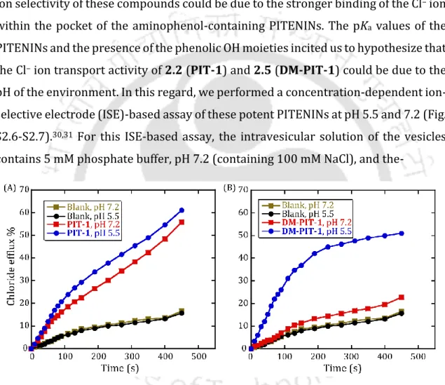
Among all the transporters 2.2 (PIT-1) and 2.5 (DM-PIT-1) were chosen to carry further experiments. Anion and cation selectivity studies across the LUVs indicated high selectivity for Cl- ion and no significant change on changing the metals outside suggesting the metals are not involved in the mechanistic pathway. Considering the pKa of 2.5 (DM-PIT-1) (6.43), it was found that 2.5 (DM-PIT-1) has 20-fold superior Cl- ion transport ability in slightly acidic condition (pH 5.5) than under physiological conditions. Hence, 2.5 (DM-PIT-1) can be used as a switch ON Cl– ion transporter upon the alternation of pH within a narrow window of the physiological pH. The ion transport properties of the 2.2 (PIT-1) and 2.5 (DM-PIT-1) in presence of FCCP and valinomycin exhibited that the synthetic transporters are involved in H+/Cl- transport. The U-Tube experiment and the cholesterol dependency assay authenticates the carrier mediated transport behaviour of the transporters. The MTT
XV
Figure 2.2. Structure of the PITENIN compounds (A). Comparisons of ion transport activity of 2.5 across EYPC/CHOL-LUVs at pH 5.5 and pH 7.2 (B). Chloride ion efflux capability of 2.5 (DM-PIT-1) across EYPC/CHOL-LUVs (C). Cell viability of in HeLacell line in presence of 2.5 (DM-PIT-1) Cell viability of 2.5 (DM-PIT-1) in HeLa in HBSS buffer with and without Cl- ion at different concentrations (C) and Western Blot analysis of HeLa cells in presence of 2.5 (DM-PIT-1) (D).
assay showed that, in the presence of 2.5 (DM-PIT-1) and 2.2 (PIT-1), the viability of cancer (HeLa, human breast cancer; MDA-MB-231) and healthy (baby hamster kidney; BHK-21) cells decreases in a dose-dependent manner. To further probe into the reason of low cell viability of HeLa cells wehave done the MTT experiment in HBSS (Hank’s balanced salt solution), having two different salt compositions (one with a Cl– ion and the other without a Cl– ion). Lower viability was noticed for cells in the Cl– ion-containing buffer as compared to the one in which there was no Cl– ion validating the fact that the decrease in cell viability is due to Cl- transport by the anionophores. The JC-1 dye experiment, western blot experiment and FACs analysis corroborated the apoptotic cell death of the cancer cells. Overall, this investigation revealed the anticancer activities of 2.2 (PIT-1) and 2.5 (DM-PIT-1), not only because of its capability to inhibit the interaction between PIP3 and AKT-PH domain but also for its transmembrane Cl– ion transport activity.
Chapter 3
3. Photoinduced Generation of the Active Chloride/Phosphate Anionophore from its Inactive Proanionophore
Ion transport-promoted unwanted death of normal cells remains a major concern for ion therapy. So, to minimize the apoptosis of the normal cells and selectively target the cancer cells, in this work photo-triggered ion transporters has been used which can give site-selective precise control over the functioning of the ion transporters. Herein, a C2 symmetric guanidinium-based macrocycle was synthesised. 13 The ion binding scaffold and the hydrophobic domain impart lipophilicity to transport the ions. To further explore the concept of a photocleavable proanionophore, the o-nitrobenzyl (ONB) group with the guanidinium-N moiety was attached. 14
Figure 3.1. Schematic representation of the photomediated generation of ionophore from ONB-linked proanionophore.
Initially, thiourea-based macrocyclic intermediate was synthesized, and then desulfurization of thiourea moiety with alkyl amines resulted in the expected guanidinium-based macrocyclic compounds. To investigate the importance of hydrophobicity in ion transportation, alkyl amines of different chain lengths (n = 4, 8, and 10) were used. Compound 3.2 showed better binding with phosphate ion and Cl- rather than NO3-. Eventually the ion transport properties of the anionophores (3.1-3.2) were explored in EYPC/CHOL⊃lucigenin using the fluorescence-based assay. The phosphate transport efficacy of compound 3.2 was higher than the compounds 3.1, 3.3, and 3.4. The higher transport proficiency of compound 3.2
XVII
could be attributed to its subtle balance of hydrophilicity and membrane residence aptitude. The anion selectivity of compound 3.2 was performed by lucigenin assay dispersing the LUVs in an aqueous of 225 mM NaxAy salt (where NaxAy = NaH2PO4, Na2SO4, NaOAc, NaClO4, and NaNO3). The selectivity studies revealed that compound 3.2 is more selective towards phosphate ions than the other tested oxyanions. To comprehend the mechanistic pathway, the cation selectivity of compound 3.2 was checked in the presence of Na+ and K+ ions. The Cl-ISE-based assay showed compound 3.2 mediated transport of phosphate/Cl─ ions across the lipid bilayers.
Additionally, when Na2SO4 was kept outside, no change in fluorescence intensity was observed due to inability of the anionophore to facilitate the SO42─/phosphate antiport process as the selectivity for SO42─ is too low for compound 3.2. This result also supports the independent nature of the transport mechanism with respect to the cations. The non-involvement of H+ in the mechanistic pathway could be assured from the enhancement of the HPTS fluorescence intensity when FCCP was added along with the transporter. The only persisting pathway is the phosphate/Cl− antiport mechanism. Moreover, the U-tube assay also propounds a similar result.
Gradually with progression of time, NaCl and NaH2PO4 steadily increases in the opposite arms of the U-Tube. This result also indicates carrier mediated pathway followed by the transporters. This outcome was moreover concluded with the help of temperature-dependent DPPC assay. The transport activities across the Tb(III)- complex-based fluorescent chemosensor (of phosphate ion) LUVs also demonstrated the phosphate transport activities of compound 3.2. The transmembrane transport of phosphate ions in the presence of compound 3.2 using giant unilamellar vesicles (GUVs) was deployed. Furthermore, 31P NMR experiments were done to provide undeniable proof of the phosphate/Cl− antiport process carried by compound 3.2.
After incubation with compound 3.2, the 31P peak decreased which can only be feasible after the phosphate transport, mediated by the transporter 3.2. The molecular dynamics (MD) simulation studies with compound 3.2 and phosphate ion embedded inside a preequilibrated dipalmitoyl phosphocholine (DPPC)/water lipid bilayer system studies revealed the retention of the compound within the bilayer and expulsion of phosphate ion from the complex suggesting the successful transport of the phosphate ion by the compound. To assess the photo-triggered cleavage of proanionophore 3.4 and in-situ generation of anionophore 3.2 the 1H NMR-based
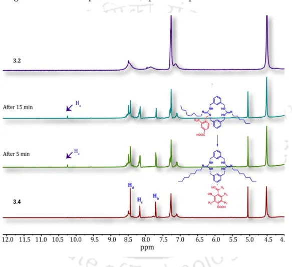
study was performed. After photo irradiation with an LED lamp (365 nm), a new peak was seen at δ = 10.25 ppm, which implies the formation of 4-formyl-3- nitrosobenzoic acid, a plausible result of the photocleavage of the proanionophore 3.4. The successful generation of the anionophore was also studied in the membrane environment. The ion transport activities of proanionophore 3.4 were recorded before and after photo irradiation. It was perceived that the transport efficacy increased by 60%. Consequently, by both the NMR and fluorescence-based methods, the resurgence of the anionophore 3.2 from proanionophore 3.4 was be affirmed.
Figure 3.2. Photoinduced release of anionophore 3.2 from proanionophore 3.4 was monitored by 1H NMR titration experiment in DMSO-d6 solvent.
The perturbation of chloride-phosphate ion homeostasis in the cancer cells could induce apoptosis or other cell death pathways. The MTT-based cell viability assay revealed that compound 3.2 induced moderate toxicity (IC50 = 20 µM) to the HeLa cells. However, the proanionophore 3.4 showed negligible toxicity even after treatment with 60 µM concentration. Interestingly, photo irradiation (UV 365 nm, 5 min) of proanionophore 3.4 promoted moderate toxicity to the HeLa cells (IC50 = 36
XIX
µM). Whereas only photo irradiation (UV 365 nm, 5 min) without the treatment of proanionophore 3.4 did not significantly alter the viability of the HeLa cells. Hence, the lower viability after the photo irradiation could be due to cleavage of proanionophore 3.4 and in-situ generation of active anionophore 3.2 under the cellular environment. Overall, these results successfully established that photoinduced disruption of chloride-phosphate ions homeostasis induces cancer cell death.
Chapter 4
4. Targeted Delivery of Pro-anionophore: Photo-induced transport of Cl- ion Across the Lipid Bilayer
Photocleavable proanionophores have attracted interest due to their controlled biological activities. In this work, the “photocleavable targeted-proanionophore”
strategy was incorporated into the domain of artificial ion transporters and a cancer cell targeting proanionophore that undergoes photo-controlled release of an active anionophore to promote Cl− ion transport-mediated cell death developed.
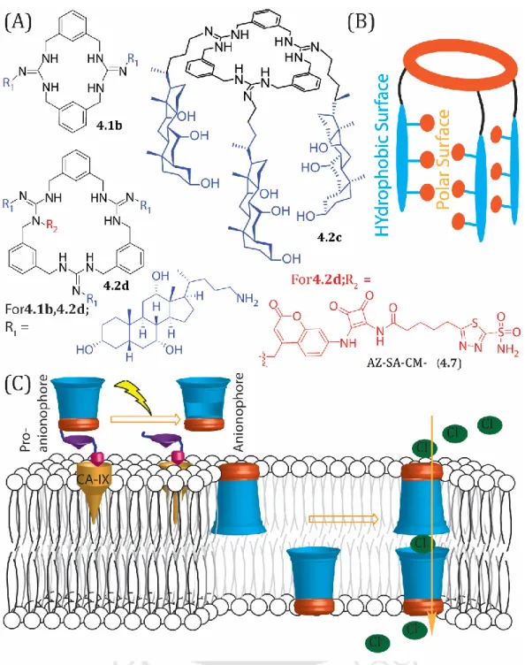
photocleavable targeted-proanionophore fusings with three important bits. 15 The first is the macrocycle, the primary ion recognition scaffold, the cholic amine, the hydrophobic domain accountable for membrane insertion. The last is the acetazolamide grafted photoactivable moiety, which could be inserted selectively into the targetted cancer cells, and then photo- release would generate the active anionophore. Acetazolamide is a known carbonic anhydrase IX (CA-IX) inhibitor that is overexpressed in certain types of cancers. CA-IX is directly associated with the hallmarks of cancer like invasion, angiogenesis, proliferation, and adaptation to microenvironmental stresses (acidosis, hypoxia, deprivation of nutrients). Various studies demonstrated that CA-IX inhibitor diminishes tumor cell proliferation and induces apoptosis. However, only the solitary use of CA-IX inhibitor may not be effectual, and its use along with the apoptosis-inducing anionophore may have an operative therapeutic effect.
Figure 4.1. Transport of the guadinium-based macrocycle anionophores across LUVs.
The desired guanidine-based macrocyclic compounds were synthesized from thiourea-based macromolecules following reported protocols. The cancer cell targeting photolabile moiety, AZ-SA-CM-Br (4.7) was affixed to the macrocyclic compound 4.2c to achieve the photocleavable targeted-proanionophore 4.2d.
Initially, the anion recognition properties of the compounds were investigated using UV-titration with Cl− ions. The binding of the compounds with Cl− follows a 2:1 host/guest stoichiometry as obtained from the BindFit analysis, suggesting that two anionophores are necessitated in the binding event. Next the ion transport
XXI
properties of the macrocyclic anionophores were evaluated using EYPC/CHOL- LUVs⊃Lucigenin. Compound 4.2c showed the highest Cl− ion transport properties among the tested ones. Compound 4.2d, on the contrary, showed limited Cl− ion transporting ability, which could be due to its structural properties. Meanwhile, it was disclosed from the dose-dependent experiment that ‘n’, known as the ‘Hill coefficient’ and referring to the number of species involved in the transport process showed a value of 2.0, implying the assembly of two molecules of anionophore 5.2c (host/guest = 2:1) is the active structure for the Cl− ion transport process. The anionophore showed highest anion selectivity for Cl- among the Na2SO4, NaHCO3, NaOAc, NaClO4, and NaCl salts. The transport efficiency was kept unaltered when the external cations were varied (M+ = Li+, Na+, K+, Rb+ and Cs+) eliminating the prospect of M+ transportalong with Cl− ions. The result of Cl- ion transport efficiency mediated by the anionophore in presence of FCCP evinces that the potent anionophore was unable to carry H+ across the lipid bilayer. The enhancement of the Cl- transport in presence of valinomycin and 4.2c as seen in EYPC/CHOL⊃lucigenin validates the antiport transport mechanism followed by 4.2c. To delve into the mode of transport adopted by the anionophore, a temperature-dependent anionophore activity was investigated using the model lipid bilayers of DPPC which showed no significant change in the Cl− ion transport rate at 25 °C and 45 °C suggesting that the transport properties of anionophore 4.2c might not be via carrier pathway but via pore formation across the lipid bilayer.16 In addition, the CHOL dependent anionophore activity measurements revealed that the rate of Cl− transportation by anionophore 4.2c was not significantly affected by the concentration of CHOL within the lipid bilayers, which also suggested the bi-molecular pore formation could be the operating pathway for the transport of Cl− ion for anionophore 4.2c. Photo-induced regeneration of anionophore 4.2c from AZ-SA-CM-linked proanionophore 4.2d (405 nm, 2 W cm−2) was investigated following the increase in fluorescence intensity of the photoproduct which is fluorescent for different time intervals (0-30 min).
Concurrently, the Cl− ion transport activity of the regenerated active anionophore 4.2c was scrutinized by the Cl-ISE-based assay. The increase in Cl− ion efflux efficiency suggests the in-situ generation of active anionophore. Similar results were obtained with the help of fluorescence-based experiment using EYPC/CHOL⊃HPTS.
Now to check the therapeutic potency of the anionophore 4.2c, the cell viability was checked in HeLa, T47D and HEK 293 cells. After UV irradiation the cell viability was seen lowest in the HeLa cells as the photoproduct contains the acetazolamide derivative which synergistically brings about cancer cell death along with the anionophoric activity of the anionophore 4.2c. Due to presence of 7-amino coumarin group in the photoproduct, ROS generation in the cancer cells also increased as measured using ROS-sensitive DCFDA dye.
Figure 4.2. Proposed regeneration pathway for the active anionophore from the proanionophore under UV irradiation (405 nm) (A). Cl- ion transport activity of the proanionophore under UV irradiation (405 nm) using the HPTS-based fluorescence assay (C).
5. Conclusion
Accordingly in this thesis four different scaffolds were designed and synthesised and their curative properties were investigated thoroughly. In Chapter 1 the importance of several anionophores and their several therapeutic applications were discussed in detail. Subsequently in Chapter 2 a new class of anionophore was developed, their Cl- ion ability was studied and their anticancer properties were investigated. To further achieve selectivity over the cancer cells, light as a stimulus was used in Chapter 3 and the generation of the active anionophore from the inactive anionophore was studied along with the Cl- ion arbitrated phosphate ion transport ability of the anionophore. Chapter 4 demonstrated the construction of photocleavable targeted proanionophore to better target the cancer cells and induce
XXIII
cancer cell death due to enhanced production of ROS upon UV irradiation on the proanionophores. Overall, in the thesis anionophoric properties of a number of synthetic transporters were investigated with the help of well-known experiments and their biological applications were examined.
6. References
(1) Akhtar, N.; Biswas, O.; Manna, D. J. C. C. Biological applications of synthetic anion transporters. Chem. Commun. 2020, 56 (91), 14137-14153.
(2) Davis, J. T.; Gale, P. A.; Quesada, R. J. C. S. R. Advances in anion transport and supramolecular medicinal chemistry. Chem. Soc. Rev. 2020, 49 (16), 6056-6086.
(3) Saha, T.; Hossain, M. S.; Saha, D.; Lahiri, M.; Talukdar, P. J. J. o. t. A. C. S. Chloride- mediated apoptosis-inducing activity of bis (sulfonamide) anionophores. J. Am. Chem.
Soc. 2016, 138 (24), 7558-7567.
(4) Lacerda-Abreu, M. A.; Russo-Abrahão, T.; de Queiroz Monteiro, R.; Rumjanek, F.
D.; Meyer-Fernandes, J. R. J. B. e. B. A.-R. o. C. Inorganic phosphate transporters in cancer: Functions, molecular mechanisms and possible clinical applications. 2018, 1870 (2), 291-298.
(5) Song, W.; Li, D.; Tao, L.; Luo, Q.; Chen, L. J. A. P. S. B. Solute carrier transporters:
the metabolic gatekeepers of immune cells. Acta Pharmacologica Sinica 2020, 10 (1), 61-78.
(6) Edelman, A.; Saussereau, E. J. A. d. P. O. O. d. l. S. F. d. P. Cystic fibrosis and other channelopathies. Archives de Pediatrie 2012, 19, S13-16.
(7) Li, H.; Valkenier, H.; Thorne, A. G.; Dias, C. M.; Cooper, J. A.; Kieffer, M.; Busschaert, N.; Gale, P. A.; Sheppard, D. N.; Davis, A. P. J. C. s. Anion carriers as potential treatments for cystic fibrosis: transport in cystic fibrosis cells, and additivity to channel- targeting drugs. Chem. Sci. 2019, 10 (42), 9663-9672.
(8) Sheppard, D. N.; Davis, A. P. Pore-forming small molecules offer a promising way to tackle cystic fibrosis. Nature Publishing Group: 2019.
(9) Carreira-Barral, I.; Rumbo, C.; Mielczarek, M.; Alonso-Carrillo, D.; Herran, E.;
Pastor, M.; Del Pozo, A.; García-Valverde, M.; Quesada, R. J. C. C. Small molecule anion transporters display in vitro antimicrobial activity against clinically relevant bacterial strains. Chem. Commun. 2019, 55 (68), 10080-10083.
(10) Elie, C. R.; David, G.; Schmitzer, A. R. J. J. o. M. C. Strong antibacterial properties of anion transporters: a result of depolarization and weakening of the bacterial membrane. J. Med. Chem. 2015, 58 (5), 2358-2366.
(11) Haynes, C. J.; Busschaert, N.; Kirby, I. L.; Herniman, J.; Light, M. E.; Wells, N. J.;
Marques, I.; Félix, V.; Gale, P. A. J. O.; chemistry, b. Acylthioureas as anion transporters: the effect of intramolecular hydrogen bonding. Org. Biomol. Chem.
2014, 12 (1), 62-72.
(12) Biswas, O.; Akhtar, N.; Vashi, Y.; Saha, A.; Kumar, V.; Pal, S.; Kumar, S.; Manna, D.
J. A. A. B. M. Chloride ion transport by PITENINs across the phospholipid bilayers of vesicles and cells. ACS Appl. Bio Mater. 2020, 3 (2), 935-944.
(13) Biswas, O.; Mazumder, P.; Naskar, A.; Srimayee, S.; Kumar, S.; Patra, N.; Manna, D. Photoinduced generation of the active chloride-phosphate anionophore from its inactive proanionophore. ChemRxiv. 2022.
(14) Salunke, S. B.; Malla, J. A.; Talukdar, P. J. A. C. I. E. Phototriggered Release of a Transmembrane Chloride Carrier from an o‐Nitrobenzyl‐Linked Procarrier. Angew.
Chem. 2019, 58 (16), 5354-5358.
(15) Mora, N. L.; Bahreman, A.; Valkenier, H.; Li, H.; Sharp, T. H.; Sheppard, D. N.;
Davis, A. P.; Kros, A. J. C. s. Targeted anion transporter delivery by coiled-coil driven membrane fusion. Chem. Sci. 2016, 7 (3), 1768-1772.
(16) Behera, H.; Madhavan, N. Anion-Selective Cholesterol Decorated Macrocyclic Transmembrane Ion Carriers. J Am Chem Soc 2017, 139 (37), 12919-12922. DOI:
10.1021/jacs.7b07479.
Chapter 1
Synthetic Anion Transporters and their Potential
Biological Applications
1 1.1. Introduction
The interchange of materials across cells is a very crucial phenomenon. This happening ensures to maintain the optimum osmotic balance across the cells which is crucial in maintaining many cellular activities of the cells 1, 2, 3. However due to the semi-permeable nature of the plasma membrane, ionic substances (solutes) can’t pass through the membrane. Nevertheless, neutral substances like water and carbon dioxide easily traverses through the lipid bilayer 4,5. Therefore, in case of solutes, aid of the membrane spanning proteins in carrying the ions through the hydrophobic bilayer or to provide a hydrophilic passage for the ion transport is very much necessary. In case of ion channels numerous non-covalent interactions with the ions, assist in the movement of ions through them. Anions, specially play a pivotal role in maintaining cell volume, pH, osmotic pressure and they also help in cell signalling processes. Hence any mutation of the genes encoding the transmembrane proteins or any structural rearrangement can result in stunted ion transport across the lipid bilayer which gives rise to a number of diseases like QT syndrome, cystic fibrosis, Isaac’s syndrome, Bartter’s syndrome among others and are collectively known as
‘Channelopathies’.
Figure 1.1. Therapeutic prospects of anionophores.
During the last two decades anion receptor chemistry received the much- revered attention. Several potent anion recognising scaffolds were recognized and
later similar moieties also exhibited ion transport properties. With the help of several biophysical experiments their ion transport behaviour across model membranes were explored which gave very promising results. Hence the concept of using the anion receptor like molecules which can mimick the natural transmembrane proteins came. In some of the cases the transporters were used in channelopathy related conditions like cystic fibrosis.
1.2. Different modes of transport of the ion transporters
The much-coveted ion transport by the synthetic transporters can transpire either by the carrier-mediated pathway or by forming channels. In carrier mediated pathway, the lipophilic natural transporters commute ions across the hydrophobic lipid bilayer. While in case of channels, the transporters self-assemble due to the presence of non-covalent interactions and form an immobile membrane spanning hydrophilic system assisting in the movement of the polar ion across them. Carriers usually transport more selectively than the channel transporters. Additionally, the transport activity of the natural carrier molecules found in the biological system is not gated whereas the activity of the channel systems is gated like in response to ligands or mechanical stress or change in membrane potential.
Figure 1.2. Different modes of ion transport.
The transporters based on their direction of transport and the number of ions involved can be classified into uniport, symport and antiport transporters. If there is an involvement of a single molecule then they are termed as uniport transporters.
When two ions are being transported in the same direction, they are known as symporters while the transport of two ions in the reverse directions are known as antiport transporters. Symport and antiport transporters maybe collectively called
3
co-transporters. Uniport transporters result in the accumulation of transmembrane potential due to the one-sided movement of the ion. Whereas symport and antiport transport leads to electroneutral transport where no net build of charge takes place due to the coequal movement of ions across the bilayer. Moreover, the transport of the ions across the cells can occur by passive transport or active transport. In passive transport, movement of the ions follows the electrochemical gradient (from high concentration towards low concentration) while in active transport, the transport happens opposite the concentration gradient from high concentration towards low concentration which requires energy expenditure in the form of ATP hydrolysis for primary active transport. Now to transport the ions across the biological membrane binding them is a prerequisite. This binding process occurs with the help of many non-covalent interactions like electrostatic interactions including the charge-charge, charge-dipole, dipole-dipole and hydrogen bonding interactions.
1.3. Therapeutic applications of ion transport
Therefore, the synthetic ion transporters have been used for many therapeutic targets. In cancer cell several of the ion channels and ion pumps are upregulated which help in cell migration, cell adhesion, cell proliferation and also in aberrant tumor growth.6, 7, 8 Cancer cells have a high concentration of Cl- ions inside (~120 mM) and the outside concentration is quite low (5-15 mM).9,10 Due to the abnormal ionic gradient in the cancer cells, it evades the apoptosis process. Apoptosis is a strictly monitored cell death process which removes the detrimental cells from our body. Hence the disruption of this carefully maintained ionic gradient in the tumor cells with the help of synthetic transporters can trigger cancer cell death. Besides due to the slightly acidic tumor cell extracellular composition, the uptake efficiency of the weak base chemotherapeutic drugs alleviates and the cancer cell develop drug resistance against the chemotherapeutic drugs. Synthetic ionophores have been shown to deacidify several cell organelles along with the tumour microenvironment owing to which the multidrug resistance of the cancer cells can be curbed.
Figure 1.3. Schematic representation of the anionophore mediated intrinsic pathway of apoptosis
In addition, the synthetic anionophores have been also used in channelopathy conditions like cystic fibrosis.11 Selective transport of Cl- and HCO3- across the epithelial cells through CFTR. Any mutation in the gene encoding CFTR can lead to disrupted transport of the ions causing the genetic disease of cystic fibrosis. Though there are some medicinally approved drugs for treating CF like lumacaftor and ivacaftor, however due to the large number of mutations possible in CF, the efficiency of the drugs greatly diminishes.12 As synthetic anionophores don’t target a particular gene, enzyme or protein and solely depend on the selective transport of the anions, they can be effectively used either independently or in combination with drugs that target CF.13 However low cytotoxicity towards the normal cells is the main prerequisite of the synthetic transporters to be used in the disease condition.
Though preliminarily the undivided attention of using the synthetic anionophore has been given to trigger cancer cell death and to be used in CF condition, later it has been found that potent Cl- ion transporters can also show excellent antibacterial properties.14, 15, 16, 17 Antibacterial resistance is one of the leading problems in the world today which greatly restricts the use of antibiotics.
After investigating the mechanism of action in most of the cases it has been established that the anion transporters work by membrane depolarisation method and as a consequence making it hard for the bacteria to develop resistance against them. Hence, by the same the same argument the synthetic anionophores should show activity against the drug resistant bacteria also. The synthetic transporters used for these purposes should be very less toxic towards the human cell lines.
Though a direct correlation couldn’t be obtained in several cases, in many cases potent anionophores have shown very good antibacterial properties.
5 1.4. Ion transport and cell death
The concentration of the various ion in the cells is precisely maintained with the assistance of natural ion channels ion pumps. Specifically, the concentration of chloride, phosphate, calcium, sodium and potassium is very much crucial for normal cell functioning. The alteration in the concentration of these ions can be responsible for the inception of apoptosis.18
Calcium predominantly acts as a messenger and plays a vital role in many cellular functions like cell differentiation, cell proliferation, cell death among others.19, 20 Escalation in the cytosolic calcium concentration can help in the further advancement of apoptosis across all stages. For example, elevated cytosolic calcium concentration can result in excess mitochondrial calcium uptake which in turn can stimulate the intrinsic pathway of apoptosis. Modification of the calcium concentration in the endoplasmic reticulum can induce mitochondria independent apoptotic pathway. Oxygen and glucose deficiency triggers calcium dependent caspase 12 activation and cleavage which is the main reason for the cell death in such cases. Surge in cytosolic calcium concentration in turn also activates several endonucleases which are DNA degrading. The calcium concentration in the cytosol, endoplasmic reticulum and mitochondrial are maintained by different calcium permeable ion channels located in the plasma membranes or in the membrane of the cell organelles. These channels’ expression and normal functioning contribute to the changed calcium concentration by tuning Ca+2 influx rate, Ca+2 uptake and release from cellular organelles and affect various processes like apoptosis.
K+ ion is present in high abundance in the cells and is one of the principal ions involved in maintaining cell volume. Decrease in cytoplasmic K+ concentration initiates the activation of nucleases and caspases, activating apoptosis.21, 22 Whereas excessive K+ concentration in the extracellular medium impedes both the intrinsic and extrinsic pathway of apoptosis. As K+ has been identified as the pro-apoptotic factor, the K+ ion channels effluxing the ion outside the cell has proved to be good therapeutic targets to modulate apoptosis.
Apart from K+, other monovalent ions like Cl- and Na+ play a very determining role both in maintaining cell volume and apoptosis. Rise in the intracellular Na+ levels happen in early apoptotic events.23 Besides it has been seen that the concentration of sodium inside the cells, directs K+ efflux in the event of apoptosis. Na+ ions are
transported into the cells with the help of integral membrane proteins selective to Na+ ions. During hypoxemia conditions the voltage dependent sodium channels gets activated which in turn initiates the caspase-3-dependent apoptosis process. This activation has been proved by using a sodium channel blocker tetrodoxin which diminishes this caspase-3 mediated activation of apoptosis. While a sodium channel modulator (veratridine) promotes the neuronal apoptotic pathway. A sodium channel blocker named saxitoxin averts apoptosis in Jurkat-T-cells by blocking sodium influx. In various cancers of lung, breast, prostate, colon, skin and others abnormal expression of sodium channels were found.
Apoptotic volume decrease (AVD) is one of the hall marks of apoptosis and apart from potassium ions, chloride ions are also responsible for AVD.24, 25 In addition to the K+ ions, Cl- ions also leave the cell during AVD. The Cl- ions efflux rates increase in reception of apoptotic stimuli. Checking this efflux of Cl- ions have shown to control AVD and in turn apoptosis. The chloride transport across the biological membrane is mainly carried with the help of chloride selective ion channels. They help to maintain the cell volume, cell cycle, help in acidification of cell organelles and apoptosis. The Cl- ion channels are however not solely selective towards the Cl- ions, hence the final effects of the transport is not the singular transport of the Cl- ions. However, considering the fact that Cl- ion is the most abundant anion found in the biological system, it can be assumed that it is the most prevalent ion passing through the ion channel. Volume -regulated anion channels (VRACs) are the most explored Cl- ion channel for their involvement in AVD. Cellular swelling alerts VRACs which initiates K+ and Cl- efflux from the cell accompanying osmotically directed H2O efflux from the cell, thereby managing the cell swelling. With regards to various apoptotic stimuli, the VRACs gets activated.
Phosphorous is a very important element found in the biological system.
Mammals acquire inorganic phosphate (Pi) through diet with the help of absorption in the small intestine. Pi is a constituent of high energy molecules like ATP, ADP, GMP among others and also an elemental component of various phospholipids and RNA and DNA.26 This Pi is very much associated with vital cell processes like energy metabolism with the help ATP by carrying energy, in the kinase signalling event, etc.
It is also linked to various important phosphorylation and dephosphorylation processes of the body. The Pi concentration is maintained in the body of a healthy