There are numerous reports on the synthesis of AuNPs using 'green' method (microbes) in the literature. Chapter Three: Sonocatalytic UV Light-Mediated Synthesis of Gold Nanoparticles Using Bacopa monnieri Leaf Extract.
Schematic representation of stable AuNPs synthesis using Boccopa monnieri Leaf Extract
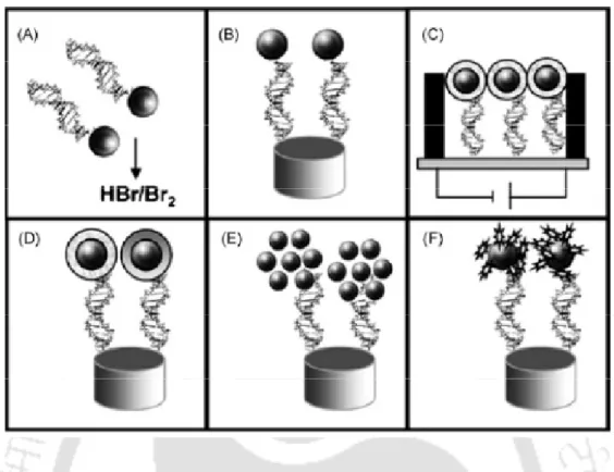
Tentative mechanisms involved in the functionalization of (a) PCL, (b) GL, and (c) PCL–GL composites onto AuNPs
Sonocatalytic UV Light Mediated Synthesis of Gold Nanoparticles Using Boccopa monneri Leaf Extract
Microwave Mediated Synthesis of Gold Nanoparticles using Plant and Fruit extracts
Sonocatalytic Synthesis of Gold Nanoparticles and Functionalization with PCL, GL and PCL-GL composites
Nanotechnology
- Richard Feynman’s idea of nanotechnology
- Concept of ‘nano’ and comparison
- General purpose of nanotechnology
- Nanotechnology– a revolution technology
- Nanotechnology time line
The following Table 1.2 describes the timeline characteristics of both pre-modern and modern era discoveries and milestones in nanotechnology. SUNY Albany launched the first college-level education program in nanotechnology in the United States, the College of Nanoscale Science and Engineering.
Nanotechnology and its applications
- Electronics and information technology
- Sustainable energy
- Environmental remediation
- Future transportation
- Nanobiosystems, medical, and health
To power mobile electronic devices, researchers are developing thin-film solar electric panels that can be mounted on computer cases and flexible piezoelectric ones. When illuminated with ultraviolet light, they emit a wide spectrum of bright colors that can be used to locate and identify specific cell types and biological activities.
Nanomaterials and its types
- Metallic nanomaterials
- Polymeric nanomaterials
- Ceramic nanomaterials
- Composite nanomaterials
- Ceramic nanocomposites
- Metallic nanocomposites
- Polymeric nanocomposites
- Properties of nanomaterials
- Optical properties
- Electrical properties
- Mechanical properties
- Magnetic properties
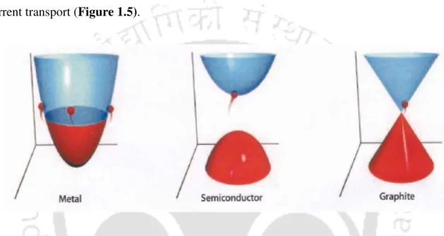
Applications based on the optical properties of nanomaterials include optical detector, laser, sensor, imaging, phosphor, display, solar cell, photocatalysis, photoelectrochemistry, and biomedicine (Susie et al., 2006). Electrical behavior of nanotubes (Reproduced with permission from Collins PG, Avouris PH et al., Scientific American.

Gold nanoparticles
- Gold: The Yellow Metal
- Gold deposits
- The first usage of gold
- Quest for gold and gold production
- Gold for human well-being: food, drinks and medicine
- Gold Nanoparticles (AuNPs)
- The first use of gold nanoparticles
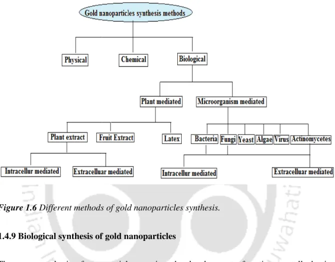
- Synthesis of gold nanoparticles
- Biological synthesis of gold nanoparticles
Gold is not only valued because of its rarity, but also because of its very unique properties and is represented as Carat (Catherine et al., 2012). In cultures such as ancient Egypt, which deified the Sun, gold represented its earthly form (Catherine et al., 2012).
Plant mediated synthesis of nanoparticles
- Intracellular synthesis of gold nanoparticles by plants
- Extracellular synthesis of gold nanoparticles
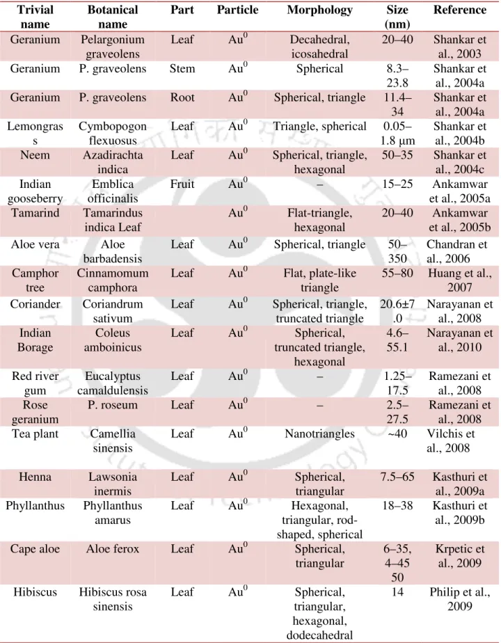
The presence of various secondary metabolites, enzymes, proteins and/or other reducing agents with electron transfer compounds are usually involved in the synthesis of gold nanoparticles by plant components. There are many reports involved in the synthesis of AuNPs are listed in Table 1.6. As per the concern about gold nanoparticles, we first reported using Calotropis procera Latex (Das et al., 2011).
Synthesis of nanoparticles from microorganisms
- Synthesis of nanoparticles from bacteria
- Synthesis of nanoparticles from Fungi
- Synthesis of nanoparticles from Yeast
- Gold nanoparticles synthesis by algae
- Synthesis of nanoparticles using Actinomycetes
- Synthesis of nanoparticles using viruses
In recent years, fungi such as Fusarium oxysporum (Mukherjee et al., 2002), Colletotrichum sp. Shankar et al., 2003), Trichothecium sp., Trichoderma asperellum, T. Tobacco mosaic virus (TMV) was used as a template for the synthesis of iron oxides by oxidative hydrolysis, co-crystallization of CdS and PbS, and synthesis of SiO2 by condensation sol-gel. Peptides such as A7 and J140, which have the ability to nucleate ZnS and CdS nanocrystals were expressed as pVIII fusion proteins in the crystalline capsid of the virus, bacteriophage M13 (Table 1.14).

Synthesis of gold nanoparticles using physical methods
- Sonolysis
- Thermolytic reduction
- Photo irradiation
- Laser ablation
- Solvothermal synthesis
- Seed growth
- Microwave reduction
AuNPs have been synthesized in a single step and a facile procedure by γ-irradiation using bovine serum albumin protein as a stabilizer ( Akhavana et al., 2010 ). UV radiation is another parameter that can improve the quality of gold nanoparticles, including when used in synergy with micelles or seeds (Mallick et al., 2001). Femtosecond laser ablation of gold results in significantly smaller nanoparticles than during nanosecond laser ablation (Haustrup et al., 2011).
Synthesis of gold nanoparticles using chemical methods
- Synthesis of gold nanoparticles using Citrate
- Synthesis of gold nanoparticles using Lactic acid
- Synthesis of gold nanoparticles using glycerol
- BSA craving method
- Two Phase Reduction: Burst-Schiffrin Method
- Hexadecylaniline reduction
- Polyol Reduction
- Sonochemical method
- Chemical Vapour Deposition (CVD)
The formation of gold nanoparticles can be visually observed by the appearance of a deep wine-red color during the course of the reaction (Nirmala et al., 2006). Spherical gold nanoparticles with an edge-to-edge length of 1.5 nm are obtained (Selvakannan et al., 2004). The reaction mechanism involves two phases: (I) the conversion of the inorganic precursor to the intermediate phase in a stepwise sequence, (1) progressive dilution of the inorganic starting material, and (2) precipitation of the intermediate phase; (II) the conversion of the intermediate phase into the metal particles in a stepwise reaction involving (1) dissolution of the intermediate phase, (2) reduction in solution, (3) evolution of the volatile products, and (4) spontaneous nucleation and growth of the metal particles (Kim et al., 2004).
Synthesis of gold nanoparticles using polymers
- Dextrose Reduction
- Gelatin Reduction
- Poly caprolactone reduction
- Polyacrylamide reduction
- Chitosan reduction
The mixed solution is shaken vigorously for 30 s and left undisturbed for 2 h at 80°C, at which point a red AuNPs-gelatin solution is obtained (Zhang et al., 2009). The light yellow color transparent solution is converted to the characteristic red rose color, indicating the formation of AuNPs (Angshuman et al., 2007). The AuNPs prepared with chitosan as a protective agent have a high monodispersity (Huang et al., 2004).
Applications of gold nanoparticles
Non-analytical applications of gold nanoparticles
- Tissue engineering
- Cancer therapy
- Manipulation of cells and biomolecules
Therefore, it is better to make a fibrous tissue covering the surface of the implant to obtain a smooth surface. More than 90% of human bone cells from a suspension adhered to a nanostructured metal surface (Adamopoulos et al., 2007). Furthermore, due to the large aspect ratios, the remanent magnetization of these nanowires can be high.
Analytical applications of gold nanoparticles
- Enzymatic biosensor based on gold nanoparticles
- Application of gold nanoparticles for genosensors
- Application of gold nanoparticles for electrocatalytic chemosensors
- Multicolor optical coding for biological assays
- Application of gold nanoparticles for signal amplification
An extremely important challenge in direct electrochemistry of proteins is the establishment of satisfactory electrical communication between the active site of the enzyme and the electrode surface (Mena et al., 2005; Andrey et al., 1997). Au nanoparticles were self-assembled on the thiol tail groups of the silicate network and grown with hydroxylamine. Biotinylated gold nanoparticles were used as signal amplification probes to improve the detection limit of 50 ng/ml of the streptavidin detection system without signal enhancement, and the calibration curve determined for net frequency changes showed good linearity over a wide range of 1 ng/ml to 10 µg/ml for quantitative analysis of the target molecule streptavidin (Kim et al., 2007).
Commercial exploration of gold nanoparticles
Evident Technologies Semiconductor quantum dots with amine or carboxyl groups on the surface, emission from 350 to 2500 nm.
Overall view of nanotechnology and gold nanoparticles
Instead, it can be achieved either by "growing" new structures through some combination of chemical catalysts and synthetic enzymes or by building them through new techniques based on the patterning and self-assembly of nanoscale materials into designs useful defaults. Even more revolutionary would be the fabrication of nanoscale machines and devices to be incorporated into micro and macro systems. Likewise, magnetic memory materials, which form the basis of hard drives, have achieved greater memory density as a result of nanoscale structuring to exploit new magnetic effects at nanodimensions.
Introduction
Materials and methods
- Materials
- Preparation of leaf extract
- Test for flavonoids and phenolic compounds
- Synthesis of AuNPs using Mentha arvensis plant extract
- Characterization of AuNPs
- UV-visible spectroscopy
- Transmission electron microscope (TEM) studies
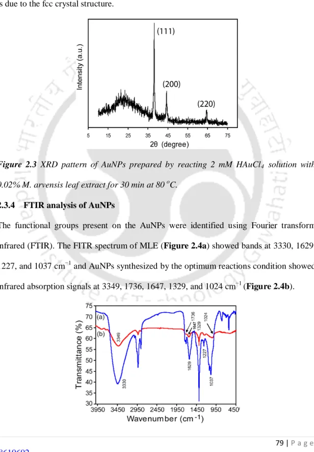
- X-ray Diffractrogram (XRD) analysis of AuNPs
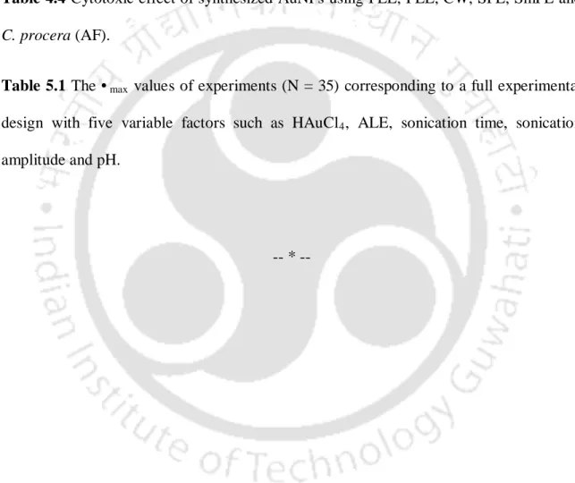
- FTIR analysis of AuNPs
- Statistical analysis
All experiments were repeated five times and the data were expressed as mean ± SD (n = 5) and absorbance recorded in a UV-Vis spectrophotometer (Cary 100 BIO, Varian, CA, USA) from 400 to 700 nm. The AuNPs solution was centrifuged at 20,000 rpm for 10 min and the resulting pellet was resuspended in 5 mL of distilled water and freeze-dried (model 1-4, Christ Gefriertrocknungsanlagen GmbH, Osterode Am Harz, Germany) for 14 h. Experiments with quantitative data were done in replicates of four independent experiments and the results were expressed as Mean ± Standard Deviation (n = 5).
Results
- UV-visible spectroscopy
- Transmission electron microscope (TEM) studies
- X-ray Diffractrogram (XRD) analysis of AuNPs
- FTIR analysis of AuNPs
- EDX analysis
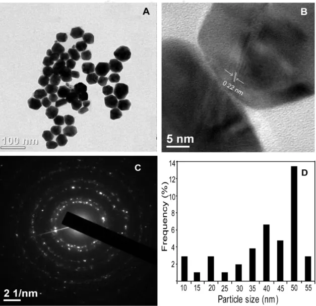
TEM monographs of AuNPs synthesized with optimal reaction conditions reveal that the nanoparticles were hexagonal and almost circular in shape (Figure 2.1A). The crystalline nature of the AuNPs was confirmed by the clear lattice fringes in the high-resolution image and the typical selected area electron diffraction (SAED) pattern with bright circular rings corresponding to the (2 2 0) and (2 2 0) planes ( Figure 2.2C). The energy dispersive X-ray spectroscopy (EDX) profile of MLE-synthesized AuNPs showed a strong gold signal along with very weak carbon and oxygen peaks (Figure 2.5).

Discussion
The clear lattice fringes of 0.22 nm in a high-resolution image revealed that the growth of the AuNPs occurred preferentially on the (1 1 1) plane (Figure 2.2B). The average size of the synthesized nanoparticles determined by TEM analysis was found to be 39±15 nm (Figure 2.2D). FTIR and EDX analysis together suggested that the flavonoids and phenolic compounds are likely to be adsorbed on the surface of the AuNPs.
Conclusions
Introduction
This type of surface functionalization of AuNPs has not been systematically reported for most of the plant extracts previously used for the green synthesis of AuNPs, but we observed a unique surface functionalization phenomenon with BLE, which facilitates these AuNPs for biomedical application such as drug delivery and molecular imaging.
Materials and methods
- Materials
- Preparation of Bacopa monnieri Leaf Extract
- Synthesis of AuNPs using B. monnieri
- Characterization of AuNPs
- UV-visible spectroscopy
- Transmission electron microscope (TEM) studies
- X-ray Diffractrogram (XRD) analysis of AuNPs
- FTIR analysis of AuNPs
- Cyto compatibility tests of AuNPs
- Statistical analysis
A few drops of AuNPs solution were spread on a microscope slide to cover the total surface and dried in a hot air oven (model LDO-150F, Daihan Labtech Co. Ltd.) at 50 oC and placed on the sample platform. The elemental composition of the AuNPs was studied using a scanning electron microscope (LEO 1430 VP) equipped with an INCA Oxford EDX facility (Oxford, UK), at an acceleration voltage of 10 ke V. To investigate the proliferation of AuNPs on cancer cells studies, monocultures of the HeLa and MCF-7 cell lines were incubated with increasing concentrations of filter (0.2 micron) sterilized AuNPs for 24 hours and cell viability was estimated using MTT dye conversion assay.
Results
- UV-visible spectroscopy analysis
- Characterization studies of AuNPs
- Transmission Electron Microscope (TEM)
- X-ray Diffractrogram (XRD) analysis of AuNPs
- FTIR analysis of AuNPs
- TGA analysis
- Cyto-compatibility assay
TEM monographs showed that the AuNPs were mainly spherical in shape with a narrow range of 3-45 nm (Figure 3.2a, 3.2b). High-resolution TEM image revealed clear fcc (0.23 nm), indicating that the growth of the AuNPs occurred preferentially on the (1 1 1) plane (Figure 3.2c). The TGA spectrum of AuNPs occurs over a wide temperature range (225-580 oC), which revealed the significant weight loss (5%) of AuNPs (Figure 3.5).
Discussion
Second, these clusters act as nuclear centers and catalyze the reduction of the remaining metal ions. The result is the autocatalytic growth of the gold cluster and reaches a particular size and is treated as particle development. Additionally, the XRD pattern of AuNPs showed three prominent Bragg reflections that were indexed based on the fcc structure of gold.
Conclusions
These functional groups (N-H, N=H, C-CH3 and OH) are attributed to the bioactive molecules present in BLE, which facilitated the reduction of Au3+ ions to Auo and covered the AuNPs during the synthesis process. The TGA spectrum of AuNPs occurs in a wide temperature range (225-580 o .. C) that revealed the significant weight loss (5%) of AuNPs. This clearly indicated that the bioactive molecules were coated on the AuNPs and completely degraded due to the high temperature.
Introduction
In Thailand, leaves are used to prevent malodor as it has antibacterial activity against obligate oral anaerobes responsible for halitosis (Niranjan et al., 2002). The biochemical profiling showed that it has a high content of sugar (fructose, glucose and sucrose), vitamin C, sorbitol and mannitol, which are good reducing agents (Young et al., 2009). The fleshy part contains various types of saponins containing sesquiterpenoligoglycoside (Kasai et al., 1986), hederagen saponins (Huang et al., 2003), dammaran-type triterpenoids (Kuo et al., 2005) and triterpenoid saponins.
Materials and methods
- Materials
- Preparation of plant leaf extracts
- Preparation of fruit extracts
- Synthesis of AuNPs using plant extracts
- Synthesis of AuNPs using F. esculentum Leaf extract (FLE)
- Synthesis of AuNPs using P. betle Leaf extract (PLE)
- Synthesis of AuNPs using fruit extracts
- Synthesis of AuNPs using coconut water (CW)
- Synthesis of AuNPs using S. indicum fruit extract (SFE)
- Synthesis of AuNPs using SmFE (SmFE)
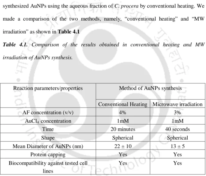
- Synthesis of AuNPs using C. procera latex aqueous faction (AF)
- Characterization of AuNPs
- UV-visible spectroscopy
- Transmission Electron Microscope (TEM) studies
- X-ray Diffractrogram (XRD) analysis of AuNPs
- FTIR analysis of AuNPs
- EDX analysis
- Biochemical test for flavonoids and phenolic compounds
- Effect of pH and salt concentration on stability of AuNPs
- Cytotoxicity assay
- Statistical analysis
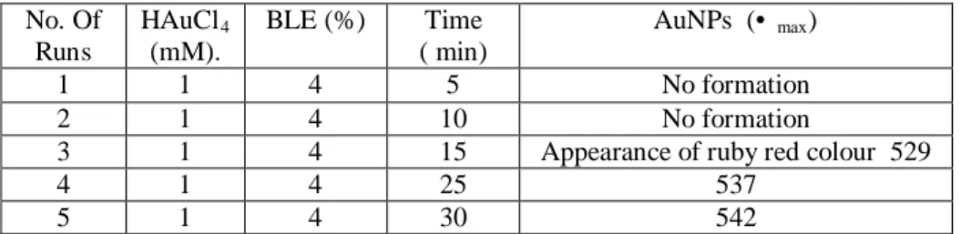
Coconut water (CW) was mixed with different concentrations of mM) HAuCl4 and the final volume was made up to 2 ml with distilled water. The synthesis of AuNPs was performed by changing the SFE at 0.5 mM HAuCl4 and the final volume was made up to 2 ml with double distilled water. This process was repeated three times and the resulting pellet was resuspended in 1 ml of distilled water.
Results
- Optimization of plant and fruit extracts for AuNPs synthesis .1 Optimization of FLE
- Optimization of PLE
- Optimization of SFE
- Optimization of SmFE
- Optimization of aqueous fraction (AF) of C. procera latex for AuNPs synthesis for AuNPs synthesis
- Optimization of HAuCl 4
- Optimization of HAuCl
- Optimization of HAuCl 4
- Optimization of HAuCl
- Optimization of time for AuNPs synthesis
- Optimization of time for AuNPs synthesis using FLE
- Optimization of time for AuNPs synthesis using PLE
- Optimization of time for AuNPs synthesis using coconut water
- Optimization of time for AuNPs synthesis using SFE
- Optimization of time for AuNPs synthesis using C. procera (AF)
- Characterization of AuNPs
- Transmission Electron Microscope (TEM) analysis of AuNPs
- High Resolution Transmission Electron Microscope analysis
- Selected area electron diffraction (SAED) pattern
- Histograms of the AuNPs
- X-ray Diffraction pattern
- FT-IR analysis of AuNPs
- Elemental composition
- Effect of P H
FT-IR spectra of AuNPs synthesized with optimized conditions indicated the presence of the biomolecules on the AuNPs. We performed a set of experiments to find out the effect of pH on the stability of AuNPs. The possible mechanisms of phenolic compounds and flavonoids mediating the synthesis of AuNPs are shown in Figure 4.18.
Discussion
UV-visible spectral analysis of the reaction products exhibited sharp surface plasmon resonance (SPR) peaks, indicating the successful formation of AuNPs. FT-IR spectra of AuNPs synthesized with 0.4% FLE and 16 s MW irradiation time indicated the presence of the bioorganic molecules on the AuNPs. FT-IR spectra of AuNPs synthesized using SFE with optimized conditions indicated the presence of the biomolecules on the AuNPs.
Conclusions
This showed that the synthesized AuNPs had insignificant toxicity on the cancer line, providing an opportunity for application in delivery and molecular imaging. This indicated that the plant and fruit extracts formed a nontoxic coating on the surface of AuNPs. This showed that the synthesized AuNPs had insignificant toxicity on the cancer line, providing an opportunity for application in delivery and molecular imaging.
Introduction
The anti-inflammatory effect of the plant is attributed to andrographolide, the main active constituent of the plant (Madav et al., 1996). It is also considered to be used as an antiphlogistic antipyretic, detoxification agent, analgesic and an agent for the treatment of acute infections of the respiratory organs and the urinary system and the gastrointestinal tract (Nazimudeen et al., 1978). For the rapid synthesis of AuNPs, we used sonication (sonics, VC 505 Vibra Cell), which has the advantage of forming the acoustic bubble and generating enormous heat (Suslick et al., 1990), thereby affecting the nucleation process of AuNPs.
Materials and Methods
- Materials
- Preparation of leaf extract
- Test for flavonoids and phenolic compounds
- Synthesis of AuNPs using A. paniculata plant extract (ALE)
- Effect of amplitude and pH on the synthesis of AuNPs
- Functionalization of AuNPs with PCL, GL, and PCL–GL
- Characterization of AuNPs
- Ultraviolet-visible (UV-vis) spectroscopy
- Transmission electron microscopy (TEM)
- X-ray diffraction (XRD), Fourier transform infrared (FT-IR) spectroscopy and energy dispersive X-ray (EDX) spectroscopy
- Cytotoxicity studies
- Statistical analysis
Both PCL and GL, 0.25% of each, separately, were added to the 5 mL of dimethylformamide (DMF) and 5 mL of the preheated water, respectively, and the total volume of each reaction mixture was adjusted to 20 mL with distilled water containing ALE (3.5%) and HAuCl4 (1 mmol/L) contained. The resulting pellet was resuspended in 3 ml of distilled water and centrifuged at 20,000 rpm for 20 min. Finally, each resulting pellet was redispersed in 5 ml of double distilled water and freeze-dried in a freeze-drying damper (Christ Gefriertrocknungsanlagen GmbH Model 1- 4) for 16 hours.
Results
- Formation of spherical AuNPs from ALE in the ultrasonication process under optimum conditions