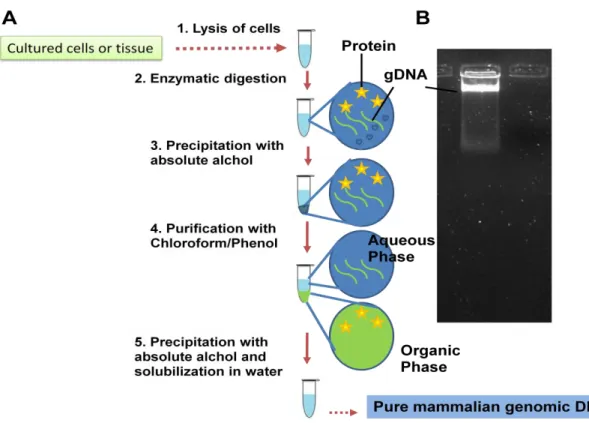
Incubation of cells with digestion buffer containing protease-K, SDS to release genomic DNA from DNA-protein complex. Genomic DNA is analyzed on 0.8% agarose gel, and a good preparation of genomic DNA gives an intact band with no visible smearing (Figure 10.2). Generation of fragments of appropriate size - Next step generation of genomic DNA into appropriate small fragments.
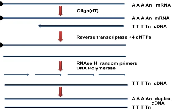
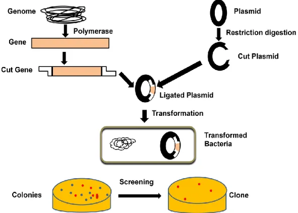
Restriction digestion: Genomic DNA can be digested with a frequent DNA cutting enzyme such as EcoR-I, BamH-I or sau3a to generate random size DNA fragments. Cloning into suitable vector - The suitable vector for genomic library preparation can be selected based on the size of the genomic DNA fragment and the carrying capacity of the vector (Table 10.1). Joint initiative of IIT and IISc – Funded by MHRD Page 8 of 65 Figure 11.3: Isolation of mRNA from total cell lysate.
Cloning of cDNA into the vector-The cDNA is ligated into the suitable vector to generate clone
Cloning of cDNA into the vector - The cDNA is ligated into the appropriate vector to generate a clone.
Identification and Isolation of a gene
- DNA sequence-This properties can be use to search both genomic library and cDNA library to identify the gene
- Expression of a particular protein with immunogenic epitope-This property can be partially useful to screen genomic library due to truncation of a full gene or no expression
- Enzymatic activity- This property exploits the ability of a protein fragment to exhibits enzymatic activity. It is useful for the screening of cDNA library but not much for
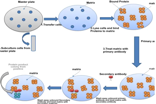
- Preparation of Replica plate- As original genomic or cDNA library is precious and will be consumed in later stage, all procedure is performed with the replica plate
- Blotting-The clone is transferred on a nitrocellulose membrane with retaining identical pattern of colonies on master plate. The cells on the membrane are lysed and released
- Development of blot (Autoradiography)-The position of labeled probe is detected by autoradiogram.The position of signal on membrane can be matched with the master plate
- Preparation of Replica plate- As original genomic or cDNA library is precious and will be consumed in later stage, all procedure is performed with the replica plate
- Blotting-The clone is transferred on a nitrocellulose membrane to get similar pattern of colonies on master plate. The cells on the membrane are lysed and released protein is
- Treatment with primary antibody-The membrane is incubated with antibody having immunoreactivity towards a particular protein. The primary antibody will binds to the
- Treatment with secondary antibody-The membrane is incubated with secondary antibody recognizing primary antibody. Secondary antibody is labeled with an enzyme
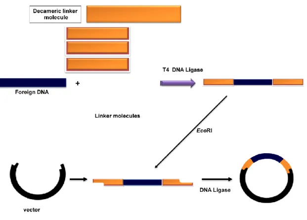
- Linker Molecule-An amplified foreign DNA may have restriction enzymes at their terminus to give cohesive end to facilitate ligation into the vector. But in cases when
- Polymerase- The first requirement of a molecular cloning to produce or isolate DNA of interest from the genomic sequence. One of the easiest process to achieve it is by
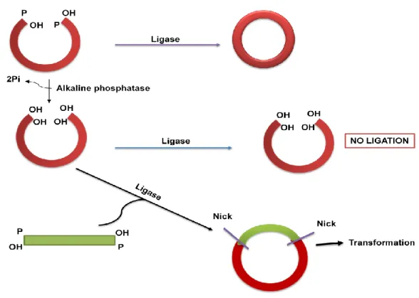
- Alkalike phosphatase- Digested linear plasmid containing cohesive ends on both sites with phosphate has a tendency to recircularize (Figure 15). Removing terminal
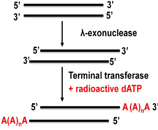
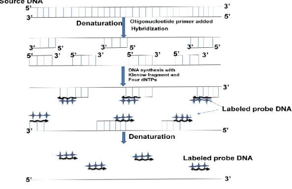
Joint initiative of IIT and IISc – Funded by MHRD Page 15 of 65 Figure 12.1: Screening of a library with a radioactive probe by colony hybridization. Random primer method- In this method, a random primer is used to anneal to the template and then a PCR reaction is performed in the presence of the radiolabeled nucleotide. Terminal Transferase- In this method, a terminal transferase enzyme will label the probe at the ends of the last nucleotide of the probe (Figure 12.3).
The probe is incubated with the probe at the ends up to the last nucleotide of the probe (Figure 12.3). Spot development (Autoradiography) – the position of the marked probe is detected by the autoradiogram. The position of the signal on the diaphragm can be matched with the autoradiogram of the main plate. The position of the signal on the membrane can be matched to the main plate to obtain the location of the corresponding colony. The primary antibody will bind to the target protein due to exclusive specificity against the antigen (Figure 12.4).
The secondary antibody will bind to the primary antibody and will allow the location of the primary antibody to be detected. Development of blot-The position of secondary antibodies is detected by performing enzymatic activity. The position of signal on membrane can be matched with the main plate to get location of corresponding colony. This is now incubated with EcoRI-digested vector in the presence of DNA ligase to obtain circular clone (Figure 13.2).
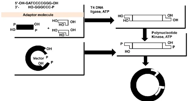
Adapter molecules are double-stranded molecules 8–10 nucleotides in length flanked by DNA sequence to provide cohesive ends (Figure 13.3). The adapter molecule containing the chimeric DNA is incubated with BamH-I digested vector in the presence of DNA ligase to obtain circular clone (Figure 13.3). As discussed in Figure 13.1, it is an essential step to generate clone containing foreign DNAdiscussed in Figure 13.1, it is an essential step to generate clone containing foreign DNA in a vector.
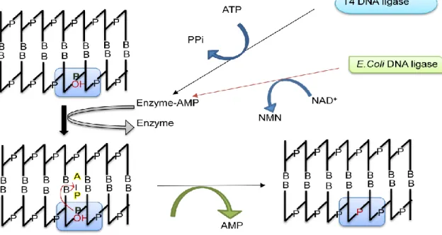
AMP is released and a phosphodiester bond is formed between the 3" and 5" ends to close the nick (Figure 13.4). Alkaline phosphatase-digested linear plasmid containing cohesive ends at both phosphate sites is prone to recircularization (Figure 13.5). Alkaline phosphatase removes the 5'-terminal phosphate groups and in this condition only in the presence of inserted DNA, as it will supply a phosphate group at both ends to facilitate the ligation reaction (Figure 13.5).
Polymerase Chain Reaction (Part-I)
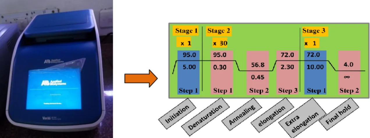
Denaturation: This is the first step in which the double stranded DNA template is denatured to form two single strand by heating at 95°C for 15-30 secs
Annealing: This is the annealing step where primers can bind to template DNA at a lower temperature (usually 50-65°C), the annealing time is 15-30 seconds and depends on the length and bases of the primers. In general, the annealing temperature is about 3-5°C below the melting temperature (Tm) of the pair of primers used. Elongation: This is the synthesis step where the polymerase performs the synthesis of a new strand in the 5' to 3' direction using primer and deoxyribonucleoside triphosphates.
- After the cycles are completed, the reaction is held at 70-74°C for several minutes to allow final extension of the remaining DNA to be fully extended
- The reaction is complete and the resulting amplified nucleic acids are held at a low temperature (~4°C) until analysis
- Primer length: Oligonucleotides between 18-24 bases is the ideal length which is long enough for adequate specificity and short enough for primers to bind easily to the
- GC Content: The number of G's and C's in the primer as a percentage of the total bases should be 40-60%
- GC clamp: As GC forms a stronger bond than AT, the number of GC content at the 3‟
- Primer secondary structures: Primer secondary structures arise as a result of intra or intermolecular attraction within the primer or with other primers which eventually reduce
Instrumentation: The thermal cycler is the instrument that performs amplification via the polymerase chain reaction (Figure 15.2). Typically the three main events are repeated for 30-40 cycles to obtain detectable amount of product at the end of the cycles. The cycler changes the temperature of the block in discrete, pre-programmed steps using the peltier effect.
In PCR, two primers are required to bind to each of the single-stranded DNA (obtained after denaturation) flanking the target sequence. Two restriction enzyme sites are added to the 5' end of each primer to facilitate cloning. Primer length: Oligonucleotides between 18-24 bases are the ideal length that is long enough for sufficient specificity and short enough for primers to bind easily to the enough for sufficient specificity and short enough for primers to bind easily to the template at the annealing temperature.
Primer Melting Temperature (Tm): Primers with melting temperatures in the range of 52-58oC generally give the best results. The two primers should be prepared so that their Tm difference should not exceed 2°C, otherwise poor annealing efficiency will result. GC Content: The number of Gs and Cs in the primer as a percentage of the total bases should be 40-60%.
GC clamp: Since GC forms a stronger bond than AT, the number of GC content is at 3‟. Primer Secondary Structures: Primer secondary structures are formed as a result of intra- or intermolecular attraction within the primer or with other primers that eventually reduce the intermolecular attraction within the primer or with other primers that eventually reduce amplification efficiency as the availability of single-stranded primers will be limited for PCR . Primer and template homology: Primers must be designed such that there is no homology within the template other than the target site.
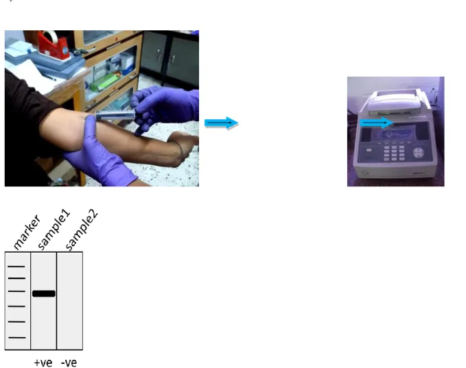
Analysis of PCR results: When the PCR cycle is completed, the amplified product is loaded into the agarose gel and observed after ethidium bromide staining under UV light source (Figure 15.3).
Polymerase Chain Reaction (Part-II)
- Post entry into the host should give phenyotypic changes-Another important feature is that vector DNA should give additional phenotypic changes in the host cell so that
- Muliple cloning site with unique restriction site- A short stretch of DNA on vector DNA containing restriction site for possible site for insertion of foreign DNA is desirable
- High copy number-A high copy number is desirable as it gives high amount of DNA after growing host cells
- Selection marker- Selection marker in the form of either antibiotic resistance gene or enzymatic gene is essential to give phenotypic changes in host after entry of the plasmid
PCR in infectious diseases: PCR technology has become the basis for a wide range of clinical diagnostic tests for a variety of infectious agents, including viruses and bacteria (Figure 16.2). Knowing the gene associated with the disease in a person prone to these disorders has the potential to control the problem much in advance. PCR in Plant Science: There are various fields of plant science that require the use of PCR technology to achieve their goals.
PCR in tissue culture: Used in the analysis of DNA and specific genes in plant cells at different stages of regeneration during in vitro culture together with RAPD (Random Amplification of Polymorphic DNA) technology. PCR in veterinary parasitology: Because of its speed and sensitivity compared to antibody-based diagnosis, PCR is used in almost all aspects of biological work, including veterinary clinical diagnosis. A sample of blood, hair root, or tissue left at a crime scene can be used to identify a person by PCR by comparing the crime scene DNA with the suspect's DNA or with a DNA database of previous convictions (Figure 16.3).
PCR in Research Applications: Biological research requires molecular biological techniques as starting material and so on, which cannot be achieved without the use of PCR. Introduction: In the steps of cloning discussed in previous lectures, a vehicle DNA is needed to carry foreign DNA to generate a recombinant construct so that it allows easy amplification of chimeric DNA in the host. Criteria for a good vector: The vector DNA has two main responsibilities: (1) the ability to carry foreign DNA, (2) the ability to replicate in the host.
After entry into the host should give phenotypic changes - Another important feature is that vector DNA should give additional phenotypic changes in the host cell, that is, vector DNA should give additional phenotypic changes in the host cell, so that recognition of transformed cells will be easier. Selection Marker - Selection marker in the form of an antibiotic resistance gene or an enzymatic gene is essential to produce phenotypic changes in the host upon entry into the plasmid. The origin of replication is derived from pMB1 and a detail of the construction of pBR322 with multiple steps of recombination is provided in the following article.
In the similar advance, a multiple cloning sites were added to facilitate easy cloning without disrupting antibiotic resistance gene. In addition, two enzymes of MS can be used to insert the foreign DNA without disturbing plasmid sequences. The bacterial culture is collected by centrifugation at the bottom and resuspended in the solution I containing 50 mM glucose, 25 mM TrisHCl pH 8.0, 10 mM EDTA pH 8.0.
Deproteination: Resulting supernatant containing plasmid DNA and protein is treated with phenol: chloroform: isoamyl alchol mixture to remove protein in the
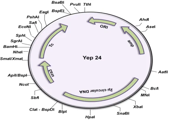
Vector as extrachromosomal DNA- These vector remains in eukaryotic cell as extrachromosomal DNA and express the protein
Integration Vector- These vectors carry an integration site to facilitate recombination medited integration into the chromosomal DNA of the host cell
Integration vector – These vectors have an integration site to facilitate recombination-mediated integration into the chromosomal DNA of the host cell. They all have some similar features, such as the presence of an MCS, a transfer vector (the origin of replication for E.coli and yeast) and the presence of a selectable marker. Episomal Vector - A yeast episomal vector is constructed by combining a bacterial plasmid with either a yeast 2µ origin of replication or an autonomous replication sequence (ARS).
The yeast vector containing ARS is a high copy number plasmid, but they are unstable in the absence of selection pressure. In yeast, integration plasmids contain the target sequence for integration into chromosomal DNA, selection marker, and bacterial origin of replication. Before delivery to the yeast, the vector is digested with the unique restriction endonuclease to produce linear DNA to increase transformation efficiency and integration.
In most cases, integration is performed in such a way that the yeast chromosomal DNA remained intact, and integration may not affect yeast growth. However, in an alternative approach, part of the yeast's chromosomal DNA is replaced by the vector DNA through homologous recombination. YAC vector is like a chromosome as it has ARS sequences, centromere sequence and telomere at the two ends to provide stability.
It has an ampicillin resistance gene (Ampr) for selection in e.coli and an e.coli origin of replication for propagation in bacteria. In addition, it has ARS for replication, CEN for centromere function and URA3, TRP1 for selection in yeast. The gene of interest will be inserted into the cloning site located next to the promoter.
It has a polyhedron termination sequence downstream of the cloning site to stop transcription of the cloned gene.