This page intentionally left blank
M olecular P lant –
M icrobe i nteractions
Edited by
Kamal Bouarab
Département de Biologie Université de Sherbrooke Sherbrooke
Québec Canada
Normand Brisson
Department of Biochemistry Université de Montréal Montréal
Québec Canada
and
Fouad Daayf
Department of Plant Science University of Manitoba
Winnipeg
Manitoba
Canada
CABI is a trading name of CAB International
CABI Head Office CABI North American Office
Nosworthy Way 875 Massachusetts Avenue
Wallingford 7th Floor
Oxon OX10 8DE Cambridge, MA 02139
UK USA
Tel: +44 (0)1491 832111 Tel: +1 617 395 4056
Fax: +44 (0)1491 833508 Fax: +1 617 354 6875
E-mail: [email protected] E-mail: [email protected]
Website: www.cabi.org
© CAB International 2009. All rights reserved. No part of this publication may be reproduced in any form or by any means, electronically,
mechanically, by photocopying, recording or otherwise, without the prior permission of the copyright owners.
A catalogue record for this book is available from the British Library, London, UK.
Library of Congress Cataloging-in-Publication Data Molecular plant-microbe interactions / edited by Kamal Bouarab, Normand Brisson and Fouad Daayf.
p. cm.
Includes bibliographical references and index.
ISBN 978-1-84593-574-0 (alk. paper)
1. Plants--Disease and pest resistance. 2. Plant-microbe relationships.
3. Fungal diseases of plants. 4. Virus diseases of plants. I. Bouarab, Kamal. II. Brisson, Normand, 1955- III. Daayf, Fouad. IV. Title.
SB750.M67 2009 632’.3--dc22
2009007330 ISBN-13: 978 1 84593 574 0
Typeset by Columns Design, Reading.
Printed and bound in the UK by the MPG Books Group, Bodmin.
The paper used for the text pages in this book is FSC certified. The FSC (Forest Stewardship Council) is an international network to promote responsible management of the world’s forests.
v
Contributors vii
Preface xi
1 Plant RNA-silencing Immunity and Viral Counter-defence
Strategies 1
Santiago Wadsworth and Patrice Dunoyer
2 Mitogen-activated Protein Kinase Cascades in Plant
Defence Responses 36
Fengming Song, Huijuan Zhang and Shuqun Zhang 3 Molecular Mechanisms of the Radical Burst in
Plant Immunity 59
Hirofumi Yoshioka, Shuta Asai,Noriko Miyagawa, Tatsushi Ichikawa, Miki Yoshioka and Michie Kobayashi 4 Disease Resistance in Arabidopsis, Starring TGA2
and also Featuring NPR1 75
Patrick Boyle, Pierre R. Fobert and Charles Després
5 Disease Resistance Genes: Form and Function 94 Melanie A. Sacco and Peter Moffett
6 Transcription Factor Families Involved in Plant Defence:
from Discovery to Structure 142
Jean-Sébastien Parent, Laurent Cappadocia, Alexandre Maréchal, Pierre R. Fobert and Normand Brisson
vi Contents 7 Cross Talk Between Induced Plant Immune Systems 163
Rocío González-Lamothe, Mohamed El Oirdi, Taha Abd El Rahman, Raphaël Sansregret, Hamed Bathily
and Kamal Bouarab
8 The Needle and the Damage Done: Type III Effectors
and the Plant Immune Response 179
Jennifer D. Lewis, Karl Schreiber and Darrell Desveaux 9 Virulence Determinants and the Global Regulation of
Virulence in Xanthomonas campestris 211
Adrián A. Vojnov, J. Maxwell Dow and Kamal Bouarab 10 Suppression of Induced Plant Defence Responses by
Fungal and Oomycete Pathogens 231
Abdelbasset El Hadrami, Ismail El Hadrami and Fouad Daayf 11 Sustainable Agriculture and the Multigenomic Model:
How Advances in the Genetics of Arbuscular
Mycorrhizal Fungi will Change Soil Management Practices 269 Erin Zimmerman, Marc St-Arnaud and Mohamed Hijri
12 Microbial Traits Associated with Actinobacteria
Interacting with Plants 288
Anne-Marie Simao-Beaunoir, Sébastien Roy and Carole Beaulieu 13 Insight into Fusarium–Cereal Pathogenesis 319
Rajagopal Subramaniam, Charles G. Nasmith, Linda J. Harris and Thérèse Ouellet
Index 337
vii Abd El Rahman, Taha, Centre de Recherche en Amélioration Végétale, Département de Biologie, Université de Sherbrooke, 2500 Boulevard de l’Université, Sherbrooke, Québec, J1K 2R1, Canada.
Asai, Shuta, Laboratory of Defense in Plant–Pathogen Interactions, Graduate School of Bioagricultural Sciences, Nagoya University, Chikusa, Nagoya 464-8601, Japan.
Bathily, Hamed, Centre de Recherche en Amélioration Végétale, Département de Biologie, Université de Sherbrooke, 2500 Boulevard de l’Université, Sherbrooke, Québec, J1K 2R1, Canada.
Beaulieu, Carole, Centre SÈVE, Département de biologie, Université de Sherbrooke, Sherbrooke, Québec, J1K 2R1, Canada; carole.beaulieu@
usherbrooke.ca
Bouarab, Kamal, Centre de Recherche en Amélioration Végétale, Département de Biologie, Université de Sherbrooke, 2500 Boulevard de l’Université, Sherbrooke, Québec, J1K 2R1, Canada; Kamal.
Boyle, Patrick, Department of Biological Sciences, Brock University, 500 Glenridge Avenue, St Catharines, Ontario, L2S 3A1, Canada.
Brisson, Normand, Department of Biochemistry, Université de Montréal, Montréal, Québec, Canada; [email protected]
Cappadocia, Laurent, Department of Biochemistry, Université de Montréal, Montréal, Québec, Canada.
Daayf, Fouad, Department of Plant Science, University of Manitoba, 222, Agriculture Building, Winnipeg, Manitoba, R3T 2N2, Canada;
Després, Charles, Department of Biological Sciences, Brock University, 500 Glenridge Avenue, St Catharines, Ontario, L2S 3A1, Canada;
Desveaux, Darrell, Centre for the Analysis of Genome Evolution and Function & Department of Cell and Systems Biology, University of
viii Contributors Toronto, 25 Willcocks Street, Toronto, Ontario, M5S 3B2, Canada;
Dow, J. Maxwell, BIOMERIT Research Centre, Department of Microbiology, National University of Ireland, Cork, Ireland.
Dunoyer, Patrice, Institut de Biologie Moléculaire des Plantes, CNRS, 67084 Strasbourg Cedex, France; [email protected] strasbg.fr
El Hadrami, Abdelbasset, Department of Plant Science, University of Manitoba, 222, Agriculture Building, Winnipeg, Manitoba, R3T 2N2, Canada.
El Hadrami, Ismail, University Cadi Ayyad, Marrakech, Morocco.
El Oirdi, Mohamed, Centre de Recherche en Amélioration Végétale, Département de Biologie, Université de Sherbrooke, 2500 Boulevard de l’Université, Sherbrooke, Québec, J1K 2R1, Canada.
Fobert, Pierre R., National Research Council Canada, Plant Biotechnology Institute, 110 Gymnasium Place, Saskatoon, Saskatchewan, S7N 0W9, Canada.
González-Lamothe, Rocío, Centre de Recherche en Amélioration Végétale, Département de Biologie, Université de Sherbrooke, 2500 Boulevard de l’Université, Sherbrooke, Québec, J1K 2R1, Canada.
Hamed, Bathily, Centre de Recherche en Amélioration Végétale, Département de Biologie, Université de Sherbrooke, 2500 Boulevard de l’Université, Sherbrooke, Québec, J1K 2R1, Canada.
Harris, Linda J., Eastern Cereal and Oilseed Research Centre, Agriculture and Agri-Food Canada, Ottawa, Ontario, K1A 0C6, Canada.
Hijri, Mohamed, Institut de Recherche en Biologie Végétale, Université de Montréal, 4101 rue Sherbrooke Est, Montréal, Québec, H1X 2B2, Canada.
Ichikawa, Tatsushi, Laboratory of Defense in Plant–Pathogen Interactions, Graduate School of Bioagricultural Sciences, Nagoya University, Chikusa, Nagoya 464-8601, Japan.
Kobayashi, Michie, Laboratory of Defense in Plant–Pathogen Interactions, Graduate School of Bioagricultural Sciences, Nagoya University, Chikusa, Nagoya 464-8601, Japan.
Lewis, Jennifer D., Department of Cell and Systems Biology, University of Toronto, 25 Willcocks Street, Toronto, Ontario, M5S 3B2, Canada.
Maréchal, Alexandre, Department of Biochemistry, Université de Montréal, Montréal, Québec, Canada.
Miyagawa, Noriko, Laboratory of Defense in Plant–Pathogen Interactions, Graduate School of Bioagricultural Sciences, Nagoya University, Chikusa, Nagoya 464-8601, Japan.
Moffett, Peter, Département de Biologie, Université de Sherbrooke, 2500 Boulevard de l’Université, Sherbrooke, Québec, J1K 2R1, Canada;
Nasmith, Charles G., Eastern Cereal and Oilseed Research Centre, Agriculture and Agri-Food Canada, Ottawa, Ontario, K1A 0C6, Canada.
Ouellet, Thérèse, Eastern Cereal and Oilseed Research Centre, Agriculture and Agri-Food Canada, Ottawa, Ontario, K1A 0C6, Canada.
Parent, Jean-Sébastien, Department of Biochemistry, Université de Montréal, Montréal, Québec, Canada.
Roy, Sébastien, Centre SÈVE, Département de biologie, Université de Sherbrooke, Sherbrooke, Québec, J1K 2R1, Canada.
Sacco, Melanie A., Department of Biological Science, California State University, Fullerton, 800 North State College Blvd., Fullerton, CA 92831-3599, USA.
Sansregret, Raphaël, Centre de Recherche en Amélioration Végétale, Département de Biologie, Université de Sherbrooke, 2500 Boulevard de l’Université, Sherbrooke, Québec, J1K 2R1, Canada.
Schreiber, Karl, Department of Cell and Systems Biology, University of Toronto, 25 Willcocks Street, Toronto, Ontario, M5S 3B2, Canada.
Simao-Beaunoir, Anne-Marie, Centre SÈVE, Département de biologie, Université de Sherbrooke, Sherbrooke, Québec, J1K 2R1, Canada.
Song, Fengming, National Key Laboratory for Rice Biology, Institute of Biotechnology, Zhejiang University, Hangzhou, Zhejiang 310029, China; [email protected]
St-Arnaud, Marc, Institut de Recherche en Biologie Végétale, Université de Montréal, 4101 rue Sherbrooke Est, Montréal, Québec, H1X 2B2, Canada.
Subramaniam, Rajagopal, Eastern Cereal and Oilseed Research Centre, Agriculture and Agri-Food Canada, Ottawa, Ontario, K1A 0C6, Canada; [email protected]
Vojnov, Adrián A., Instituto de Ciencia y Tecnolgía Dr. Cesar Milstein, CONICET; [email protected]
Wadsworth, Santiago, Institut de Biologie Moléculaire des Plantes, CNRS, 67084 Strasbourg Cedex, France.
Yoshioka, Hirofumi, Laboratory of Defense in Plant–Pathogen Interactions, Graduate School of Bioagricultural Sciences, Nagoya University, Chikusa, Nagoya 464-8601, Japan; [email protected] Yoshioka, Miki, Laboratory of Defense in Plant–Pathogen Interactions,
Graduate School of Bioagricultural Sciences, Nagoya University, Chikusa, Nagoya 464-8601, Japan.
Zhang, Huijuan, National Key Laboratory for Rice Biology, Institute of Biotechnology, Zhejiang University, Hangzhou, Zhejiang 310029, China.
Zhang, Shuqun, Department of Biochemistry, University of Missouri- Columbia, MO 65211, USA.
Zimmerman, Erin, Institut de Recherche en Biologie Végétale, Université de Montréal, 4101 rue Sherbrooke Est, Montréal, Québec, H1X 2B2, Canada; [email protected]
This page intentionally left blank
xi Recent developments in molecular biology and in the burgeoning omics have brought about a great deal of new data in all areas of plant sciences. However, the use of these data towards their exploitation into higher performance plants has proved to be a slow process. For example, although producing genomics and proteomics data has become routine in a large number of laboratories around the world, functional genomics studies to understand the meaning of the accumulating data still lag behind. Carrying out these studies in such important areas as plant development, photosynthesis or plant responses to abiotic stress, bounces within a large window of complexity and difficulty levels. These levels escalate when more than one organism is involved, in either a mutually beneficial or an antagonistic interaction with the plant. However, navigating through such a network of interactions makes the journey more exciting, at least from the view of a plant pathologist. Studying molecular plant–microbe interactions is very stimulating indeed. There is no guarantee that a microbe, even from the same species or race within it, would act exactly the same way in its coevolution with the host plant. The same applies to the plant regarding ‘upgrading’ its arsenal to fight external threats.
This creates a certain dynamism that fuels new discoveries, and which scientists in the molecular plant–microbe interactions field value so much. In such a dynamic discipline, it is useful to revisit the field more often than, say, every 20 years. In this volume, authors of world repute in different aspects of molecular plant–microbe interactions have agreed to contribute a chapter about their research and their views on the current developments in the fields of plant defences, pathogen counter-defences and mutually beneficial plant–
microbe interactions.
This book explores recent discoveries in the area of molecular plant–
microbe interactions. It focuses mainly on the mechanisms controlling plant disease resistance and the cross talk among the signalling pathways involved, and the strategies used by fungi and viruses to suppress these defences.
xii Preface Furthermore, two chapters related to the role of symbionts during their interactions with plants are included.
We would like to thank all the contributors for their hard work towards meeting the deadlines for this volume.
The Editors Kamal Bouarab Normand Brisson
Fouad Daayf
© CAB International 2009. Molecular Plant–Microbe Interactions (eds Bouarab et al.) 1
1 Plant RNA-silencing Immunity and Viral Counter-defence Strategies
S
antiagoW
adSWorth andP
atriced
unoyer Institut de Biologie Moléculaire des Plantes, Strasbourg, FranceAbstract
RNA silencing is a conserved eukaryotic process mediated by small RNA molecules that inhibit gene expression at the transcriptional, mRNA-stability or translational level through sequence-specific interactions. Diverse roles have been identified for RNA silencing such as genome defence against mobile DNA elements or downregulation of specific factors during plant and animal development. In plants, RNA silencing plays a crucial role in antiviral defence by inhibiting viral accumulation and sometimes preventing systemic infection.
As a counter-defence mechanism, viruses have evolved a set of anti-silencing strategies, of which the most common is the production of viral suppressors of RNA silencing (VSRs). Here we review the different strategies underlying VSRs action including prevention of viral-derived small (vs)RNAs synthesis, vsRNAs sequestration or inhibition of vsRNA-guided effector complexes. We will also underline the consequences of this molecular arms race on the evolution of both viral and host genomes.
1.1 Introduction
RNA silencing is an ancient eukaryotic process involved in the control of gene expression. It is triggered by double-stranded (ds)RNA and causes a sequence- specific shut down of the expression of genes with sequences identical or highly similar to the initiating dsRNA (Fire et al., 1998; Wesley et al., 2001). RNA silencing may act at both the RNA and the DNA levels. Mechanisms of silencing at the RNA level (called post-transcriptional gene silencing (PTGS) in plants or RNA interference (RNAi) in animals) include mRNA cleavage or translational repression. RNA silencing at the DNA level involves DNA and/or histone methylation and subsequent transcriptional gene silencing (TGS) through hetero-
2 S. Wadsworth and P. Dunoyer chromatin formation and maintenance (Bartel, 2004; Jones-Rhoades et al., 2006). All these manifestations of RNA silencing rely on the action of small RNA (sRNA) molecules of 21–24 nucleotides (nt) in length, which originate from the processing of the dsRNA trigger (Hamilton and Baulcombe, 1999; Elbashir et al., 2001). During PTGS, these sRNA molecules control stability or regulate translation of their mRNA targets by guiding endogenous effector complexes (Hammond et al., 2000; Bartel, 2004; Jones-Rhoades et al., 2006).
Originally, PTGS was first observed in attempts to overexpress an endogenous gene by transforming petunia plants with an extra copy of the gene of interest, which led to the extinction of both endogenous and transgenic copies (Napoli et al., 1990). Subsequently, the inhibition of gene expression was confirmed in the nematode Caenorhabditis elegans by injecting sense or antisense copies of the target mRNA (S-PTGS and AS-PTGS, respectively) (Fire et al., 1991; Guo and Kemphues, 1995). Nowadays, the strongest and most commonly used mechanism to inhibit gene expression in eukaryotic cells is PTGS triggered by an inverted-repeat construct (IR-PTGS) that produces a dsRNA molecule with a hairpin-like structure (Beclin et al., 2002; Giordano et al., 2002).
Besides its essential role in plant and animal development, the first biological role attributed to RNA silencing in plants was defence against viral infections (Lindbo et al., 1993; Ratcliff et al., 1997; Anandalakshmi et al., 1998; Brigneti et al., 1998; Kasschau and Carrington, 1998; Hamilton and Baulcombe, 1999). Attempts to overexpress an endogenous gene from recombinant viral vectors did not result in enhanced protein accumulation but led to the specific degradation of the corresponding mRNA (Ruiz et al., 1998).
This phenomenon, called virus-induced gene silencing (VIGS), probably triggered by the dsRNA intermediates formed during viral replication, was suggested to be a manifestation of an RNA silencing-based antiviral defence response (Ratcliff et al., 1999). Currently, several indications suggest that RNA silencing is the primary immune system against viruses in plants: (i) sRNAs derived from a viral genome (viral-derived small RNAs or vsRNAs) are invariably detected during infection; (ii) mutant plants affected in the silencing pathways are hypersusceptible to viral infections; and (iii) as a counter-defensive strategy, plant viruses express proteins that are able to suppress the antiviral silencing response (Li and Ding, 2006; Ding and Voinnet, 2007). Indeed, back in 1998, two different viral proteins, previously identified as important pathogenicity determinants, were characterized as effective suppressors of RNA silencing (Anandalakshmi et al., 1998; Brigneti et al., 1998). Since then, expression of viral suppressors of RNA silencing (VSRs) has emerged as a major counter- defence mechanism against the antiviral RNA silencing response (Li and Ding, 2006; Ding and Voinnet, 2007). After a short presentation of the core mechanism of RNA silencing and its different pathways in the model plant Arabidopsis thaliana, we first discuss how viruses induce the antiviral silencing response in plants. We then review the different strategies underlying VSRs action as well as their effect on endogenous silencing pathways and on the evolution of both viral and host genomes.
1.2 Plant RNA Silencing Pathways
The core mechanism
The initial step of RNA silencing depends on the recognition of a dsRNA molecule by an RNase III-like enzyme called Dicer (or DCL, for Dicer-like in plants) that processes the trigger into sRNA duplexes of 21–24 nt in length with 2 nt 3' overhangs (Hammond, 2005). These sRNA are divided in two main classes in both plants and animals: small interfering RNAs (siRNAs) and microRNAs (miRNAs).
siRNAs are processed from long, perfectly based-paired dsRNAs, whereas miRNAs originate from primary miRNA transcripts (pri-miRNAs) that form imperfect intramolecular hairpin-like structures. A single Dicer enzyme in worms and humans produces both miRNAs and siRNAs, whereas in Drosophila they are produced by Dicer-1 and Dicer-2, respectively (Hammond, 2005). In plants, Dicer-like proteins are even more diverse. For instance, A. thaliana encodes four Dicer-like enzymes (DCL 1–4). Whereas most mature miRNAs synthesis relies on DCL1 (Bartel, 2004), populations of 21, 22 and 24 nt-long siRNAs are synthesized from dsRNA through DCL4, DCL2 and DCL3 activity, respectively (Brodersen and Voinnet, 2006; Vazquez, 2006; Chapman and Carrington, 2007).
sRNAs are then incorporated into argonaute (AGO)-containing effector complexes termed RNA-induced silencing complexes (RISCs) and RNA-induced initiation of transcriptional silencing complexes (RITS) and, as part of these complexes, direct sequence-specific PTGS or TGS, respectively (Hammond et al., 2000; Ekwall, 2004). During PTGS, the outcome of RISC activity is either cleavage and/or translational inhibition of the target mRNA and this probably relies on the degree of complementarity with the sRNA and on the different cellular factors involved. Proteins of the AGO family (ten different members in Arabidopsis) have been crucially implicated in the functions of both RISC and RITS complexes. AGO proteins contain at least one single strand (ss)RNA- binding PAZ domain and a PIWI domain that confers the endonucleolytic (or slicer) activity. So far, slicing activity in Arabidopsis has been only demonstrated for AGO1, AGO4 and AGO7 (Baumberger and Baulcombe, 2005; Qi et al., 2006; Montgomery et al., 2008) and the former was shown to direct both miRNA- and 21 nt-long siRNA-mediated target cleavage without requiring further protein partners (Baumberger and Baulcombe, 2005; Dunoyer et al., 2007). Upon unwinding of the sRNA duplex and loading into the AGO protein, the selected strand (guide strand) guides RISC to target all RNA molecules presenting sequence complementarity to the incorporated sRNA, whereas the non-selected strand (passenger strand) is degraded. Interestingly, although the imperfect complementary strand of the miRNA, called miRNA star (miRNA*), is also normally degraded, a recent study suggests a potential biological role for miRNA*s in Drosophila (Okamura et al., 2008). Finally, during their synthesis, a plant methyltransferase enzyme called HEN1 protects all classes of sRNAs from uridylation and subsequent degradation through addition of a methyl group on the 2' hydroxy group at their 3' end termini (Yang et al., 2006).
4 S. Wadsworth and P. Dunoyer A third class of sRNA that do not depend on Dicer but rather on AGO-like proteins for their biosynthesis are called the piwi-associated interfering RNAs (piRNAs). However, as so far evidence of these 26–30 nt-long RNAs is confined exclusively to the germline of fruit flies and vertebrates (Zamore, 2007), they will not be covered in the present chapter.
Endogenous Arabidopsis silencing pathways
In plants, most miRNAs seem to function primarily like siRNAs: they are incorporated into an AGO1-containing RISC that retrieves and cleaves cellular mRNAs (Llave et al., 2002), many of which encode transcription factors (TF) involved in important developmental processes (Rhoades et al., 2002). Based on those observations, it has been proposed that miRNAs ensure clearance of regulatory transcripts from specific daughter cell lineages and thereby enable cell differentiation and tissue identity, an idea that has now received experimental validation in plants (Kidner and Martienssen, 2004; Parizotto et al., 2004).
In addition to the four DCL and ten AGO paralogues, the Arabidopsis genome encodes six RNA-dependent RNA polymerases (RDR). RDR proteins participate in mechanisms that account, in plants, nematodes, fungi and other organisms for sRNA amplification through de novo dsRNA synthesis from ssRNA template molecules (a process also known as ‘transitivity’; Himber et al., 2003). Recent studies revealed that several pathways involving specific DCL/AGO/RDR combinations produce a highly diverse set of sRNAs acting in PTGS or TGS. For instance, RDR6 (also known as SDE1 or SGS2; Dalmay et al., 2000; Mourrain et al., 2000) has been implicated, together with DCL4 and DRB4 (a double-stranded RNA binding protein (dsRBP)), in the production of 21 nt-long trans-acting siRNAs (tasiRNAs) that require AGO1 or AGO7 functions to mediate post-transcriptional silencing of genes controlling heteroblasty and leaf polarity (Xie et al., 2005; Adenot et al., 2006; Fahlgren et al., 2006; Hunter et al., 2006). tasiRNAs derive from non-coding, single- stranded transcripts that are, upon miRNA-guided cleavage, converted into dsRNA by RDR6 giving rise to siRNAs produced in a specific 21 nt phase registry (Peragine et al., 2004; Vazquez et al., 2004; Yoshikawa et al., 2005).
RDR2 is required for the production of 24 nt-long DCL3-dependent siRNAs (called repeat-associated siRNAs or rasiRNAs; Xie et al., 2004). In yeast, rasiRNAs interact with AGO to form the RITS complex. In plants, a hypothetical RITS-like complex, containing AGO4 and/or AGO6 and a plant- specific RNA polymerase called PolIVb, guides RNA-directed DNA methylation (RdDM), leading to TGS of repeated DNA loci and mobile elements (Herr et al., 2005; Kanno et al., 2005; Matzke and Birchler, 2005; Pontier et al., 2005; Zaratiegui et al., 2007). Another class of endogenous 24 nt siRNAs, named natural-antisense-transcript siRNAs (nat-siRNAs), arise from dsRNA formed by two stress-induced overlapping transcripts through a complex pathway involving the action of DCL2/RDR6/PolIV (Borsani et al., 2005). As yet, no AGO has been assigned to the nat-siRNA pathway.
Spreading of RNA silencing
In plants and in some animals, an outstanding property of RNA silencing is that its effects can extend beyond the sites of its initiation, owing to the movement of signal molecules. These silencing signals must have RNA components that account for the nucleotide sequence-specificity of their effects.
In plants, a first movement process involves cell-to-cell trafficking through the plasmodesmal channels connecting plant cells (Himber et al., 2003). For instance, in A. thaliana, tissue-specific expression of an inverted repeat leads to the production of a signal molecule that normally spreads and directs the cleavage of target mRNA over ten to 15 cells. This ‘short-range’ cell-to-cell silencing movement is independent of both the presence of the targeted RNA in the recipient cells and the RNA-dependent RNA polymerase activity of RDR6 and is mediated by DCL4-dependent 21 nt-long siRNAs (Himber et al., 2003; Dunoyer and Voinnet, 2005). This short-range silencing signal can be further amplified to give extensive cell-to-cell spread, ultimately invading the entire leaf lamina, through reiteration of short-distance signalling events. This second process is coined ‘long-range’ cell-to-cell silencing movement. It requires, in cells that have received the short-range signal, the production of secondary 21 nt-long siRNAs from homologous transcripts converted into new dsRNA through the activity of RDR6. This transitivity process re-amplifies the initial pool of primary 21 nt-long siRNAs (Himber et al., 2003). The secondary siRNAs, produced in a DCL4-dependent manner (Moissiard et al., 2007), are generated from both 5' and 3' regions of the sequence initially targeted by the primary siRNAs resulting in silencing amplification (Dalmay et al., 2000).
A third silencing movement process, known as the systemic ‘long-distance’
movement, triggers silencing of the cognate messenger in tissues remotely located from the initiation zone. This other non-cell-autonomous silencing phenomenon was first described, through grafting-experiments, in solanaceous plant species where it follows a strict source-to-sink pattern, indicating phloem- mediated transport (Palauqui et al., 1997; Voinnet et al., 1998). Although the nature of the systemic signal is still unknown, previous work has suggested that it is distinct from the cell-to-cell signal (Himber et al., 2003). For instance, treatment with non-toxic cadmium concentrations prevented systemic but not cell-to-cell silencing of a reporter transgene indicating that this two-stage process can be pharmacologically uncoupled (Ueki and Citovsky, 2001).
Interestingly, a tight correlation between the accumulation of 24 nt-long siRNA and the onset of silencing in systemic leaves was shown even if, so far, there is no clear evidence that this molecule represents the systemic signal per se (Hamilton et al., 2002; Himber et al., 2003). RDR6, together with RDR2, PolIVa, DCL3 and to a lesser extent AGO4, have been critically implicated in the perception of the long-distance silencing signal whereas the genetic requirements for its production remain elusive (Schwach et al., 2005; Brosnan et al., 2007).
6 S. Wadsworth and P. Dunoyer
1.3 RNA Silencing as the Antiviral Immune System in Plants
RNA viruses trigger sequence-specific RNA degradation
An early observation linking antiviral defence and a sequence-specific mechanism operating at the RNA level in plants came from the observation that transgenic tobacco plants expressing an untranslatable version of the Potato virus Y (PVY) or Tobacco etch virus (TEV) coat protein, became resistant to the virus from which the transgene was derived (Lindbo et al., 1993; Smith et al., 1994). This resistance was either effective in the inoculated leaves or was induced in emerging tissues, in which case it was called ‘recovery’. Interestingly, recovered plants became resistant to later challenges with the same virus or with heterologous recombinant viruses carrying a fragment of the initial viral genome, whereas other, unrelated, viruses established normal systemic infection indicating the sequence specificity of this phenomenon (Ratcliff et al., 1997).
Subsequently, some studies showed that recovery does not require homology between the viral and plant genomes. For instance, wild-type (wt) plants infected with a recombinant Potato virus X (PVX) expressing the green fluorescent protein (GFP) gene were resistant to later challenges with a recombinant Tobacco mosaic virus (TMV) carrying the same GFP sequence, but not to TMV carrying an unrelated GUS insert (Ratcliff et al., 1999). This RNA-mediated resistance explained, at least partly, the principle of ‘cross- protection’, whereby attenuated strains of a given virus are used to immunize crops against severe strains of the same virus (Sequeira, 1984). Finally, the process known as VIGS whereby recombinant RNA viruses can trigger silencing of endogenous mRNAs in wt plants provided that they share homology with exon sequences of host nuclear genes is also a consequence of antiviral silencing (Kumagai et al., 1995; Ruiz et al., 1998). This leads to low levels of both viral and endogenous mRNA indicating that viruses are both trigger and targets of RNA silencing.
In the beginning was dsRNA
The finding that viral-derived siRNAs (vsRNAs) of both positive (+) and negative (−) polarities accumulate in infected plants confirmed unambiguously the antiviral role of RNA silencing (Hamilton and Baulcombe, 1999). Since dsRNA is known to trigger RNA silencing, vsRNAs were proposed to be Dicer products resulting from the processing of dsRNA intermediates that transiently accumulate during RNA virus replication. This was confirmed by the cloning and sequencing of relative equal amounts of vsRNAs coming from both the (+) and (−) strands of Cucumber yellows closterovirus (CuYV) and Turnip mosaic potyvirus (TuMV) infected plants (Yoo et al., 2004; Ho et al., 2006). However, this was not the case for all virus families. Indeed, recent studies described an asymmetric accumulation of vsRNAs that preferentially derived from discrete hotspots present within the (+)-stranded RNA genome of tombusviruses and carmoviruses (Molnar et al., 2005; Ho et al., 2006). Structural analyses of
these regions revealed that they correspond to stem loop-like structures formed by intramolecular base pairing that can be recognized and processed by plant Dicers to produce the vsRNAs (Plate 1). Moreover, as this non-uniform distribution of vsRNAs was also observed in the case of TMV and PVX, it suggests that this feature might be a general characteristic of (+)-strand RNA viruses (Molnar et al., 2005). Interestingly, Itaya and colleagues have shown that during potato spindle tuber viroid (PSTVd) infection, viroid-derived siRNAs were predominantly generated from discrete regions of the (+) strand genome, suggesting that highly structured RNA is also the primary substrate for DCL activity during viroid infection (Itaya et al., 2007).
Plate 1
Plate 1 shows vsRNA production has different Dicer requirements regarding the nature of the viral genome. In the pathway shown in Plate 1(a) DCL4 is the primary Dicer to process RNA viruses and is replaced by DCL2 if the former is not functional. In the case of DNA viruses in the pathway shown in Plate 1(b), DCL1 may facilitate the access of viral double-stranded (ds)RNA structures to the other three Dicers. vsRNAs of 21, 22 and 24 nt in length are produced by DCL4, DCL2 and DCL3, respectively. DCL3-dependent vsRNAs may inhibit DNA virus accumulation through DNA/histone methylation of their genomic DNA. In the pathway shown in Plate 1(c) aberrant (ab) viral mRNA lacking a cap or a poly A tail serve as substrate to produce de novo dsRNA, through the action of host-RNA dependent RNA polymerase, that will be further processed to produced vsRNAs. After being stabilized through HEN1-dependent 2′
O-methylation, vsRNAs are unwound by an ATP-dependent RNA helicase and then incorporated into an AGO-containing RISC. AGO1 is presented as the major antiviral slicer but other AGO paralogues are likely to be involved. The RISC complex is then directed to the viral mRNAs sharing sequence complementarity with the incorporated-guide strand, while the non-incorporated- passenger strand is degraded. Targeted viral mRNAs are then degraded following RISC mediated cleavage. Alternatively viral mRNAs may be translationally repressed, but this possibility is yet to be demonstrated. DCL proteins are represented in association with a dsRNA-binding protein.
In the case of plant DNA viruses, vsRNAs may be produced by two different mechanisms, depending on the nature of the viral genome. The 5' end of the polycistronic transcript of the dsDNA Cauliflower mosaic pararetrovirus (CaMV), called the ‘35S leader’ region, represents the major source of vsRNAs due to its extensive secondary structure that serves as Dicer substrate. The fact that such a structure had not been counter-selected is probably explained by its biological role during the viral life cycle (Moissiard and Voinnet, 2006). In the case of ssDNA viruses, such as geminiviruses, it has been suggested that vsRNAs may arise from dsRNA formed by pairing of sense and antisense transcripts produced during the transcription of their circular genomes (Chellappan et al., 2004) (Plate 1).
Finally, another possible source of vsRNAs might rely on the activity of endogenous RDRs to produce new Dicer substrates from viral transcripts,
8 S. Wadsworth and P. Dunoyer similarly to what occurs during S-PTGS. Indeed, high replication rates of viruses can produce aborted viral transcription products that will be recognized as aberrant mRNAs, a known template for RDRs (Plate 1). The involvement of Arabidopsis RDRs during antiviral defence has already received experimental support. For instance, tobacco plants with reduced RDR6 activity are more susceptible than wt plants to a broad set of viruses (Qu et al., 2005; Schwach et al., 2005). Arabidopsis rdr6 mutant displays hypersensitivity to Cucumber mosaic virus (CMV) infection but not to TMV or Tobacco rattle virus (TRV) (Dalmay et al., 2000; Mourrain et al., 2000). Similarly, Tobacco mutants with an impaired RDR1 activity were found to be hypersusceptible to potex- and tobamoviruses (Xie et al., 2004). Interestingly, overexpression of RDR1 only restored resistance to the latter suggesting a potential redundancy between RDRs members during antiviral defence (Yu et al., 2003; Yang et al., 2004).
However, the involvement of RDRs in this antiviral response has to be carefully interpreted. Indeed, at least in the case of RDR6, its activity does not seem to be required for the production of vsRNAs per se and for the initiation of silencing in infected leaves but is rather involved to limit the spread of viruses in young emerging tissue (Qu et al., 2005; Schwach et al., 2005), in a process probably related to its role in the perception of the systemic silencing signal (see sections ‘Spreading of RNA silencing’ and ‘Ready to repel the invader here and there: the systemic aspects of antiviral silencing’ of this chapter).
Dicing: transforming your enemy into a weapon
Identification of the antiviral Dicer in Arabidopsis was not straightforward as no individual DCL mutant displayed enhanced susceptibility to virus infection, with the possible exception of Turnip crinkle virus (TCV) on dcl2 mutant plants (Xie et al., 2004). This observation strongly suggested functional redundancy among DCLs in plant antiviral immunity (Brodersen and Voinnet, 2006).
Accordingly, hypersusceptibility to several RNA viruses was recently found to occur only upon inactivation of both DCL4 and DCL2 (Bouche et al., 2006;
Deleris et al., 2006; Fusaro et al., 2006; Diaz-Pendon et al., 2007). DCL4 is the primary antiviral Dicer and produces 21 nt-long vsRNAs (Plate 1). However, upon DCL4 inactivation, DCL2 rescues antiviral silencing by generating 22 nt-long vsRNAs that are normally below detection limits in wt infected plants, indicating the surrogate role of DCL2 in antiviral defence.
By contrast, no significant contribution was found for DCL1 and DCL3 in immunity against RNA viruses. Indeed, in the triple dcl2/dcl3/dcl4 mutant background, the production of vsRNAs by the miRNA-specific DCL1 was shown to be almost negligible and similar viral accumulation was observed for CMV and TCV between wt and dcl1 mutant plants (Deleris et al., 2006).
DCL3-dependent 24 nt-long vsRNAs are only significantly produced when DCL4, alone or in combination with DCL2, is inactivated, further supporting the hierarchical access of the four Arabidopsis DCLs to the viral dsRNA substrates. Moreover, presumably because DCL3 products normally guide transcriptional silencing at the DNA level, these 24 nt-long vsRNAs are not
able to mediate degradation of homologous transcripts as evidenced by lack of DCL3-directed VIGS of the endogenous phytoene desaturase (PDS) gene:
infection of recombinant TRV carrying a fragment of the PDS gene (TRV- PDS) leads to photobleaching of the emerging Arabidopsis leaves in all single or double Dicer combination mutants, except in the dcl2/dcl4 double mutant, where only 24 nt vsRNAs are produced (Deleris et al., 2006).
Interestingly, Dicer requirements in antiviral defence change when looking at DNA viruses replicating in the nucleus. In this case, the four DCLs seem to cooperate to mediate antiviral defence (Blevins et al., 2006; Moissiard and Voinnet, 2006). In wt plants infected by CaMV and Cabbage leaf curl geminivirus (CaLCuV), vsRNAs accumulated mainly as 21 and 24 nt products of DCL4 and DCL3, respectively, indicating that these two were the prevalent Dicers (Plate 1). Similarly to what occurs with RNA viruses, DCL2 activity was mostly evident following DCL4 inactivation, further supporting its subordinate role in antiviral silencing. Increased susceptibility to CaMV was only observed when DCL4, DCL2 and DCL3 were simultaneously inactivated, suggesting that 24 nt-long vsRNA may trigger transcriptional silencing of viral DNA genomes that replicate in the nucleus as minichromosomes (Moissiard and Voinnet, 2006). Thus DCL3 has an antiviral function during DNA virus infection. Finally, inactivation of DCL1 led to a general reduction of CaMV- derived 21 nt and 24 nt-long vsRNA accumulation, whereas in the triple dcl2/
dcl3/dcl4 mutant background, production of DCL1-dependent vsRNAs was almost negligible. It was proposed that DCL1 acts early in the dicing pathway by excising the 35S leader region (the major source of CaMV-derived vsRNAs) from the primary transcript to facilitate its subsequent processing by DCL4 and DCL3 (Moissiard and Voinnet, 2006) in a process reminiscent of the nuclear and DCL1-dependent pri-miRNA to precursor miRNA (pre-miRNA) conversion step (Plate 1). This DCL1 facilitating activity was also recently shown to be similarly involved in the processing of inverted repeat transcripts produced during IR-PTGS (Dunoyer et al., 2007). It probably does not affect hairpin structures present within RNA viruses because they are cytoplasmic.
Beside their intrinsic affinity for various dsRNA substrates, the different Dicer requirements for production of vsRNAs from RNA or DNA viruses cannot be exclusively explained by the subcellular localization of viral dsRNA and DCLs. Indeed, reporter gene fusion experiments showed that, for instance, DCL4 accumulates exclusively in the nucleus (Hiraguri et al., 2005) yet it is the main antiviral Dicer for cytoplasmically replicating RNA viruses. Therefore, either the localization data are inaccurate or, may be more interestingly, DCLs might relocalize during viral infection. This question will hopefully be addressed in the near future.
Setting up antiviral silencing complexes
The existence of an antiviral RISC in plants was not obvious as dicing of viral dsRNA is, in principle, sufficient to inhibit virus replication. However, VIGS experiments with RNA or DNA recombinant virus carrying a fragment of endogenous gene trigger symptoms that phenocopy those of the corresponding
10 S. Wadsworth and P. Dunoyer null mutants. This observation indicated that vsRNAs are able to inhibit the expression of homologous cellular transcripts, at least in trans, through RISC- guided cleavage (Ruiz et al., 1998; Blevins et al., 2006). But the effect of vsRNA-loaded RISC directly on viral RNA (in cis) was still to be demonstrated.
First indications of the existence of an antiviral RISC comes from TRV- mediated VIGS experiments. Equivalent amount of 21, 22 or 24 nt-long vsRNAs were detected in dcl2/dcl3, dcl3/dcl4 and dcl2/dcl4 mutant plants, respectively. However, only the latter exhibited hypersusceptibility and elevated viral titres suggesting that dicing per se of the viral RNA is not sufficient to mediate antiviral defence (Deleris et al., 2006). A more direct evidence for antiviral RISC activity came from the study of recovered plants following in-fection by an attenuated strain of Cymbidium ringspot tombusvirus (CymRSV) (Pantaleo et al., 2007). Previous experiments showed that these virus-infected plants contained vsRNAs, which predominantly originated from folded regions of the (+) strand of the viral RNA (Szittya et al., 2002; Molnar et al., 2005).
Therefore a vsRNA-guided RISC was expected to target cleavage mainly at a symmetrical position in the (−) strand. These cleavage products were indeed detected and carried non-templated U residues at the predicted vsRNA-directed cut sites, a known signature of RISC-mediated cleavage (Pantaleo et al., 2007).
Several lines of evidence indicate that, among the ten Arabidopsis AGOs, at least AGO1 associates with the antiviral RISC (Plate 1). As stated above, AGO1 is one of the three Argonaute proteins for which slicer activity has been demonstrated (Baumberger and Baulcombe, 2005). Hypomorphic ago1 mutant was found hypersusceptible to CMV infection and accumulates higher amounts of viral RNAs than wt plants (Morel et al., 2002). Moreover, Arabidopsis FLAG-tagged AGO1 coimmunoprecipitates vsRNAs from plants infected with CMV and Turnip yellow mosaic virus (TYMV) (Zhang et al., 2006). Finally, both CymRSV vsRNAs and cellular miRNAs cofractionate in two protein complexes that are likely to correspond to free AGO1 and partially or fully assembled RISC (Pantaleo et al., 2007).
Importantly, in addition to perfect complementarity to the ( − ) strand, cloned vsRNAs from infected plants show also partial complementarity to the (+) strand viral RNAs from which they derived (Szittya et al., 2002; Molnar et al., 2005). In plants, RISC can mediate mRNA cleavage when there is perfect or near perfect base pairing between targeted mRNAs and the sRNA (Llave et al., 2002) or translational repression when there is partial complementarity (Aukerman and Sakai, 2003; Chen, 2004). Therefore, it is conceivable that besides RNA degradation, vsRNAs can also mediate translational inhibition of their targets. Another argument for the possible involvement of RISC-mediated translational repression during antiviral defence comes from a recent study showing that even miRNAs and siRNAs perfectly complementary to their targets also trigger translational repression (Brodersen et al., 2008). Finally, whereas AGO1 binds preferentially sRNAs with a 5' terminal uridine, AGO2, AGO4 and AGO7 recruit sRNAs with a 5' terminal adenosine, and AGO5 sRNAs that initiate with cytosine (Qi et al., 2006; Mi et al., 2008; Montgomery et al., 2008; Takeda et al., 2008). The involvement of these other Argonaute proteins during antiviral immunity will deserve careful attention.
Ready to repel the invader here and there: the systemic aspects of antiviral silencing
Obviously, plants did not elaborate the diverse and sophisticated signalling systems described above (see section ‘Spreading of RNA silencing’) for the purpose of long-distance silencing of transgenes. The link with antiviral defence first became apparent from the striking similarities between the timing and pathways of systemic silencing and virus movement in plants (Cruz et al., 1996, 1998; Voinnet et al., 1998). Because viruses were found to be potent triggers of RNA silencing within infected cells, it was speculated that non-cell autonomous silencing could represent the systemic arm of this response, whereby transmission of a virus-induced-silencing signal ahead of the infection front would prime silencing in naïve cells that are yet to be infected.
Consequently, movement of the pathogen into those cells would be delayed or precluded (Voinnet et al., 1998). This hypothesis received support from elegant VIGS experiments. Recombinant PVX virus carrying a fragment of the ribulose bisphosphate carboxylase small subunit (rbcs) endogenous gene was inoculated on lower leaves of wt tobacco plants. Systemic spread of silencing in the upper leaves was monitored through the appearance of the characteristic chlorotic phenotype due to rbcs silencing. By using movement- deficient mutants of this recombinant virus, which are restricted to the inoculated leaves, the authors showed that a PVX-derived silencing signal was able to reach non-inoculated, systemic leaves. Moreover, systemic silencing was only apparent when replication competent PVX was used as an inoculum, supporting the idea that the observed signal, or at least part of it, has an RNA component because PVX has no DNA phase in its biology (Voinnet et al., 2000).
As mentioned previously (see section ‘Spreading of RNA silencing’), an intact RDR6 activity is a prerequisite for efficient perception (Schwach et al., 2005; Brosnan et al., 2007) of the systemic silencing signal. Moreover, RDR6 has been involved in defence against several viruses by inhibiting viral accumulation in newly emerging leaves and excluding virus from the apical growing point (Qu et al., 2005; Schwach et al., 2005). These observations strongly suggest that the virus-induced silencing signal primes an RDR6- mediated silencing mechanism that inhibits viral accumulation and impairs viral spread in naïve cells that have perceived this signal. How this RDR6-mediated mechanism exactly operates is still an open question, as the nature of the systemic signal remains unknown. Assuming the signal is an sRNA, it could be used by RDR6 as a primer in order to convert viral RNA de novo into dsRNA in cells that have just been infected. Alternatively, if the signal is a long ssRNA, it might be used directly as an aberrant template by RDR6 for synthesis of the complementary strand. Similarly to what occurs for transgene-derived production of primary and secondary siRNAs (see section ‘Spreading of RNA silencing’), this new dsRNA would then be processed by DCL4 into 21 nt-long vsRNAs. Consistent with the fact that 21 nt are the prominent, if not exclusive, sRNA involved in cell-to-cell silencing movement (Dunoyer et al., 2005, 2007), these vsRNAs would then move ahead of the infection front and incorporate
12 S. Wadsworth and P. Dunoyer into an antiviral RISC to ensure that a potent antiviral response is mounted in cells that are about to be infected, in a process similar to immunization.
1.4 Viral Suppressors of RNA Silencing
A brand new set of arms: identification, diversity and ubiquity of VSRs
Studies of viral synergisms provided deeper insights into RNA silencing as an antiviral defence mechanism. In 1991, Vance and colleagues showed that coinfection with the potyvirus PVY promoted up to tenfold more PVX RNA accumulation compared to a single PVX infection in tobacco plants (Vance, 1991). Later on, expression of the potyviral P1/HcPro protein, either transgenically or from a recombinant virus, was shown to induce dramatic hypersusceptibility to PVX, TMV or CMV, prompting the idea that this viral protein potentially inactivates a general antiviral defence system (Pruss et al., 1997). This was indeed confirmed when HcPro, simultaneously with the CMV 2b protein, was shown to inhibit RNA silencing (Anandalakshmi et al., 1998;
Brigneti et al., 1998; Kasschau and Carrington, 1998). The two proteins were thus defined as the first virus-encoded silencing suppressors (VSRs). Since then, many more viral proteins have been shown to suppress silencing and have been identified from virtually all types of phytoviruses (Voinnet et al., 1999;
Voinnet, 2005; Li and Ding, 2006; Ding and Voinnet, 2007) (see Table 1.1).
Interestingly, most of them had been previously defined as pathogenicity factors or virulence determinants. The ubiquity of this viral counterstrategy further reinforced the importance of RNA silencing as a general antiviral defence in plants.
Two main techniques are used to identify VSRs (Moissiard and Voinnet, 2004). The first one is based on expression of the candidate viral protein from a recombinant viral vector. This usually leads to enhanced disease symptoms if the protein displays silencing suppression activity. Alternatively or concurrently, inoculation of the recombinant virus on silenced transgenic plants can lead to silencing reversion if the candidate protein is a VSR. Initial studies involved PVX as expression vector because it appeared to be neutral on its own in this reversal assay (Brigneti et al., 1998). However, another approach (see below) revealed that PVX encodes a VSR, the P25 protein (Voinnet et al., 2000) raising the possibility of an additive or a synergistic effect of P25 on the originally tested suppressors. This also underlines the importance of the choice of the viral vector used in the experiment.
The second approach relies on transient expression through transferred DNA (T-DNA) of a recombinant Agrobacterium culture where the candidate suppressor is codelivered with a transgene construct that triggers RNA silencing (either S-PTGS or IR-PTGS) of a stably integrated reporter transgene (most commonly encoding GFP). In the absence of silencing suppression activity, the reporter mRNA is rapidly degraded whereas in the presence of a VSR its accumulation is usually stabilized (Llave et al., 2000; Voinnet et al., 2000;
Dunoyer et al., 2002; Hamilton et al., 2002; Pfeffer et al., 2002; Takeda et
unity and Viruses13
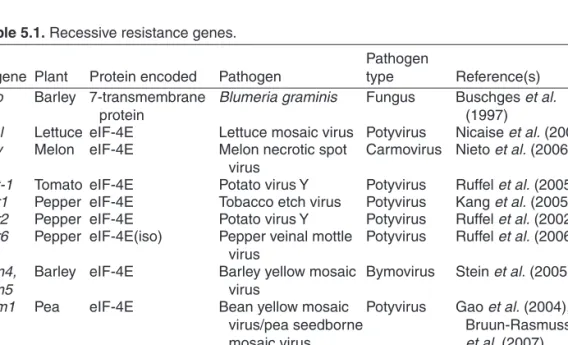
Virus type Viral family Virus VSRs Other functions Reference(s)
Positive-strand
RNA viruses Aureusvirus Pothos latent virus P14 Merai et al. (2005)
Carmovirus Turnip crinkle virus P38 Coat protein Thomas et al. (2003) Closterovirus Beet yellows virus P21 Replication enhancer Reed et al. (2003), Lu et al.
(2004), Chiba et al.
(2006) Citrus tristeza virus P20 Replication enhancer
P23 Nucleic acid binding CP Coat protein Grapevine leaf roll-associated
virus 2
P22 Crinivirus Sweet potato chlorotic mosaic
virus
P22 Kreuze et al. (2005)
Comovirus Cowpea mosaic virus S protein Small coat protein Liu et al. (2004) Cucumovirus Cucumber mosaic virus, Tomato
aspermy virus
2b Host specific movement Brigneti et al. (1998), Zhang et al. (2006), Diaz-Pendon et al. (2007)
Furovirus Soil-borne wheat mosaic virus 19K Te et al. (2005)
Hordeivirus Barley yellow mosaic virus γb Replication enhancer, movement, seed
transmission, pathogenicity determinant
Yelina et al. (2002)
Pecluvirus Peanut clump virus P15 Movement Dunoyer et al. (2002)
Polerovirus Beet western yellows virus, Curcubit aphid-born yellows virus
P0 Pathogenicity determinant Pfeffer et al. (2002), Baumberger et al. (2007), Bortolamiol et al. (2007)
Potexvirus Potato virus X P25 Movement Voinnet et al. (2000)
Potyvirus Potato virus Y, Tobacco etch
virus, Turnip mosaic virus HcPro Movement, polyprotein processing, aphid
transmission, pathogenicity determinant
Anandalakshmi et al.
(1998), Brigneti et al.
(1998), Kasschau and Carrington (1998)
Continued
14S. Wadsworth and P. Dunoyer
Virus type Viral family Virus VSRs Other functions Reference(s)
Sobemovirus Rice yellow mottle virus P1 Movement, pathogenicity
determinant Voinnet et al. (1999) Tobamovirus Tobacco mosaic virus, Tomato
mosaic virus P122,
P130 Replication Kubota et al. (2003), Csorba et al. (2007)
Tobravirus Tobacco rattle virus 16K Liu et al. (2002)
Tombusvirus Tomato bushy stunt virus, Cymbidium ringspot virus, Carnation Italian ringspot virus
P19 Movement, pathogenicity
determinant Voinnet et al. (1999), Silhavy et al. (2002) Tymovirus Turnip yellow mosaic virus P69 Movement, pathogenicity
determinant Chen et al. (2004)
Vitivirus Grapevine virus A P10 Chiba et al. (2006)
Negative-strand RNA viruses
Teniuvirus Rice hoja blanca virus NS3 Unknown Bucher et al. (2003),
Hemmes et al. (2007) Tospovirus Tomato spotted wilt virus NSs Pathogenicity determinant Bucher et al. (2003) Double-stranded
RNA viruses
Phytoreovirus Rice dwarf virus Pns10 Cao et al. (2005)
Single-stranded DNA viruses
Begomovirus African cassava mosaic virus (Kenyan strain)
AC2 Transcriptional activator protein (TrAP)
Voinnet et al. (1999), van Wezel et al. (2002), Chellappan et al. (2005), Trinks et al. (2005), Zrachya et al. (2007), Glick et al. (2008)
African cassava mosaic virus
(Cameroon strain)) AC4
Tomato yellow leaf curl virus C2 V2 Tomato leaf curl virus C2 Mungbean yellow mosaic virus C2
Curtovirus Beet curly top virus L2 Transcription factor Wang et al. (2005) Double-stranded
DNA viruses Caulimovirus Cauliflower mosaic virus P6 Replication factor Love et al. (2007) Table 1.1. – Continued
al., 2002; Bucher et al., 2003). These rapid assays allowed the identification of more than 30 VSRs from many distinct virus types (see Table 1.1). Moreover, a single type of virus might encode several distinct VSRs as was found with Citrus tristeza virus (Lu et al., 2004).
Strikingly, VSRs are highly divergent in terms of sequence and structure, and represent different strategies to suppress silencing, providing a compelling example of evolutionary convergence (Plate 2). The small size of viral genomes and the low number of encoded proteins has also forced viruses to usually cumulate several functions into a single protein. In line with this observation, VSRs often display other functions during the virus life cycle. For instance, TCV- and TMV-encoded silencing suppressors are the coat protein (CP) of the virion (Qu et al., 2003; Thomas et al., 2003) and the p122 subunit of the viral replicase, respectively (Csorba et al., 2007). Other VSRs are also often encoded by novel, overlapping genes contained within more ancient ones.
These new genes are usually created by ‘overprinting’, whereby an existing coding sequence is translated from a different open reading frame. This evolutionary process creates isolated VSR lineages in virus phylogenies. These overlapping VSR genes include the poleroviral P0, geminiviral AC2 and AC4, tombusviral P19 and P14, cucumoviral 2b or tymoviral P69 proteins (Li and Ding, 2006). VSR have also been isolated from fungus, insect and mammalian viruses and their activity have been demonstrated to be retained in cross- kingdom analyses indicating that they are likely to be targeting key steps in the RNA silencing pathways (Delgadillo et al., 2004; Dunoyer et al., 2004; Li et al., 2004; Reavy et al., 2004; Segers et al., 2006; Hemmes et al., 2007;
Schnettler et al., 2008). The corollary of this observation is that RNA silencing also represents an antiviral defence mechanism in other eukaryotic organisms (Li et al., 2002; Wang et al., 2006; Segers et al., 2007). However, in the case of vertebrates’ VSRs, this assumption is yet to be firmly demonstrated as identification of these proteins were obtained in non-vertebrate systems such as plants or insect cells and not characterized during the context of natural infection (Bucher et al., 2004; Delgadillo et al., 2004; Li et al., 2004).
Plate 2
Plate 2 shows different strategies for suppression of RNA silencing. One strategy to suppress silencing is to avoid vsRNA production as exemplified with potyviral HcPro that inhibit dsRNA processing probably through direct binding of the dsRNA trigger, thereby blocking access to DCLs. VSRs can also directly block Dicer activity as shown in the case of the TCV P38 that inhibits DCL4 or PVX P25 that impairs production of DCL3-dependent 24 nt-long siRNA.
Inhibition of DCL4 by P38 revealed the redundant antiviral function of DCL2, which generates 22 nt-long siRNA. The antiviral activity of the 22 nt vsRNA is also compromised by P38 through an unknown mechanism. Inhibition of HEN1-mediated protection of vsRNA, for instance by HcPro or TMV P122, leads to destabilized and subsequent degra dation of vsRNAs. Another commonly used silencing suppression strategy is shown by beet yellows virus (BYV) P21 and CymRSV P19 that both bind small RNA duplexes. Although P21 does not
16 S. Wadsworth and P. Dunoyer show any size specificity, P19 acts as a head-to-tail homodimer that specifically measures 21 bp duplexes that are the product of DCL4. This binding impairs their subsequent incorporation into AGO1-containing RISC. Alternatively, African cassava mosaic virus (ACMV) AC4 sequesters single stranded small RNA molecules after unwinding of the duplex thereby preventing RISC- mediated cleavage of targeted RNA. AGO1 activity can also be inhibited through direct interaction with the VSR as shown with CMV 2b or through stimulation of its degradation rate through an ubiquitin-mediated proteolysis pathway as recently found for the poleroviral P0 protein. The DCL4-dependent 21 nt-long siRNA are the prominent if not exclusive cell-to-cell silencing signal that move ahead of the infection front and incorporate into an antiviral RISC to set up a potent antiviral response, similarly to immunization. Sequestration of the 21 nt siRNA duplexes by P19 precludes cell-to-cell movement of the silencing signal ahead of the infection front. Although the nature of the systemic silencing signal is still unknown, P25 and 2b have been shown to prevent the synthesis, spread or perception of this signal ahead of the infection front.
Finally, how DCL3-dependent 24 nt-long vsRNA are produced during cytoplasmically replicating virus infection is an open question. Either some viral RNA enters the nucleus or DCL3 is delocalized from the nucleus to the cytoplasm during viral infection.
Many ways of invading your enemy’s territory: viral suppression strategies The diverse genomic and evolutionary origins of VSRs, supporting the idea that they appeared independently in each virus family, is the basis of the increasing diversity found in their mode of action.
Suppression of siRNA accumulation
The simplest way for a virus to avoid the RNA silencing response is to inhibit the process from the beginning, by preventing Dicer from accessing the viral dsRNA trigger(s). For instance, transgenic expression of the potyviral HcPro has been shown to inhibit the DCL-dependent processing of dsRNA (Mallory et al., 2002; Dunoyer et al., 2004). Indeed, when coexpressed with an inverted-repeat transgene designed to silence the Arabidopsis endogenous gene chalcone synthase (CHS, CHS-RNAi line), suppression of silencing was correlated with the accumulation of unprocessed CHS dsRNA. It is noteworthy that HcPro mainly inhibits the accumulation of the DCL4-dependent 21 nt-long siRNA and has a much reduced effect on the DCL3-dependent 24 nt-long siRNA. Given that HcPro is a cytoplasmic protein, this observation is in contra- diction with the presumed nuclear localization of DCL4. Hence, the stronger effect of HcPro on 21 nt- rather than 24 nt-long siRNA accumulation, is probably explained by the fact that biogenesis of the former most likely occurs in the same subcellular compartment as the VSR, whereas biogenesis of the latter occurs in the nucleus (Li et al., 2006; Pontes et al., 2006). Interestingly, this inhibition of Dicer processing was only partial, as substantiallevels of 21
nt-long CHS siRNAs were still detected (Dunoyer et al., 2004). However, despite those residual siRNA levels, degradation of the CHS mRNA was prevented, suggesting that in addition to its effect on Dicer activity, HcPro also inhibits the activity of RISC. This proposal is consistent with the effect of HcPro on miRNA-guided cleavage of endogenous transcripts or with its sRNA binding capacity (see below).
In contrast to HcPro, P1 from Rice yellow mottle virus (RYMV) and the PVX P25 suppressor specifically impair 24 nt-long siRNA production during S-PTGS (Hamilton et al., 2002; Himber et al., 2003). Moreover, PVX-infected wt plants only accumulate 21 nt-long vsRNAs suggesting that this DCL3- specific inhibition by P25 also occurs during viral infection (Schwach et al., 2005). In line with this observation, P25 was shown to be localized in the nucleus from early time points of the infection (Samuels et al., 2007). However, the fact that 24 nt-long vsRNAs are detected in the absence of P25 still raises the question of how DCL3 gains access to the cytoplasmic viral dsRNA (Plate 2). Moreover, how HcPro, P1 and P25 interfere with the processing of dsRNA by Dicers is also unsolved. As HcPro and P25 have been shown to display RNA binding property in a non-sequence-specific manner, one possibility is that these proteins directly bind to the viral dsRNA triggers thereby blocking access to DCLs (Urcuqui-Inchima et al., 2000; Kasschau and Carrington, 2001; Leshchiner et al., 2006). Alternatively, these VSRs can directly inhibit Dicer activity, as recently observed with the TCV P38 protein (Plate 2). Indeed, when dcl2 mutant plants were found more susceptible to TCV infection, the initial thought was that DCL2 was the main antiviral Dicer in plants (Xie et al., 2004). Supporting this idea, DCL2-dependent 22 nt-long vsRNAs were the predominant TCV-derived sRNAs to accumulate in wt infected plants. However, the finding that wt plants yield 21 nt instead of 22 nt-long vsRNA when infected with the VSR-deficient TCV (TCV-∆P38) indicated that DCL2 only substitute s DCL4 when its activity is compromised by P38 (Deleris et al., 2006). As the 22 nt-long sRNAs were not competent to mediate viral RNA cleavage in the presence of P38, this suggests that P38 also impairs the activity of the DCL2- dependent siRNAs (Deleris et al., 2006), a property probably related to its sRNA binding capacity (Merai et al., 2006).
One point to keep in mind when analysing sRNA levels in transgenic or infected plants is that we are looking at steady state levels at a given time point, which takes into account rate of production and rate of degradation.
Study of the Peanut clump virus (PCV) P15 protein in the CHS-RNAi system indicated that this VSR triggers a strong reduction in CHS siRNA accumulation without interfering with dsRNA processing, suggesting that this protein likely acts downstream of Dicers (Dunoyer et al., 2004). Reduced siRNA accumulation levels may result from a reduced incorporation into RISC, which may cause their instability. Alternatively, as P15 has been sh