Mycobacterium tuberculosis Transcription Factor EmbR
Regulates the Expression of Key Virulence Factors That Aid in Ex Vivo and In Vivo Survival
Suresh Kumar,aMehak Zahoor Khan,aNeha Khandelwal,bChen Chongtham,aBiplab Singha,aAnkita Dabla,aDebashree Behera,d Archana Singh,c Balasubramanian Gopal,dG. Aneeshkumar Arimbasseri,aSiddhesh S. Kamat,b Vinay Kumar Nandicooria*
aNational Institute of Immunology, New Delhi, India
bDepartment of Biology, Indian Institute of Science Education and Research, Pune, India
cAcademy of Scientific and Innovative Research (AcSIR), Ghaziabad, India
dMolecular Biophysics Unit, Indian Institute of Science, Bangalore, India
ABSTRACT
Mycobacterium tuberculosis encodes
;200 transcription factors that modu- late gene expression under different microenvironments in the host. Even though high-throughput chromatin immunoprecipitation sequencing and transcriptome sequencing (RNA-seq) studies have identi
fied the regulatory network for
;80% of transcription factors, many transcription factors remain uncharacterized. EmbR is one such transcription factor whose in vivo regulon and biological function are yet to be elucidated. Previous in vitro studies suggested that phosphorylation of EmbR by PknH upregulates the embCAB operon. Using a gene replacement mutant of embR, we investigated its role in modulating cellular morphology, antibiotic resist- ance, and survival in the host. Contrary to the prevailing hypothesis, under normal growth conditions, EmbR is neither phosphorylated nor impacted by ethambutol resistance through the regulation of the embCAB operon. The embR deletion mu- tant displayed attenuated M. tuberculosis survival in vivo. RNA-seq analysis sug- gested that EmbR regulates operons involved in the secretion pathway, lipid me- tabolism, virulence, and hypoxia, including well-known hypoxia-inducible genes devS and hspX. Lipidome analysis revealed that EmbR modulates levels of all lyso- phospholipids, several phospholipids, and M. tuberculosis-speci
fic lipids, which is more pronounced under hypoxic conditions. We found that the EmbR mutant is hypersusceptible to hypoxic stress, and RNA sequencing performed under hypoxic conditions indicated that EmbR majorly regulates genes involved in response to acidic pH, hypoxia, and fatty acid metabolism. We observed condition-speci
fic phosphorylation of EmbR, which contributes to EmbR-mediated transcription of several essential genes, ensuring enhanced survival. Collectively, the study estab- lishes EmbR as a key modulator of hypoxic response that facilitates mycobacterial survival in the host.
IMPORTANCE
Mycobacterium tuberculosis modulates its transcriptional machinery in response to dynamic microenvironments encountered within the host. In this study, we identi
fied that EmbR, a transcription factor, plays important roles in modulating cellular morphology, antibiotic resistance, and survival in the host. We found that EmbR undergoes condition-speci
fic phosphorylation for its activation. Together, the study establishes a key role of EmbR as a transcriptional activator of genes belong- ing to multiple pathways, viz., virulence, secretion, or polyketide synthesis, that aid in mycobacterial survival during hypoxia and within the host.
KEYWORDS
transcription, transcription factors, hypoxia, mycobacteria, tuberculosis, granuloma, EmbR, Mycobacterium tuberculosis
EditorChristina L. Stallings, Washington University School of Medicine in St. Louis Copyright© 2022 Kumar et al. This is an open- access article distributed under the terms of theCreative Commons Attribution 4.0 International license.
Address correspondence to Vinay Kumar Nandicoori, [email protected].
*Present address: Vinay Kumar Nandicoori, CSIR-Centre for Cellular and Molecular Biology, Hyderabad, India.
The authors declare no conflict of interest.
Received14 January 2022 Accepted4 April 2022 Published26 April 2022
RESEARCH ARTICLE
D espite its existence since antiquity, tuberculosis still ranks among the foremost kill- ers of the 21st century. Upon infection, Mycobacterium tuberculosis is phagocy- tosed by the alveolar macrophages, often remodeling the site of infection into granu- loma (1). M. tuberculosis can survive in the latent state inside the granuloma in 90% of the cases or form active disease in;10% of the infected individuals. The ability to sur- vive under various host-induced stresses, such as reactive nitrogen intermediates (RNI), reactive oxygen species (ROS), nutrient starvation, and hypoxia, requires more pro- found insight into mechanisms adopted by M. tuberculosis. In response to these stresses, the bacilli induce transcriptional modulation, resulting in the secretion of viru- lence factors, metabolic changes, induction of stress response genes, etc. This helps in adaptation and persistence inside the host, resulting in the infection’s establishment (2–7). Thus, it is imperative to investigate the effectors of these signaling pathways that aid in bacillary survival inside the host. These effectors’expression is often regu- lated by a plethora of transcription factors (TFs) (8–10).
M. tuberculosis genome encodes
;214 TFs, which regulate gene expression under various conditions and stages of its life cycle. Studies have found
;16,000 binding events from 154 TFs from the M. tuberculosis genome, underlining the importance of transcriptional regulation in M. tuberculosis. A total of 5,400 of 15,980 TF binding sites (approximately one-third) are present within the 220-bp promoter window, which has resulted in 47,200 TF
–promoter interactions (11, 12). TFs undergo a range of posttrans- lational modi
fications (PTMs) that orchestrate their transcriptional activity (13).
Phosphorylation is a more common and prominent PTM than sumoylation, methyla- tion, acetylation, glycosylation, etc. M. tuberculosis encodes 11 serine threonine protein kinases (STPKs) that phosphorylate various TFs (14, 15).
embR (Rv1267c) is one such transcription factor, belonging to the family of Streptomyces antibiotic regulatory proteins (SARP) that is located in the same operon, upstream of the pknH gene (Rv1266c) (16). The crystal structure of EmbR showed three distinct domains: N-terminal OmpR/PhoB-like DNA binding domain (DBD), consisting of winged-like structure for the binding; middle central all-helical bacterial transcrip- tional activation domain (BTAD), hypothesized to aid in the DNA binding; and the C- terminal fork head-associated (FHA) domain (Fig. 1a) (17). FHA domain is essential for its interaction and phosphorylation by PknH and results in enhanced transcription of embCAB genes (18
–20). In addition to PknH, EmbR is phosphorylated in vitro by PknA, PknB, PknE, and PknF (21, 22). The augmented transcription of embC increases the lip- oarabinomannan/lipomannan (LAM/LM) ratio, modulating the arabinogalactan (AG) layer (Fig. 1b) (19). Furthermore, mutations in embCAB and embR have been linked to ethambutol (EMB) resistance in various clinical isolates, highlighting the importance of EmbR-mediated regulation in M. tuberculosis virulence and pathogenesis (23, 24).
In this study, we aimed to validate previously known indirect in vitro evidence with the help of a gene replacement mutant and asked the following questions. (i) What is the biological function of EmbR? (ii) What are the genes that EmbR regulates? (iii) Does EmbR promote the survival of M. tuberculosis inside the host? Our results show that EmbR deletion mutant displayed attenuated M. tuberculosis survival inside the murine peritoneal macrophages and in vivo. Scanning electron microscopy (SEM), transmission electron microscopy (TEM), and expression analysis suggested that EmbR modulates cellular length and cell wall ultrastructure but not through embCAB. Among all the in vitro stress experiments, mutant demonstrated reduced survival, speci
fically in modi-
fied Wayne
’s hypoxia model. RNA sequencing data showed downregulation of operons involved in the secretion pathways, lipid metabolism, virulence, and hypoxia establish- ment in the mutant. Lipidome analysis suggested that EmbR plays a crucial role in cell wall lipid composition during normoxia and hypoxia. Furthermore, RNA sequencing of hypoxic cultures indicated condition-speci
fic regulation of several essential genes, including those involved in fatty acid metabolism, response to acidic pH, and hypoxia.
Interestingly, while we could not detect phosphorylation of EmbR under normal
growth conditions, it is robustly phosphorylated under acidic and hypoxic conditions.
FIG 1 EmbR is phosphorylated in vitro but not in vivo under regular growth conditions. (a) Schematic depicting domain architecture of EmbR in M. tuberculosis. (b) Model demonstrating previously known roles of EmbR.
Phosphorylation of EmbR by PknH results in enhanced transcription of embCAB operon, modulating AG layer biosynthesis. OM, outer membrane; AG, arabinogalactan layer; PG, peptidoglycan layer; IM, inner membrane. (c) Schematic representation of the pDuetEmbR-kinase vectors used for the dual expression ofembRandM. tuberculosis STPKs. Vectors with different STPKs in MCS II with MBP (Maltose Binding Protein) tag andembRwith N-terminal hexa- His tag were transformed in the surrogate hostE. coliBL21 cells, and the expression of EmbR- and MBP-tagged kinases was induced with 0.1 mM isopropyl-b-D-thiogalactopyranoside (IPTG). (d) Western blot analysis of lysates confirming coexpression of EmbR and STPKs with anti-His (upper) and anti-MBP (lower), respectively. (e) His-EmbR was immunoprecipitated (IP) using Ni21 affinity agarose beads from various pDuet-EmbR-Kinase strains. The pulldowns were probed witha-p-Thr (upper) to assess EmbR phosphorylation and witha-EmbR anda-His antibodies as controls.
(f) Table summarizingfive phosphopeptides obtained from PknB- and PknH-targetedE. coliEmbR using MS/MS. T-164 and T-198/T-209 are unique to PknH and PknB, respectively. T-22 and T-275 are common target sites from both PknB and PknH. PSM, peptide-spectrum match; PEP, posterior error probability; PRS, pattern recognition for spectra; aa, amino acid. (g)M. tuberculosis RvDembR::F-embRstrain was electroporated with pNit-PknB, pNit-PknD, pNit-PknE, and (Continued on next page)
EmbR Modulates Virulence Factor Expression mBio
Together, this study identi
fies novel downstream targets of EmbR and establishes EmbR as a crucial regulatory protein in determining M. tuberculosis virulence and pathogenesis.
RESULTS
EmbR is phosphorylatedin vitrobut notin vivounder regular growth conditions.
Based on the in vitro kinase assays, EmbR has been demonstrated to be a substrate for serine/threonine protein kinases PknA, PknB, PknE, PknF, and PknH (20
–22). However, the target phosphorylation sites for these kinases have not been identi
fied. We sought to utilize Escherichia coli as the surrogate host and a previously developed pDUET vec- tor system to identify the target sites. Either MBP (Maltose Binding Protein) or MBP- tagged full-length or kinase domains of all 11 M. tuberculosis STPKs were cloned into the second MCS (multiple cloning site). EmbR was cloned into the unique HindIII site in the
first MCS, and the constructs were transformed into E. coli BL21 Codon Plus strain (Fig. 1c). We observed robust expression of EmbR and MBP or MBP-tagged kinases for 8 out of 11 kinases (Fig. 1d). To evaluate the phosphorylation status of EmbR, His- tagged EmbR was pulled down with the help of Ni
21af
finity beads and probed with
a-p-Thr and
a-EmbR antibodies. In
a-EmbR blots, we observed higher-molecular-mass band(s) when EmbR was coexpressed with PknB, PknD, and PknH. Consonantly,
a-p- Thr blots indicated that this higher-molecular-mass band(s) corresponds to pEmbR (Fig. 1e), suggesting that PknB, PknD, and PknH robustly phosphorylate EmbR. The phosphorylation sites on EmbR for kinases PknB and PknH were identi
fied by excising EmbR band, followed by trypsinization and subjecting the tryptic peptides to liquid chromatography coupled to mass spectrometric (LC-MS) analysis. We identi
fied
five target sites on EmbR for PknB and PknH, of which T164 is a unique target site for PknH, while PknB exclusively phosphorylated T198 and T209. T22 and T275 were phosphoryl- ated by both PknB and PknH (Fig. 1f; see also Fig. S1 and S2 in the supplemental mate- rial). Subsequently, we sought to delineate the role of PknB- and PknH-mediated phos- phorylation on the function of EmbR. Toward this end, we
first sought to establish if EmbR is indeed phosphorylated in vivo. M. tuberculosis Rv
DembR mutant was coelec- troporated with pF-F-embR and pNit-PknB/PknD/PknH constructs. Expression of ki- nases was induced, and FLAG-tagged EmbR was immunoprecipitated, resolved, and probed with
a-FLAG and
a-p-Thr antibodies. To our surprise, we did not detect any speci
fic band corresponding to pEmbR (Fig. 1g). FLAG-EmbR was excised from the gel and trypsinized, and the peptides were subjected to LC-MS analysis to con
firm the results further. LC-MS experiment did not detect any phosphorylated peptides, sug- gesting that EmbR is not phosphorylated in M. tuberculosis under regular growth conditions.
Deletion of EmbR does not impactM. tuberculosisgrowthin vitro.
EmbR comes from an in vitro nonessential gene in Himar1 transposon mutagenesis (25, 26). To investigate the role of EmbR to modulate embCAB operon expression and its impact on the pathogen
’s survival in vivo, we sought to generate an embR gene replacement mutant. Linearized allelic exchange substrate (AES) was electroporated into recombin- eering-pro
ficient M. tuberculosis H37Rv strain (Rv; note that genotype is used as the strain name throughout the manuscript). Recombinants obtained were con
firmed for the replacement of embR with hyg
rselectable marker at its native locus by PCRs with three sets of primers using the genomic DNA isolated from wild-type Rv and Rv
DembR mutant (Fig. 2a and b). pF-embR-HA construct was electroporated into Rv
DembR mu- tant to generate Rv
DembR::embR complementation strain. Western blot analysis of lysates prepared from Rv and Rv
DembR strains further con
firmed the deletion of embR.
Notably, the expression of EmbR in the Rv
DembR::embR mutant was similar to that of
FIG 1Legend (Continued)
pNit-PknF. The resulting strains were induced for the kinase overexpression with 5mM isovaleronitrile (IVN), and the WCLs were probed witha-FLAG antibody. The lysates were immunoprecipitated (IP) with FLAG-M2 beads; 1/10 of the IP was probed witha-FLAG, and the remaining 9/10 of the IP was probed witha-p-Thr antibodies.
endogenous expression (Fig. 2c). Next, we evaluated the impact of embR deletion on the in vitro survival of M. tuberculosis. The cultures from Rv, Rv
DembR, and Rv
DembR::
embR strains were grown in nutrient-rich 7H9 medium and limited Sauton
’s medium.
No signi
ficant differences were observed in Rv
DembR strain compared with Rv and Rv
DembR::embR strains (Fig. 2d and e), which led us to conclude that the deletion of EmbR does not affect the in vitro growth of mycobacteria.
EmbR-mediated changes in mycobacterial cell wall architecture are not through embCAB.
Electrophoretic mobility shift assays with the promoter regions of embC, embA, and embB and puri
fied EmbR and pEmbR suggested that (i) it binds to the pro- moter region and (ii) the binding is improved upon its phosphorylation (21). Since EmbA and EmbB proteins are involved in the biosynthesis of arabinogalactan (AG), a signi
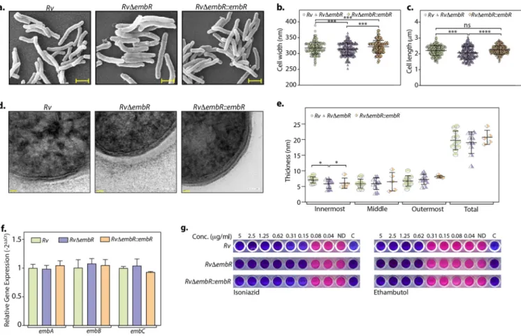
ficant cell wall component, we evaluated the impact of EmbR deletion on the cel- lular architecture. Toward this end, we performed scanning electron microscopy (SEM) and observed signi
ficant changes in cell width and length upon deletion of EmbR
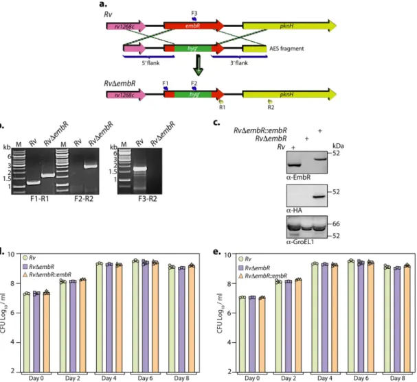
FIG 2 Generation and characterization ofRvDembRmutant. (a) Pictorial representation of the genetic organization ofembR loci inRvandRvDembRmutant. Thefigure depicts the replacement ofembRwithhygr. Primer sets used for validating the generation of mutant strain are indicated. (b) The deletion ofembRat its native locus was confirmed with the help of PCRs using different sets of primers. Thefirst panel shows PCR amplicons with a gene-specific primer set (F1-R1) inRv(1,147 bp) andRvDembRmutant (1,728 bp). The second panel shows amplicons (F2-R2) expected only inRvDembRmutant (2,549 bp), and the third panel shows amplicons (F3-R2) expected only inRv(1703 bp). M represents 1-kb gene ruler ladder. (c) A total of 30mg of WCLs prepared from Rv, RvDembR, and RvDembR::embR strains was resolved, transferred to the nitrocellulose membrane, and probed witha-EmbR (upper),a-HA (middle), anda-GroEL1 (lower) antibodies. (d and e)Rv, RvDembR, and RvDembR::embRstrains were inoculated at an OD600of;0.1 in 7H9-ADC (d) and Sauton’s (e) medium. The bacillary survival was monitored by CFU enumeration at indicated time points. Data are presented as mean CFU log10/mL 6 standard deviations (SD) and are representative of two biologically independent experiments, each performed in triplicates (n= 3).
EmbR Modulates Virulence Factor Expression mBio
(Fig. 3a to c). We then evaluated the cell wall ultrastructure of Rv, Rv
DembR, and Rv
DembR::embR strains using transmission electron microscopy (TEM). The results dem- onstrated that while the mycolic acid layer (outermost) and the arabinogalactan (mid- dle) were comparable in all three strains, peptidoglycan (innermost) was marginally thinner upon deletion of EmbR (Fig. 3d and e). Next, we investigated whether cell length and width changes are due to putative regulation of embCAB. To validate this, we examined the role of EmbR in regulating the transcription of embCAB genes by per- forming quantitative real-time PCR (qRT-PCR) using cDNA obtained from Rv and Rv
DembR strains. We did not observe any discernible difference in the transcript levels of embCAB genes, suggesting that EmbR does not regulate this operon
’s expression under regular growth conditions (Fig. 3f). Previous studies suggested a link between mutations in embCAB and embR genes to EMB resistance. Thus, we assessed the impact of embR deletion on EMB resistance by determining MIC values for isoniazid (control) and EMB in Rv, Rv
DembR, and Rv
DembR::embR strains. The MIC values obtained for both INH and EMB were comparable in all three strains, emphasizing that EmbR does not play a role in antibiotic resistance to these antibiotics (Fig. 3g). Together, these results suggest that even though EmbR modulates cell morphology, it does not regu- late embCAB genes and affect EMB susceptibility.
Deletion of EmbR attenuates intracellular survival ofM. tuberculosis.
To evalu- ate the role of embR in the ex vivo and in vivo survival of M. tuberculosis, we performed studies in peritoneal macrophages and a murine infection model. Peritoneal macro- phages were infected with Rv, Rv
DembR, and Rv
DembR::embR strains, and CFU were enumerated at various time points postinfection. Compared with Rv and Rv
DembR::
FIG 3 EmbR does not influence mycobacterial cell wall architecture. (a to c) Cultures ofRv,RvDembR, andRvDembR::embRstrains were grown until an OD600of;0.6 in 7H9-ADC medium andfixed. (a) Cell morphology was observed through SEM at 15,000. Scale bar, 1.0mm. Cell width (b) and length (c) were quantified by the Smart Tiff program and plotted. (d) Fresh cultures ofRv,RvDembR, andRvDembR::embRstrains were grown until an OD600of;0.6, fixed, and processed for TEM. Cell wall architecture was observed at 200 kV, 50,000. (e) Quantification of innermost, middle, outermost, and total layer thickness. (f) Gene expression ofembCABoperon was quantified inRvandRvDembRstrains using qRT-PCR. (g) MICs of isoniazid and ethambutol were determined inRv,RvDembR, andRvDembR::embRstrains as described in Materials and Methods.
FIG 4 Deletion of EmbR in M. tuberculosis attenuates its ability to survive in the host. (a) Murine peritoneal macrophages isolated from BALB/c mice were infected withRv,RvDembR, and RvDembR::embRstrains at an MOI of 1:10. At indicated time points, the cells were lysed with 0.05% SDS and bacillary survival was monitored. Data are represented as mean CFU log10/mL6 SD and are representative of two independent biological experiments, each performed in triplicates (n= 3). (b) Schematic representation of mouse infection experiment. (c) BALB/c mice (n= 6) were infected with 100 CFU/mouseRvandRvDembRstrains, and the bacterial deposition was determined in the lung homogenates (n= 2) at day 1 postinfection (p.i.). Mean log10CFU obtained forRvandRvDembRstrains were 6.07 and 5.66 at 4th week and 6.16 and 4.76 at 8th week. (d) Mean log10CFU obtained forRvandRvDembRstrains was 3.99 and 3.07 at 4th week and 3.74 and 3.32 at 8th week in infected animals’spleens. Data are represented as mean CFU log10/mL6SD. *,P, 0.05;**,P, 0.005;***,P,0.0005. (e) Lungs and spleen of infected mice depicting gross pathology. (f) Total granuloma score (mean6SD) in hematoxylin-eosin-stained lung sections of animals infected with M. tuberculosisstrains at 8 weeks p.i. Each data point in panels c, d, and f represents data value from one infected animal.
EmbR Modulates Virulence Factor Expression mBio
embR strains, we observed 5-fold reduction in the survival in the Rv
DembR mutant at 96 and 120 h postinfection (Fig. 4a), which suggested that EmbR promotes mycobacte- rial survival within the host. To investigate the role of EmbR in the survival of the patho- gen in vivo, we performed murine infections in mice with Rv and Rv
DembR strains through aerosol routes. The CFU present in the lung and spleen were enumerated 1 day and 4 and 8 weeks postinfection (Fig. 4b). The bacillary load at day 1 suggested ef
ficient and equivalent deposition of all three strains (Fig. 4c). Disease progression was assessed by examining lungs and spleen bacillary load 4 and 8 weeks postinfection. While the bac- illary load at day 1 was comparable for Rv and Rv
DembR strains, the mutant showed com- promised survival at 4 and 8 weeks (Fig. 4c). Furthermore, we observed a signi
ficant reduction in the bacillary load at 8 weeks in the mutant compared with the wild type, indicating that EmbR plays a critical role in the chronic phase of infection. The splenic bacillary load in Rv
DembR mutant was also observed to be lower than that of the wild type, suggesting reduced dissemination of bacilli in the animals infected with mutant strain (Fig. 4d). Consistent with the above-described observations, we also found a reduced number of granulomas by gross pathology in the lungs of mice infected with Rv
DembR mutant (Fig. 4e). However, these differences were not re
flected in the granu- loma score obtained from the histopathological analysis of lung sections, probably due to high variation and possibly fewer replicates (Fig. 4f). Collectively the data strongly support that EmbR may play a role in modulating the survival at later stages of infection, suggest- ing a correlation with bacterial virulence.
EmbR regulates the expression of genes involved in virulence, secretion, polyketide synthesis, and hypoxia.
Thus far, results have negated the prevailing hy- pothesis on (i) how EmbR transcriptional activity is regulated and (ii) how it modulates the cellular processes. Since EmbR is a transcription factor, we performed a global tran- scriptomic analysis of Rv and Rv
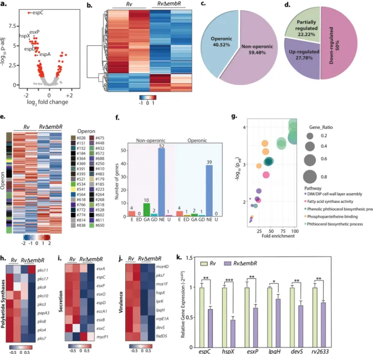
DembR strains using Illumina-based RNA sequencing from two independent biological replicates. Differential gene expression (DEG) analysis was performed using DESeq2 and is depicted in the volcano plot, where the red dots represent genes that are differentially expressed (1.5-fold change; adjusted P value [Padj] of
,0.1) (Fig. 5a and Table S1a). Heat map generated for both biological repli- cates for each sample represents a complete set of up- and downregulated genes in Rv and Rv
DembR mutant (Fig. 5b). Deletion of EmbR resulted in a total of 116 DEGs, of which 90 genes were downregulated, indicating EmbR was a plausible transcriptional activator (Table S1a). In consonance with qRT-PCR data presented in Fig. 4c, we did not detect embCAB operon genes or those involved in arabinogalactan or lipoarabino- mannan biosynthesis among the DEGs. Next, we predicted the operonic arrangement of these 116 DEGs using the Rockhopper tool (27) and found that 47 (40.52%) DEGs are part of 36 predicted operons (Fig. 5c and Table S1b).
Although not all the genes of these operons met the thresholds set for DEGs, we examined if all the genes in a given operon show a similar trend in the expression level changes. Interestingly, 50% of these operons (18) show decreased transcript levels for all the genes in the operon, while all the genes in 10 operons (27.78%) show increased transcript levels in the mutant (Fig. 5d and e). Next, we investigated how many of these genes are essential for the bacillary survival in vitro based on published data (25) and found that among the 116 genes that are differentially expressed, only eight are anno- tated as essential for in vitro growth (Fig. 5f). Interestingly, the six most signi
ficantly enriched biological processes that were found among the upregulated genes are related to lipid metabolism (Fig. 5g and Table S1c), explaining altered cellular length and cell wall architecture of the mutant strain. Since we observed that the mutant strain shows attenuated survival in vivo, we assessed the expression levels of virulence- associated genes. Indeed, the volcano plot (Fig. 5a) contains several virulence-associ- ated genes that are downregulated in the mutant strain.
Further analysis of the data con
firmed that several genes associated with processes
required for virulence, such as lipid metabolism and secretion, are downregulated in
the mutant strain (Fig. 5h to j). Interestingly, among the virulence factor genes, we
found vital hypoxia regulators devS and hspX, suggesting a role of EmbR in adaptation
to hypoxic conditions. Subsequently, we selected six highly downregulated Rv
DembR mutants associated with hypoxia and virulence to validate the transcriptome sequenc- ing (RNA-seq) data. qRT-PCR was performed to determine the expression pro
file of espC, hspX, esxP, lpqH, devS, and Rv2633 genes in Rv and Rv
DembR mutant (Fig. 5k). The qRT-PCR data were in accordance with the RNA sequencing data. Together, the RNA
FIG 5 EmbR modulates the expression of various polyketide synthases, virulence factors, and secretory proteins. (a) Volcano plot illustrating differentially expressed genes (DEGs) inRvcompared withRvDembRstrain. Red spots indicate the genes that show differential expression (Padj,0.1). (b) Heatmaps showing the normalized read counts of DEGs in both biological replicates of Rv and RvDembR strains. Color intensity indicates relative up- or downregulation. (c) Pie chart depicting the percentage of DEGs that are operonic and nonoperonic. (d) Pie chart depicting the percentage of operonic DEGs that are fully upregulated, fully downregulated, or partially regulated. (e) Heatmaps showing the normalized read counts of differentially regulated operons along with their numbers in both biological replicates ofRvandRvDembRstrains. Color intensity indicates relative up- or downregulation. (f) Bar graph demonstrating the number of operonic and nonoperonic DEGs that belong to essential (E), essential domain (ED), growth defect (GD), growth advantage (GA), nonessential (NE), and uncertain (U) categories. (g) The six most significantly enriched Gene Ontology (GO) biological process categories of upregulated DEGs. (h to j) Heat maps showing the normalized read counts in both biological replicates ofRv andRvDembRstrains of DEGs that are involved in polyketide synthesis (h), genes involved in virulence (i), and genes involved in the secretion pathway (j). Color intensity shows relative up- or downregulation. (k) Selected DEGs obtained from RNA-seq analysis were validated through qRT-PCR. Data were normalized with respect to 16S rRNA and plotted as mean6SD, representative of two independent biological experiments, each performed in triplicates (n= 3).***,P,0.0005;**,P,0.005;
*,P,0.05; not significant, ns.
EmbR Modulates Virulence Factor Expression mBio
sequencing data suggest that EmbR is a transcriptional modulator regulating multiple pathways important for virulence.
EmbR aids inM. tuberculosissurvival during hypoxia.
Mycobacterial adaptation to hypoxia is thought to have an essential role in disease pathogenesis. During hypoxia, two-component sensor and response regulator dosS (devS) and dosR (devR) are upregu- lated in a coordinated manner (28, 29). hspX (Rv2031), a dominant antigen required for M.
tuberculosis growth and disease pathogenesis in vivo, is also known to be induced under hypoxic stress (30). Since both devS and hspX were signi
ficantly downregulated in Rv
DembR mutant, we speculated that EmbR plays a role in combating hypoxic stress found in the granuloma. To con
firm EmbR-mediated regulation of the hspX promoter, we performed a luciferase reporter assay, wherein the hspX promoter was cloned upstream of the luciferase gene. The reporter construct was electroporated into Rv, Rv
DembR, and Rv
DembR::embR strains and luciferase activity was determined in the lysates. Luciferase activity in the lysates of Rv
DembR mutant was
;1.5-fold lower than activity in the lysates
FIG 6 EmbR positively regulates hypoxia-related genehspX.(a)Rv, RvDembR, andRvDembR::embR strains were electroporated with pSW- hspXpr-luciferase and pSW1-devSpr-luc construct, and luciferase activity was measured in the WCLs of the transformants. Experiment was performed in triplicates (n= 3). (b and c)Rv,RvDembR, andRvDembR::embRcultures were inoculated at an OD600of;0.1 and subjected to hypoxic stress in tightly sealed tubes for 20 and 40 days. The data were plotted as means6SD. The experiment was performed in triplicates (n= 3).***,P,0.0005;**,P,0.005;*,P,0.05. (b) The expression ofhspX,embA,embB, andembCgenes was assessed through qRT-PCR at the 40th day. The data were normalized with respect to 16S rRNA. (c) CFU were enumerated to quantify bacterial survival at indicated time points. (d to g) Rv, RvDembR, and RvDembR::embR strains were assessed for their susceptibility to in vitro stresses. Strains were subjected to nitrosative stress with 3 mM for 2 days (d), oxidative stress with 50mM CHP for 24 h (e), reductive stress with 1 mM DTT for 24 h (f), and surfactant stress with 0.1% SDS for 3 h (g). CFU were enumerated on 7H11 plates after subjecting cells to indicated stresses. Data are presented as mean CFU log10/mL6SD. Control sample testing in each case was performed in duplicates. Stress samples with nitrosative, oxidative, reductive, or surfactant were performed in triplicates.
of Rv (Fig. 6a). Similarly, devS promoter-driven luciferase reading was nearly
;5-fold lower in the Rv
DembR mutant than Rv and Rv
DembR::embR strains (Fig. 6a). Next, we inspected the DNA binding ability of EmbR to biotinylate hspX promoter with the help of surface plasmon resonance (SPR). However, we did not observe any signi
ficant binding of EmbR to hspX promoter under the tested conditions (data not shown), which may be due to the absence of phosphorylation in our in vitro study, the requirement of additional inter- acting partners, such as a sigma factor, or speci
fic in vivo conditions. Additionally, it is also possible that EmbR indirectly regulates hspX expression, for example, through another ac- tivator. Importantly, the expression pro
file of hspX in the Rv, Rv
DembR, and Rv
DembR::
embR strains subjected to hypoxic stress suggested a 50% decrease in the expression of hspX in Rv
DembR strain compared with Rv (Fig. 6b). Notably, there were no expression changes found in the levels of embCAB upon EmbR deletion, even during hypoxic condi- tions. The luciferase activity and hspX expression were higher in the complementation strain than in both Rv and Rv
DembR mutant, which could be due to differences in the expression levels of EmbR (Fig. 6a and b). Subsequently, we examined the impact of embR deletion on the survival of pathogens under hypoxic conditions. We observed a 5- fold reduction in the pathogen
’s hypoxic survival upon deletion of embR, suggesting that EmbR-mediated transcriptional regulation of hypoxic genes is necessary for combating the stress (Fig. 6c). Together, data suggest that EmbR modulates the transcription of mul- tiple virulence-associated genes, including hypoxia-responsive hspX, that aid in mycobac- terial growth and survival inside the host.
M. tuberculosis encounters a series of stresses within the host in vivo, including reac- tive oxygen and nitrogen intermediates, low pH, surfactant, and oxygen deprivation (20, 31, 32). Next, we set out to determine whether, in addition to hypoxia, EmbR con- fers a survival advantage to M. tuberculosis even during other host-engendered stresses. Rv, Rv
DembR, and Rv
DembR::embR strains were treated with 3 mM NaNO
2(nitrosative), cumene hydroperoxide-CHP (oxidative stress), 1 mM dithiothreitol (DTT) (reductive stress), and 0.1% SDS (surfactant), and the CFU were enumerated at de
fined time points. Except for hypoxia, none of the stresses had any impact on the viability of Rv
DembR mutant (Fig. 6d to g), indicating that EmbR plays a speci
fic role in modulat- ing the survival under hypoxic conditions.
EmbR regulates the expression of multiple essential genes during hypoxia.
Data presented above suggest that EmbR is speci
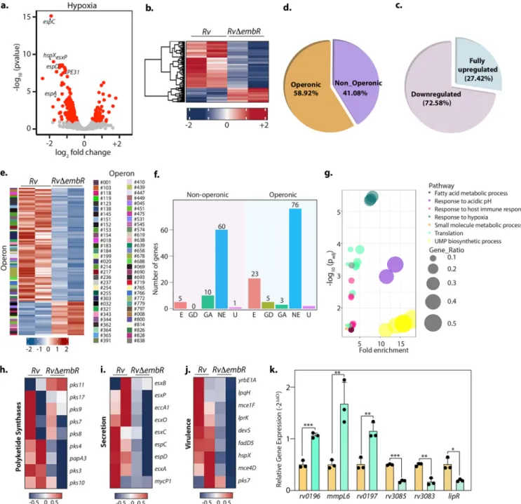
fically necessary for mycobacterial survival during hypoxic stress. Hence, we aimed to evaluate the effect of EmbR deletion on M. tu- berculosis transcriptome during hypoxia. Interestingly, EmbR regulates 185 genes during hypoxia compared to 116 genes during regular growth conditions (normoxia) (Fig. 7a and b and Table S2a). As observed during normoxic conditions, the expression of embCAB operon genes remains unchanged. Next, we assessed the operonic arrange- ment of the DEGs and found that 58.92% of DEGs (Padj
,0.1) are part of 62 predicted operons (Fig. 7c to e). The majority of the operons that have DEGs were found to be fully downregulated (72.58%) in Rv
DembR mutant compared to Rv, suggesting EmbR is a plausible transcriptional activator even during hypoxia. Importantly, unlike normoxia, where EmbR regulates expression of only 8 in vitro essential genes, deletion of EmbR impacts expression levels of 28 in vitro essential genes during hypoxia (Fig. 7f). This may explain why deletion of EmbR attenuates mycobacterial survival, speci
fically during hy- poxia. Furthermore, gene ontology analysis suggested genes related to response to acidic pH, host immunity, hypoxia, and fatty acid metabolism are highly enriched among the downregulated genes (Fig. 7g and Table S2b).
Functional categorization revealed that DEGs during hypoxia included genes involved in polyketide synthesis (Fig. 7h), secretion (Fig. 7i), and virulence (Fig. 7j). To validate the RNA-seq data, we selected three highly upregulated genes and three downregulated Rv
DembR mutants. qRT-PCR was performed to determine the expres- sion pro
file of these genes were in accordance with the RNA sequencing data (Fig. 7k).
EmbR modulates cell wall lipid composition.
In addition to modulation of expression of genes involved in the adaptation to hypoxia, the RNA sequence analysis suggested that EmbR plays a crucial role in the transcription of polyketide synthases and fatty acid
EmbR Modulates Virulence Factor Expression mBio
synthases during both normoxia and hypoxia (Fig. 5g and 7g). Polyketide synthases are mega-complex proteins that act in a concerted manner with fatty acid synthases to bio- synthesize the vast repertoire of functionally and architecturally diverse lipids and glyco- lipid conjugates present on the M. tuberculosis cell envelope. Notably, several studies
FIG 7 EmbR modulates the expression of various essential genes during hypoxia. (a) Volcano plot illustrating differentially expressed genes (DEGs) inRv compared withRvDembRstrain during hypoxia. Red spots indicate the genes that show differential expression (Padj,0.1). (b) Heat maps showing the normalized read counts of DEGs in both biological replicates ofRvandRvDembRstrains. Color intensity indicates relative up- or downregulation. (c) Pie chart depicting the percentage of DEGs that are operonic and nonoperonic. (d) Pie chart depicting the percentage of operonic DEGs that are fully upregulated and downregulated. Heat maps show the normalized read counts of differentially regulated operons and their number in both biological replicates ofRvand RvDembRstrains. Color intensity indicates relative up- or downregulation. (f) Bar graph demonstrating a number of operonic and nonoperonic DEGs that belong to essential (E), growth defect (GD), growth advantage (GA), nonessential (NE), and uncertain (U) categories. (g) The six most significantly enriched Gene Ontology (GO) biological process categories of upregulated DEGs. (h to j) Heat maps showing the normalized read counts in both biological replicates ofRvandRvDembRDEGs that are involved in polyketide synthesis (h), genes involved in virulence (i), and genes involved in the secretion pathway (j). Color intensity shows relative up- or downregulation. (k) Selected DEGs obtained from RNA-seq analysis were validated through qRT-PCR. Data were normalized with respect to 16S rRNA and plotted as means6SD. Experiment was performed in triplicates (n= 3).***,P, 0.0005;**,P,0.005;*,P,0.05; not significant, ns.
have demonstrated the importance of M. tuberculosis lipids as effectors of virulence. The polyketide synthases regulated by EmbR are present in two operons vis-à-vis pks3-pks4- papA3 and pks10-pks7-pks8-pks17-pks9-pks11.
Next, we aimed to determine whether changes in gene expression of polyketide synthases by EmbR translated into lipid composition changes. To comparatively ana- lyze the lipid pro
files, using established protocols (33), we extracted the total cellular lipids from Rv, Rv
DembR, and Rv
DembR::embR strains grown under normoxia and sub- jected them to an established LC-MS analysis (34, 35) for identi
fication and semiquanti- tative estimation of the different lipid classes. The abundances of different lipid classes for the Rv
DembR and Rv
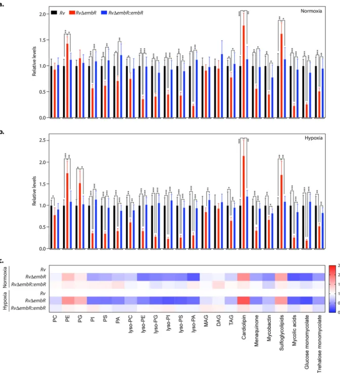
DembR::embR strain groups were normalized to the Rv group to yield relative abundances for individual groups. As represented in Fig. 8a, the dele- tion of embR resulted in signi
ficant alterations in the overall lipid content of M. tubercu- losis (Rv
DembR strain). These changes in the lipid pro
files were restored to almost wild- type levels upon complementation with embR (Rv
DembR::embR strain). Speci
fically, we found that the cellular levels of several phospholipids (PI, PS, and PA), all lysophospho- lipids, and M. tuberculosis-speci
fic lipids (menaquinone, mycobactin, mycolic acid, glu- cose monomycolate, and trehalose monomycolate) were most notably reduced upon embR deletion (Fig. 8a). Concomitant to these alterations, we also found that deletion of embR resulted in an accumulation of some cellular phospholipids (PE, PG, and cardi- olipins) and sulfoglycolipids in the Rv
DembR strain (Fig. 8a). Next, we tested whether EmbR also modulates the lipid pro
file of M. tuberculosis during hypoxia. Toward this end, Rv, Rv
DembR, and Rv
DembR::embR strains were subjected to hypoxia, and lipi- dome was measured as described earlier. We found from this lipidomics analysis that while the trend in the lipid alterations remained the same between the normoxic and hypoxic samples, the EmbR-mediated changes for the different lipid classes were more pronounced during hypoxia (Fig. 8a to c and Table S3). Altogether, the lipidomics data suggest that EmbR plays a crucial role in the biosynthesis of various lipids during nor- moxia and hypoxia.
EmbR is phosphorylatedin vivoin a condition-specific manner.
Based on the results presented above, we conclude that (i) EmbR modulates M. tuberculosis cell wall composition through altered expression of polyketide synthases and fatty acid syn- thases; (ii) EmbR modulates key hypoxia-related genes and confers a survival advant- age to M. tuberculosis during hypoxia; and (iii) RNA-seq and lipidomics analysis sug- gested that the extent of EmbR-mediated transcriptional and lipid changes are more pronounced during hypoxic conditions. Since in vitro phosphorylation of EmbR is known to upregulate DNA binding ability (19), we speculated that in vivo EmbR phos- phorylation is context speci
fic. Toward this end, Rv was grown in 7H9 and either left untreated or subjected to various stresses, such as nutrient-limiting (Sauton
’s), reduc- tive (DTT), oxidative (CHP), acidic (pH 4.5), and hypoxic. Endogenous EmbR protein was immunoprecipitated and probed with
a-EmbR and
a-p-Thr antibodies (Fig. 8a). In line with the previous results (Fig. 1g), we did not detect any phosphorylation under nor- mal growth conditions. Moreover, phosphorylation could not be detected under nutri- ent-limiting, reductive, and oxidative conditions. Interestingly, we observed robust phosphorylation of EmbR under acidic and hypoxic conditions (Fig. 9a). Next, we iden- ti
fied the target phosphorylation sites on EmbR under hypoxic conditions using previ- ously described LC-MS analysis. We identi
fied eight phosphorylation sites on EmbR (Fig. 9b and Fig. S3), namely, T22, T57, T109, T164, T189, T209, T275, and T384. All the sites that were identi
fied in vitro (Fig. 1f) were detected in vivo (Fig. 9b). Importantly, we identi
fied three additional target phosphorylation sites, T57, T109, and T384, specif- ically under in vivo hypoxic conditions.
To further investigate the functional relevance of EmbR phosphorylation, we com- pared the RNA-seq data from hypoxia and normoxia. When subjected to hypoxia, both strains exhibited differential expression of several hundred genes, 2,795 for wild type and 2,195 for Rv
DembR mutant (Fig. 9c and d and Table S4a and b). To better under- stand the precise role of EmbR in gene expression associated with hypoxia, we utilized
EmbR Modulates Virulence Factor Expression mBio
two approaches. First, we compared the genes that are regulated by EmbR in normoxia and hypoxia. Among the downregulated genes (1.5-fold change and Padj
,0.1), we found 18 mRNAs had reduced levels under both conditions, indicating that they are regulated by EmbR irrespective of the growth condition (Fig. 9e and f). Only one of the genes on the upregulated gene lists was shared by both conditions (Fig. 9e and f).
Interestingly, the fold change is signi
ficantly pronounced for 3 out of these 18 DEGs during hypoxia (Table S5a). Next, we asked what fraction of hypoxia-related genes is regulated by EmbR under normoxic and hypoxic conditions. Out of 68 genes that are
FIG 8 EmbR modulates cell wall lipid composition. (a and b) Relative abundance of the different lipid classes obtained from the total cellular lipids extracted fromRv,RvDembR, andRvDembR::embRstrains grown under normoxic (a) and hypoxic conditions (b). Lipids were analyzed by LC-MS/MS, and resulting abundances were collated as per lipid class, where PC is phosphatidylcholine, PE is phosphatidylethanolamine, PG is phosphatidylglycerol, PI is phosphatidylinositol, PS is phosphatidylserine, PA is phosphatidic acid, MAG is monoacylglycerol, DAG is diacylglycerol, and TAG is triacylglycerol. Data represent means6 SD (n= 5). (c) A heat map plot depicting relative changes of various lipid classes inRv, RvDembR, andRvDembR::embRstrains grown under normoxic and hypoxic conditions.
identi
fied as hypoxia-associated genes in PantherDB, we found that 18 are differen- tially expressed in Rv
DembR mutant under hypoxic conditions (Table S5b and c). On the other hand, only 4 of these are differentially expressed in these cells under nor- moxic conditions. These results indicate that EmbR regulates different subsets of genes
FIG 9 EmbR is phosphorylatedin vivoin a condition-specific manner. (a) Endogenous EmbR was immunoprecipitated from whole-cell lysates ofRvgrown under indicated conditions. The immunoprecipitated samples were probed with a-EmbR and a-p-Thr antibodies. (b) Table summarizing eight target phosphorylation sites on EmbR under hypoxic conditions using a mass spectrometer. (c) Volcano plot illustrating differentially expressed genes (DEGs) inRvgrown under hypoxic conditions compared with normoxic conditions. Red spots indicate the genes that show differential expression (Padj, 0.1). (d) Volcano plot illustrating differentially expressed genes (DEGs) inRvDembRstrain grown under hypoxic conditions compared with normoxic conditions. Red spots indicate the genes that show differential expression (Padj , 0.1). (e) Venn diagram illustrating the common set of downregulated and upregulated genes under normoxic conditions and hypoxic conditions (log2fold change of less than 20.6). (f) Heat map depicting genes that EmbR regulates under both normoxic and hypoxic conditions.(g) Model depicting EmbR-mediated transcriptional regulation of genes primarily involved in virulence, secretion, and polyketide synthesis. The study establishes the importance of EmbR using in vitro hypoxia,ex vivoperitoneal macrophage infection, andin vivomurine infection model.
EmbR Modulates Virulence Factor Expression mBio
under different conditions with some overlap between conditions (Fig. 9e and f).
Collectively, these results suggest context-dependent modulation of EmbR activity through phosphorylation.
DISCUSSION
SARP family of transcription factors has been associated with regulating gene expres- sion during different host-induced stress conditions and upon antibiotic treatment. One such highly conserved TF belonging to this family in M. tuberculosis is EmbR (16). Based on in vitro kinase assays and gel shift assays, PknH is reported to phosphorylate EmbR, which in turn regulates its DNA binding and expression of embCAB operon (Fig. 1b) (19
–21). However, to date, the function of EmbR in vivo has not been evaluated. In this study, we generated a gene replacement mutant (Fig. 1) of embR in M. tuberculosis and analyzed its impact on M. tuberculosis growth and survival in vitro, ex vivo, and in vivo.
Protein phosphorylation of speci
fic amino acid residues like serine/threonine or ty- rosine is one of the most common regulatory mechanisms in prokaryotes and eukar- yotes. M. tuberculosis encodes 11 STPKs that regulate its pathogenicity under various environmental stresses (36). Membrane-bound kinases play a critical role in communi- cating the external stimuli to the bacterium. EmbR is phosphorylated by multiple STPKs, including PknA, PknB, PknE, PknF, and PknH, in vitro (18
–22). However, these studies did not identify the target residues on EmbR, whether EmbR is phosphorylated in M. tuberculosis, and its functional implication. Our kinase experiments in the surro- gate host E. coli showed PknB, PknD, and PknH robustly phosphorylate EmbR. With the help of mass spectrometry, we identi
fied
five target sites for PknB and PknH, with T22 and T275 being common for both kinases. However, despite our efforts using both anti-pThr antibodies and LC-MS analysis, we could not establish that EmbR is phospho- rylated in M. tuberculosis during normal conditions. These results are contrary to those obtained by Sharma et al., where, using the anti-pThr antibody, the authors demon- strated the phosphorylation of EmbR in Mycobacterium smegmatis (19). These differen- ces could be due to differences in the host organism used or the fact that we have not overexpressed EmbR in the cell. In a recent high-throughput phosphoproteomics study, one phosphopeptide corresponding to EmbR was identi
fied (37). The absence of phosphopeptides in our targeted LC-MS analysis for EmbR could be because we did not enrich phosphopeptides before this analysis or speci
fic growth and environmental conditions. Nevertheless, our results agree with multiple high-throughput phospho- proteomic studies where phosphopeptides corresponding to EmbR have not been identi
fied under normal growth conditions (38, 39). Thus, it is likely that even if EmbR is phosphorylated in vivo, the stoichiometry of phosphorylation is very low, suggesting the phosphorylation is not a major regulatory mechanism under regular growth condi- tions (Fig. 1).
Interestingly, although the deletion of EmbR was found to be nonessential for the in vitro growth of M. tuberculosis (Fig. 2), the deletion resulted in the attenuated sur- vival of bacteria inside host peritoneal macrophages (Fig. 4). Concordantly, murine infection experiments reiterated the importance of EmbR in bacterial survival inside the host with no drastic change in the infected organelles
’histopathology (Fig. 4).
These observations demonstrate that EmbR deletion compromises the survival of M.
tuberculosis inside the host at later time points, suggesting a role in the chronic phase
of M. tuberculosis infection. The rigid mycobacterial cell wall undergoes refurbishment
inside the host and acts as one of the innate defense mechanisms against host-
induced stresses (40). The thick cell wall acts as an impermeable barrier to most antibi-
otics. Based on in vitro DNA binding experiments and PknH overexpression in M. smeg-
matis, EmbR has been suggested to regulate embCAB operon that has genes involved
in arabinogalactan biosynthesis (19). The binding of EmbR to the embC, embA, and
embB promoter is expected to modulate its transcriptional levels, affecting the cellular
morphology and cell wall architecture. Concordantly, we found signi
ficant changes in
the cell length, width, and cell wall ultrastructure. However, contrary to the prevailing
hypothesis, expression levels of embCAB were unchanged in Rv
DembR mutant (Fig. 3, 5, and 7). Mutations in embCAB operon (speci
fically embB) have been associated with EMB resistance in various clinical isolates from different countries (41
–45). In line with the qRT-PCR data of embCAB presented in Fig. 3f, we did not observe any changes in MIC of EMB upon embR deletion (Fig. 3). Collectively, our results negate the proposed hypothesis of regulation of EmbR by phosphorylation and its functional association with embCAB genes under normal growth conditions.
Mycobacterial adaptation to different environmental stimuli during infection is key to survival in the host macrophages
’dynamic microenvironment (32, 46). Adaptation to low-oxygen tension found inside the granuloma is thought to be a foremost determinant (31). However, the current understanding regarding the signals and factors that are asso- ciated with latency and reactivation is rather limited. During latency, hypoxia and nitric oxide are sensed by DosS (DevS) and DosT (DevT) kinases, which in turn enables bacterial adaptation by transcribing a set of genes through cognate response regulator DosR (DevR) (28, 29, 47). HspX is an antigen regulated by DevR and is the most abundant up- regulated protein during hypoxia (30). At later time points during hypoxia, enduring hy- poxia response (EHR) is mediated by different transcriptional regulators (31, 48).
Transcriptome pro
file of embR deletion strain revealed DEG mainly involved in virulence, secretion, and polyketide synthesis pathways, which was further con
firmed by qRT-PCRs (Fig. 5). Furthermore, we observed hypoxia-related gene downregulation, such as hspX and devS in Rv
DembR mutant. Thus, based on transcriptome pro
filing, we speculated that EmbR mediates adaptation to hypoxia by transcriptional modulation of hspX and devS. In accordance with this, we observed speci
fic upregulation of hspX promoter-driven lucifer- ase activity in Rv and Rv
DembR::embR mutant. Importantly, we found that EmbR modu- lates hspX transcripts levels. Signi
ficantly, the deletion of EmbR attenuates mycobacterial survival, speci
fically during hypoxia (Fig. 6). Furthermore, we found EmbR regulates the expression of 185 genes during hypoxia (Fig. 7).
The rather unusual cell wall of M. tuberculosis comprises three core layers made up of peptidoglycan, mycolic acids, and arabinogalactan. Several free lipids, phos- phatidylinositol mannosides, lipomannans, and lipoarabinomannans, intersperse between these core cell wall layers. In addition to maintaining cell wall and shape, the diverse repertoire of mycobacterial lipids also plays a key role in cellular signal- ing, initiation of the innate response, and antibiotic resistance. We found that EmbR positively regulates the expression of polyketide synthases and fatty acid synthases, enzymes that biosynthesize mycobacterial lipids. Consequently, 18 out of 22 com- plex mycobacterial lipids analyzed by LC-MS/MS were found to be downregulated in Rv
DembR mutant compared with Rv and Rv
DembR::embR mutant (Fig. 8). The degree of differences in lipid abundances between Rv, Rv
DembR mutant, and Rv
DembR::
embR mutant was intensi
fied during hypoxia. Interestingly, the mycobacterial cell wall undergoes cell wall remodeling to adapt to hypoxic stress. Hence, we speculate that reduced survival of Rv
DembR mutant during hypoxia is due to EmbR
’s ability to modulate the expression of polyketide synthases, fatty acid synthases, and essential hypoxia-associated genes such as devS and hspX. Sharma et al. showed that in vitro phosphorylation of EmbR by PknH increased phosphorylation of EmbR is a positive modulator of its DNA binding ability for embC, embA, and embB promoter regions (19). While EmbR is not phosphorylated under regular growth conditions (Fig. 1), it is speci
fically phosphorylated under acidic and hypoxic conditions (Fig. 9). However, this was not re
flected in vivo in reduced transcript levels of embCAB in EmbR mutant under normoxic or hypoxic conditions. The disparity in the observations between in vitro and in vivo data may be due to the additional factors/interactions of EmbR or DNA::p-EmbR stoichiometry.
Furthermore, EmbR-dependent lipidome regulation is more pronounced under hypoxic conditions (Fig. 8). Hence, it is conceivable that EmbR phosphorylation enhances its DNA binding under hypoxic conditions, altering the expression of its target hypoxia-associated genes. Concordantly, we found expression of 185 genes
EmbR Modulates Virulence Factor Expression mBio
was regulated by EmbR during hypoxia, compared to 116 DEGs during normoxia (Fig. 5 and 7). While a subset of EmbR regulons is common during either of the tested conditions, the majority of the genes (90.81%) are regulated in a condition- speci
fic manner (Fig. 9). Together, our study establishes a crucial role of EmbR as a transcriptional modulator of genes belonging to multiple pathways, viz., virulence, secretion, or polyketide synthesis, that aid in mycobacterial survival during hypoxia and within the host (Fig. 9f).
MATERIALS AND METHODS
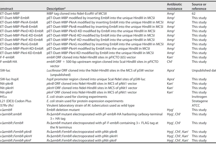
Bacterial strains and growth conditions.Table 1 describes the bacterial strains and constructs used in the study.Escherichia coliDH5a(Invitrogen) andE. coliBL21(DE3) codon plus (Stratagene) strains were used for cloning and protein purification, respectively. 7H9 medium supplemented with 10% ADC (NaCl, dextrose, bovine serum albumin, and catalase), 0.2% glycerol, and 0.1% Tween 80 was used for the liquid growth ofM. tuberculosisstrains. 7H11 agar with 10% OADC (ADC along with oleic acid) and 0.2% glycerol was used forM. tuberculosisstrain growth on plates.E. colitransformants were selected on ampicillin (100mg/mL)–kanamycin (50mg/mL)–hygromycin (150mg/mL)–chloramphenicol (34mg/mL) or apramycin (30mg/mL). M. tuberculosis recombinants were selected on kanamycin (25 mg/mL)– hygromycin (100mg/mL)–chloramphenicol (40mg/mL) or apramycin (30mg/mL). Medium components were from BD Difco, Sigma-Aldrich, and Hi-Media. Molecular grade reagents were procured from Merck, Ameresco, or Sigma; Luciferase reporter assay kit was from Promega (number E1500); restriction-modifica- tion enzymes were from NEB; and SEM chemicals were from Electron Microscopy Sciences. Oligonucleotides (Table 2) were procured from Sigma.
Generation of embRdeletion mutant inM. tuberculosis.Upstream and downstream regions (;1,000 bp each), including;200 bp ofM. tuberculosis embRloci, were amplified fromM. tuberculosis genomic DNA using Phu DNA polymerase. Amplicons were digested and ligated with OriE and coslas well ashygrandsacB, obtained from pYUB1474 (49), to generate AES (allelic exchange substrate). SnaBI- linearized AES was used to generateRvDembRmutant as described previously (50). Hygromycin-resist- ant colonies were screened to replace theembRgene at its native loci with the help of multiple PCRs and Western blots.
TABLE 1Constructs generated and strains used in this study
Construct Descriptiona
Antibiotic resistance
Source or reference
pET-Duet-MBP MBP tag cloned into NdeI-EcoRV of MCSII Ampr 62
pET-Duet-MBP-EmbR pET-Duet-MBP modified by inserting EmbR into the unique HindIII in MCSI Ampr This study pET-Duet-MBP-PknA-EmbR pET-Duet-MBP-PknA modified by inserting EmbR into the unique HindIII in MCSI Ampr This study pET-Duet-MBP-PknB-EmbR pET-Duet-MBP-PknB modified by inserting EmbR into the unique HindIII in MCSI Ampr This study pET-Duet-MBP-PknD-KD-EmbR pET-Duet-MBP-PknD-KD modified by EmbR into the unique HindIII in MCSI Ampr This study pET-Duet-MBP-PknE-KD-EmbR pET-Duet-MBP-PknE-KD modified by EmbR into the unique HindIII in MCSI Ampr This study pET-Duet-MBP-PknF-KD-EmbR pET-Duet-MBP-PknF-KD modified by EmbR into the unique HindIII in MCSI Ampr This study pET-Duet-MBP-PknG-EmbR pET-Duet-MBP-PknG modified by inserting EmbR into the unique HindIII in MCSI Ampr This study pET-Duet-MBP-PknH-KD-EmbR pET-Duet-MBP-PknH modified by EmbR into the unique HindIII in MCSI Ampr This study pET-Duet-MBP-PknI-KD-EmbR pET-Duet-MBP-PknI-KD modified by EmbR into the unique HindIII in MCSI Ampr This study
pF-F-embR embRORF cloned into NdeI-HindIII sites in pFICTO (65) vector Kanr This study
pF-embR-HA embRORF1500-bp upstream region cloned into ScaI-HindIII sites in pFICTO vector
Chlr This study
pSW-luc LuciferaseORF cloned into the NdeI-HindIII sites in the MCS of pSW vector (unpublished)
Aprar Unpublished data pSW-luc-hspX hspXpromoter region cloned into unique ScaI-NdeI sites of pSW-luc Aprar This study
pNit-pknB pknBORF cloned into NdeI-HindIII sites in MCS of pNit1 vector Kanr This study
pNit-pknH pknHORF cloned into NdeI-HindIII sites in MCS of pNit1 vector Kanr This study
pNit-pknF pknFORF cloned into NdeI-HindIII sites in MCS of pNit1 vector Kanr This study
DH5a E. colistrain used for cloning experiments Invitrogen
BL21 (DE3) Codon Plus E. colistrain used for protein expression experiments Stratagene
H37Rv (Rv) Virulent laboratory strain ofM. tuberculosisused as wild type ATCC
RvDembR EmbR deletion mutant Hygr This study
RvDembR::embR RvDembRmutant electroporated with pF-embR-HA harboring carboxy-terminal 3HA tag
Hygr, Chlr This study RvDembR::FembR RvDembRmutant electroporated with pF-F-embR containing 3FLAG tag at
the N terminus
Hygr, Chlr This study RvDembR::FembR-pknB RvDembR::FembRelectroporated with pNit-pknB Hygr, Chlr, Kanr This study RvDembR::FembR-pknH RvDembR::FembRelectroporated with pNit-pknH Hygr, Chlr, Kanr This study RvDembR::FembR-pknF RvDembR::FembRelectroporated with pNit-pknF Hygr, Chlr, Kanr This study
aORF, open reading frame.
Generation of plasmid DNA constructs and strains.pNiT-1 and pNiT-ET plasmids were a kind gift from Christopher M. Sassetti and Eric Rubin, respectively (51). pQEII-embR, used for protein expression and purification, was generated by amplifyingembRfromH37Rvgenomic DNA and cloned into pQEII vector using NdeI/HindIII sites.embRwas further subcloned into pFICTO to generate pF-F-embR (with an N-terminal 3FLAG tag). The constructs were further electroporated intoRvDembRmutant to generate RvDembR::FembRmutant. To generate embRcomplementation constructs with its native promoter, embRwithout stop codon and 500 bp immediately upstream of the gene was PCR amplified and cloned into ScaI-HindIII sites of pFICTO vector to generate pF-embR-HA (with a C-terminal 3hemagglutinin [HA] tag). pF-embR-HA construct was electroporated intoRvDembRmutant to generateRvDembR::embR mutant. For the reporter assays, the 500-bp region upstream ofhspXwas cloned in ScaI-NdeI sites of pSW1-Luciferase construct (unpublished data) using ScaI-NdeI sites to generate pSW1-hsp-luc. The con- struct was electroporated intoRv,RvDembR, andRvDembR::embRstrains.
Growth rate kinetics.Exponential-phase cultures of Rv, RvDembR, and RvDembR::embR strains grown in Middlebrook 7H9-ADC medium were seeded at an optical density at 600 nm (OD600) of;0.05 in 7H9/Sauton’s medium. OD600was monitored every 24 h, and the CFU were enumerated by serially diluting the cultures and plating them on 7H11 agar.
Scanning electron microscopy and transmission electron microscopy.Mycobacterial strains cul- tured in 7H9-ADC medium and 10-mL cultures at an OD600of;0.6 were harvested. SEM and TEM were performed as previously described (50). Briefly, for TEM, cells werefixed, gradually dehydrated, and poly- merized using Epson 812 resin;;63-nm sections were cut using an ultramicrotome (Leica) and subse- quently stained using uranyl acetate and lead citrate for visualization under a Tecnai G2 20 twin (FEI) transmission electron microscope (52). Cell length, width, and thickness were quantified with the help of Smart TIFF software and Carl Zeiss Tiff Annotation Editor.
In vitrostress susceptibility assays.Susceptibility ofRv,RvDembR, andRvDembR::embRstrains to variousin vitrostresses was determined as previously described (53). Briefly, log-phase bacillary cultures were washed with PBST80(phosphate-buffered saline and 0.05% Tween 80) and seeded at an OD600of TABLE 2List of primers used in this study
Reference no. Description Sequence (59–39)
STL261 embR-AES 59flank forward harboring AlwNI and SnaBI sites CACCTTTTCAGAAACTGTACGTACACGCCGTGATCGTCAGCATC STL262 embR-AES 59flank reverse harboring AlwNI site TTTTTTTTCAGTTCCTGTACGCAGATTAGACACGTAGG STL263 embR-AES 39flank forward harboring AlwNI site CACCTTTTCAGAGACTGTGCATGTGCAGCACGAGCGAA STL264 embR-AES 39flank forward harboring AlwNI and SnaBI sites TTTTTTTTCAGCTTCTGTACGTAAAGTCGACCAGATAGGCAAAG STL181 Hygend sequence forward forembRmutant screening CGATCCGGAGGAACTGGCGCA
VKN415 pknHend reverse forembRmutant screening harboring NotI site AGCTGCGGCCGCTCATTCCTTGTTGACTTTGTC STL53 pknHmiddle reverse forembRmutant screening harboring HindIII site AGCTAAGCTTCTATGCGGGTGGCTGCCGAGGAGC VKN277 embRmiddle forward forembRmutant screening harboring DraIII site TTTTTTTTCACAAAGTGGTCAGCAACCGCTGGATGCC
VK98 embRforward harboring NdeI site CACCCATATGGCTGGTAGCGCGACAGTG
VK99 embRreverse harboring HindIII site AAGCTTGATCTACGTGCCGCCATGCGT
BSL31 embRpromoter forward (500 bp upstream) harboring ScaI site CACCAGTACTAAAACGGCATTGTTCACCTC BSL32 embRreverse without stop codon harboring HindIII site AGCTAAGCTTCGTGCCGCCATGCGTCCC
STL265 embRforward for pDuet harboring HindIII site CACCTAAAGCTTATGGCTGGTAGCGCGACAG
STL266 embRreverse for pDuet harboring HindIII and AfII sites TTTCTTAAGCTTCTACGTGCCGCCATGCGTC
BSL400 hspXpromoter forward harboring ScaI site CCCAGTACTAAGTCAATTGACGCCAGA
BSL401 hspXpromoter reverse harboring NdeI site CCCCATATGATGCCTCCTAATCGATGG
VK900 16S rRNA qRT forward primer ACGAACAACGCGACAAACC
VK901 16S rRNA qRT reverse primer CCAGCAGCCGCGGTAA
BSL29 embCforward qRT primer AATTGTCCAGTCCCCGTTG
BSL30 embCreverse qRT primer CAAAGCCTGTAGGTTAGACCG
STL1169 embAforward qRT primer TGGTTCTACGTCGGCAACTA
STL1170 embAreverse qRT primer GTCTTTGACTTCGGTGTGCC
STL1173 embBforward qRT primer TATACGGAGAGCAGCCCAAG
STL1174 embBreverse qRT primer GCCCACATATTCCTGCAGTG
BSL375 hspXforward qRT primer ACATTATGGTCCGCGATGG
BSL376 hspXreverse qRT primer ACCGACACAGTAAGAATGCC
BSL377 esxPforward qRT primer GGCAACACGTTTTATGACGG
BSL378 esxPreverse qRT primer CATGGTGTCTAGCGAGGTC
BSL379 Rv2633forward qRT primer ACATTCACTTCCGCATCGAG
BSL380 Rv2633reverse qRT primer GTTCCACTCTTCTTCATACCCG
BSL381 espCforward qRT primer ATGTGTACTTGACTGCCCAC
BSL382 espCreverse qRT primer CCTCGCTATATATCTTCGCCG
BSL383 lpqHforward qRT primer GAGGTGAAGTCCGTTGGG
BSL384 lpqHreverse qRT primer GGTCCCAGTGATCTTGTAGTG
BSL385 devSforward qRT primer TCGAAGATCCCAAACCGTTAC
BSL386 devSreverse qRT primer AGAGTGCCGAACGATTCATC
EmbR Modulates Virulence Factor Expression mBio
;0.05 in 7H9-ADS (ADC without catalase) medium containing 50mM cumene hydroperoxide for a day.
CFU were enumerated by plating on 7H11-OADS (OADC without catalase) agar plates. For assessing the susceptibility to surfactant stress, bacterial cells were cultured in 7H9-ADC medium containing 0.05%
SDS for 3 h. The survival of strains was also examined upon the addition of 1 mM DTT for 24 h. To induce nitrosative stress, cultures were grown in 7H9-ADC medium (pH 5.5, acidified with the help of HCl) in the presence and absence of 3 mM NaNO2for 48 h. Harvested cells were washed with PBST80, and seri- ally diluted cultures were spotted on 7H11-OADC agar to enumerate CFU.
Hypoxia experiment.In vitrohypoxia stress was assessed through modified Wayne’s hypoxia model as described earlier (54). Briefly, bacterial strains were inoculated in 7H9-ADC at an OD600of ;0.1;
1.5mg/mL of methylene blue was added to a visual redox indicator. The experiment was carried out in tightly sealed glass tubes with 15% headspace at 37°C without agitation. Bacterial cells were serially diluted and plated on 7H11-OADC agar to enumerate the CFU.
Peritoneal macrophage infections.Peritoneal macrophages were isolated from BALB/c mice and cultured as previously described (55). Briefly, 4- to 6-week-old mice were injected with 4% thioglycolate solution (Difco) in the peritoneal cavity. After 96 h, the mice were euthanized, and isolated peritoneal macrophages were seeded at a density of 1 million cells/well in a 12-well tissue culture plate in RPMI 1640 with 10% fetal bovine serum. Single-cell suspensions of the mycobacterial Rv,RvDembR, and RvDembR::embRstrains were prepared to infect the peritoneal macrophages at a multiplicity of infection (MOI) of 1:10 (cell-bacteria). Cells were lysed with SDS (0.05%), and CFU were enumerated.
Murine infection experiment.Exponential-phase cultures ofRvandRvDembRmutant were washed and resuspended in the neutral saline, and single-cell suspensions were prepared. BALB/c mice (4 to 6 weeks old) were housed in the Tuberculosis Aerosol Challenge Facility (TACF) at International Centre for Genetic Engineering and Biotechnology (ICGEB), New Delhi. Mice in groups (n= 6 to 7) for each time point were challenged with the bacterial strains by aerosol to implant 100 bacilli per mouse. Bacillary deposition in the lungs was assessed 24 h postinfection. Four and 8 weeks postinfection, the bacterial survival was examined in both lungs and spleen. Thefixed tissues were also examined for histopathol- ogy and granuloma scoring by hematoxylin and eosin staining, as described previously (54).
RNA isolation and qRT-PCR analysis. Isolation of RNA was performed using TRIzol reagent (Ambion). Cell numbers equivalent to an OD of 10 forRv,RvDembR, andRvDembR::embRstrains were harvested and resuspended in TRIzol reagent. The cells were lysed using 0.1-mm zirconium beads, fol- lowing which RNA was extracted through chloroform, precipitated, and washed using 70% ethanol (pre- pared in diethyl pyrocarbonate-water). The RNA was resuspended in the nuclease-free water and further purified using the RNeasy minikit (Qiagen). The samples were treated with DNase I to remove any left- over DNA traces. cDNA was synthesized using the iScript cDNA synthesis kit (Bio-Rad) according to the manufacturer’s protocol. Gene expression with gene-specific primers (Table 2) was measured with iTaq Universal SYBR green supermix (Bio-Rad). The gene induction ratio was normalized to 16S rRNA, and results were analyzed using theDDCTmethod.
RNA-seq analysis.RNA isolated from two biological replicates of either normoxic or hypoxic sam- ples ofRvandRvDembRstrains were used to prepare the library and sequenced on the Illumina plat- form. The reads obtained were assessed for quality control using the FastQC package, followed by align- ment with theM. tuberculosisgenome and annotation downloaded from Ensembl (56) using bowtie2 (57). Htseq-count was used to count the number of reads mapped per gene (58). DEseq2 was used to get normalized counts and for analysis of differential expression (59). Figures were made using custom scripts in python3.
Gene ontology was performed for genes withPadj of,0.1 and 0.4 log2fold change using PANTHER version 14 (60). The obtained results were graphically represented using R.
Luciferase assays.Rv,RvDembR, andRvDembR::embRstrains electroporated with pSW1-hspX-luc construct and pSW1-devS-luc construct were grown in 7H9-ADC medium until mid-log phase. Cells were harvested at an OD600of;0.6, and the lysates were prepared in the lysis buffer (25 mM Tris-phos- phate, pH 7.8, 10% glycerol, 2 mM DTT, 1% Triton X-100, protease inhibitor). Luciferase assay using Luciferase assay kit (Promega E1500) was performed per the manufacturer’s protocol. Briefly, a defined volume of luciferase assay reagent (LAR) was added to1mg of whole-cell lysates from each strain, and lu- minescence was measured using a Berthold luminometer.
MIC.Resazurin microtiter assay (61) was used to determine the MIC inM. tuberculosisstrains. Briefly, antibiotics were serially diluted in 7H9-ADC medium (without Tween 80) with appropriate controls in a flat-bottom 96-well plate. Mid-log-phase bacilli fromRv,RvDembR, andRvDembR::embRstrains were diluted to an OD600of;0.006 in 7H9-ADC, and 100mL from each strain was added to the plate. A vol- ume of 20mL of resazurin solution (0.02%) was added after 5 days of incubation at 37°C. Bacterial growth was indicated by a color transition from blue to pink the following day. The antibiotic concentra- tion that prevented this color change was defined as the MIC.
Lipidomics analysis.Rv,RvDembR, andRvDembR::embRstrains were grown in 7H9-ADC either under regular growth conditions or subjected to hypoxic stress. Cells equivalent to an OD600of ;6 were washed thrice with cold, sterile PBS and resuspended in 1 mL PBS. The resuspended cells were trans- ferred into a glass vial. The cellular lipids were extracted using an established protocol (33) by adding 3 mL of 2:1 (vol/vol) chloroform-methanol mixture. Samples were vortexed and centrifuged at 2,800g (15 min at 4°C), and the organic layer was collected. The remaining aqueous layer was acidified (2% for- mic acid) and reextracted using chloroform (2 mL) to enrich phospholipids. The samples were once again centrifuged, and the organic layer was collected. The organic layers were pooled and dried under a stream of nitrogen gas at room temperature. The dried pellets were resuspended 200mL of 2:1 (vol/vol) chloroform-methanol and subjected to semiquantitative (relative quantification) information-dependent
acquisition (IDA)-mediated LC-MS/MS analysis (34, 35) on a Sciex X500R quadrupole time-of-flight (QTOF) mass spectrometerfitted with an Exion ultrahigh-performance LC system. The LC was performed on a Gemini 5U C18column (5mm, 50 by 4.6 mm; Phenomenex) coupled to a Gemini guard column (4 by 3 mm;
Phenomenex security cartridge; Phenomenex).
Phosphorylation study and mass spectrometry analysis. We used the previously described pETDuet-1 system for coexpressing EmbR andM. tuberculosisSTPKs (62). pDUET-STPK constructs, where STPKs were cloned with N-terminal MBP tag into MCS2, were described earlier (62).embRamplified usingRvgenomic DNA was cloned into duet-STPK constructs using the HindIII site in MCS-I site such that the protein was expressed with N-terminal hexa-His tag. Constructs were further transformed inE.
coliB21 DE3 Codon Plus, and the expression of His-EmbR and MBP-STPKs was confirmed by Western blotting. His-tagged EmbR was pulled down using Ni21-nitrilotriacetic acid agarose beads and processed for mass spectrometry using the in-gel digestion method (63).
For identifying EmbR phosphorylation sites inM. tuberculosis, pNit-pknB, pNit-pknD, pNit-pknH, and pNit-pknF constructs were electroporated intoRvDembR::F-embRstrain. FLAG-EmbR was immunopreci- pitation (IP), and 1/10 of the immunoprecipitated samples was probed witha-FLAG antibodies and 9/
10th of the sample was resolved on SDS-PAGE and probed witha-p-Thr antibodies. To identify EmbR pho