Molecular Biology, Biochemistry, Immunology and Physiology
Molecular Biology, Biochemistry, Immunology and Physiology
Edited by
Aaron G. Maule
Parasitology Research Group School of Biology and Biochemistry
Queen’s University of Belfast Belfast
UK and
Nikki J. Marks
Parasitology Research Group School of Biology and Biochemistry
Queen’s University of Belfast Belfast
UK
CABI Head Office CABI North American Office
Nosworthy Way 875 Massachusetts Avenue
Wallingford 7th Floor
Oxfordshire OX10 8DE Cambridge, MA 02139
UK USA
Tel: +44 (0)1491 832111 Tel: +1 617 395 4056 Fax: +44 (0)1491 833508 Fax: +1 617 354 6875 E-mail: [email protected] E-mail: [email protected] Website: www.cabi.org
©CAB International 2006. All rights reserved. No part of this publication may be reproduced in any form or by any means, electronically,
mechanically, by photocopying, recording or otherwise, without the prior permission of the copyright owners.
A catalogue record for this book is available from the British Library, London, UK.
Library of Congress Cataloging-in-Publication Data
Parasitic flatworms : molecular biology, biochemistry, immunology and physiology / edited by Aaron G. Maule and Nikki J. Marks.
p. ; cm.
Includes bibliographical references and index.
ISBN-13: 978-0-85199-027-9 (alk. paper) ISBN-10: 0-85199-027-4 (alk. paper) 1. Platyhelminthes.
[DNLM: 1. Platyhelminths. 2. Cestode Infections. QX 350 P224 2005] I. Maule, Aaron G. II. Marks, Nikki J. III. Tittle.
QL391.P7P368 2005 616.9'62--dc22
2005016094
ISBN-10: 0-85199-027-4 ISBN-13: 978-0-85199-027-9 Typeset by SPi, Pondicherry, India.
Printed and bound in the UK by Biddles Ltd, King’s Lynn.
Contributors xv
Preface xix
Access to Colour Illustrations xx
PART I PHYLOGENY,GENETICS AND TRANSCRIPTOMES
1 The Evolution of Parasitism in Flatworms 1
D.T.J. Littlewood
Introduction 1
Parasitism in the Platyhelminthes 6
The Origins of Obligate Parasitism – The Appearance of the Neodermata 7
When did the Neodermata appear? 12
The Neodermata – advantages of a new skin 13
The Radiation of the Parasitic Flatworms 15
Radiation of the Monogenea 16
Radiation of the Cestoda 20
Radiation of the Aspidogastrea 23
Radiation of the Digenea 24
Concluding Remarks 27
Acknowledgements 30
References 31
2 Genomes and Genomics of Parasitic Flatworms 37
D.A. Johnston
Introduction 38
Genome Features 40
Genome size 40
Genome composition 42
DNA methylation 42
Repeat sequences 43
Integration of host DNA sequences 46
v
Genome instability 47
Gene processing 48
Karyotype Features 50
Chromosome number 50
Ploidy 50
Sex chromosomes 52
Recombination frequency 53
Telomeres 54
Mitochondrial Genomes 54
General characteristics 54
Gene order 58
Genetic code 59
Maternal inheritance 61
Flatworm Genomics 64
Rationale 64
Gene discovery and transcriptome analysis 64
Physical mapping 66
Chromosome mapping 66
Genome sequencing 67
Concluding Remarks 67
Acknowledgements 68
References 68
3 Genetic Discrimination of EchinococcusSpecies and Strains 81 D.P. McManus
Introduction 81
Genetic Variation in Echinococcus 82
Identification of EchinococcusIsolates Using Molecular Genetic Techniques 84
RFLP/RAPD analysis 84
PCR-amplified DNA sequences 84
Mutation scanning methods 85
Microsatellite markers 85
Molecular Identification of E. granulosusStrains 85
Sheep–dog (Genotype 1) and horse–dog (Genotype 4 – E. equinus) strains 85
Cattle–dog strain (Genotype 5 – E. ortleppi) 87
Camel–dog strain (Genotype 6) 89
Pig–dog strains (Genotypes 7 and 9) 89
Cervid strains (Genotypes 8 and 10) 89
Detection of EchinococcusNucleic Acids in Clinical Samples 90 DNA Detection of Infection in Definitive and Intermediate Hosts 90
Concluding Remarks 91
Acknowledgements 92
References 92
4 Ribosomal DNA Variation in Parasitic Flatworms 96
D. Blair
Introduction 96
Nuclear Ribosomal Operons 98
Intergenic spacers 100
Internal transcribed spacers (ITSs) 102
Intra-individual variation and repeat sequences 103
Intra- and interspecific variation 107
Functional motifs 108
Secondary structure 108
Small and Large Subunit Genes 109
Mitochondrial Ribosomal Genes 115
Hybrids 116
Concluding Remarks 117
References 117
5 Genetic Studies on Monogeneans with Emphasis on Gyrodactylus 124 C.O. Cunningham and I. Mate∨jusová
Introduction 124
Difficulties in Molecular Studies of Monogenea 125
Molecular Markers for Monogenean Species 126
The rRNA genes and spacers 126
The SSU (18S) rRNA gene 127
The rRNA internal transcribed spacer (ITS) 127
The rRNA intergenic spacer (IGS) 129
Large Subunit (LSU) (28S) rRNA gene 129
Other Regions of the Monogenean Genome 130
Monogenean Species Concept and Species Complexes 130
Molecular Analysis within Monogenean Species 131
Other Genetic Studies in Monogenea 132
Concluding Remarks 133
References 133
6 The Schistosome Transcriptome 138
S. Verjovski-Almeida and R. DeMarco
Introduction 138
The Transcriptome Projects 139
Transcriptome Comparisons 139
Evolutionary Implications 141
Novel Transcripts 141
Receptors and Host–Parasite Interaction 142
Immune Evasion 142
Sex Differences 143
Genome Data 143
Novel Drug Targets 143
Vaccines 144
Trans-splicing 145
Retrotransposable Elements 145
Functional Genomics 145
Concluding Remarks 146
References 146
7 Transgenic Flatworms 149
C.G. Grevelding
Introduction 149
The Planarian Model 150
Trematodes and Cestodes 151
Transformation Techniques for Schistosomes 153
Particle bombardment 153
The PDS-1000/He particle bombardment system 155
Reporter gene activity in bombarded schistosomes 156
Stable Transformation 162
Cestodes, on the Way to Transgenesis 164
Concluding Remarks 165
Acknowledgements 166
References 166
PART II IMMUNOBIOLOGY,HOST–PARASITE INTERACTION AND CONTROL
8 Immunobiology of Schistosomes 174
S.G. Forrester and E.J. Pearce
Introduction 174
Host–Parasite Molecular Interactions 175
How Schistosomes Evade the Host Immune System 176
Skin stage parasites 176
Lung stage parasites 178
Adult parasites 178
Host Immune Response to Schistosome Infection (Th1 versus Th2) 179 Potential Players in Th2 Response Induction during a Schistosome Infection 181 Chemotherapy – An Integral Role for the Immune Response 183
Immunotherapy 183
Concluding Remarks 184
Acknowledgements 185
References 185
9 Cestode Infection: Immunological Considerations from Host
and Tapeworm Perspectives 193
D.M. McKay and R.A. Webb
Introduction 193
Cestode Parasites 194
Immunology – The Basics 195
The Intestine and Immune Responses 197
Create an inhospitable environment 197
Mobilize an active immune response 198
Specifics of the Host Response to Cestode Infection 199
Parenteral infections of cestodes 199
Host response to the oncosphere 199
Resistance to parenteral metacestodes 200
Lumenal Infections: The Immune Response to Hymenolepidid Cestodes 202 Cestode Infections and the Immune Response – Where Do We Go from Here? 203
Evasion and Subversion of the Host Response 204
The Beneficial Tapeworm 204
Concluding Remarks 205
Acknowledgements 206
References 206
10 Signal Transduction at the Host–Parasite Interface 210 T.P. Yoshino, J.J. Vermeire and J.E. Humphries
Introduction 210
Tegumental Involvement in Signal Transduction: Perception and Reality 211
Elements of Platyhelminth Signal Transduction Systems 213
Molecular and EST data 213
Platyhelminth signalling proteins 214
Platyhelminth Signal Transduction Pathways: Tegument and Beyond 218 Transforming growth factor receptor (TGFR) pathway 218
TGF-βreceptor signalling in schistosomes 219
Epidermal growth factor receptor (EGFR) pathway 220
EGFR signalling: roles in the host–parasite relationship 220
G Protein-coupled receptors (GPCRs) 222
GPCRs in parasitic platyhelminths 222
Concluding Remarks 223
Acknowledgements 223
References 224
11 Parasite Effects on the Snail Host Transcriptome 228
M. Knight and N. Raghavan
Introduction 228
Effect of Parasite Infection on the Snail Host: Towards a Molecular Understanding 230
Innate immunity 230
Biochemical involvement 231
Parasite-induced changes in gene expression 233
Parasite-induced changes in snail neurobiology 237
Concluding Remarks 238
Acknowledgements 238
References 239
12 Developments in the Chemotherapy of Parasitic Flatworms 243 G.C. Coles
Introduction 243
Parasites of Animals 244
Infections of Humans 245
Adult tapeworms 246
Larval tapeworms 246
Taenia solium 246
Echinococcus granulosus 246
Echinococcus multilocularis 247
New Anti-parasitics: Nitazoxanide 247
Mode of Action of Fasciolicides and Cestodicides 247
Resistance 248
Resistance in Fasciola 249
Resistance in monogeneans 249
Resistance in cestodes 249
Detection of resistance 250
Concluding Remarks 250
References 251
13 Drug Resistance in Schistosomes 256
T.A. Day and S. Botros
Introduction 256
Schistosomiasis Chemotherapy 257
Impact of Praziquantel 257
Challenges to Detecting and Monitoring Anti-schistosomal Resistance 258
There is a high background of drug failure 258
Methods for detecting infection are variable 258
Factors other than resistance can produce drug failure 258 It is difficult to substantiate a decreased response in the worms 259
Praziquantel’s mechanism of action is not known 259
Defining resistance 260
Cases of Drug Resistance in Schistosomes 260
Resistance to Hycanthone and Oxamniquine 260
Resistance to Praziquantel 261
Senegal 261
Egypt 262
Concluding Remarks 264
References 265
14 Praziquantel: Mechanism of Action 269
R.M. Greenberg
Introduction 269
Praziquantel Mode of Action 270
Voltage-gated Ca2+Channels 272
Schistosome and Flatworm Ca2+Currents 274
Schistosome Ca2+Channel Subunits 275
Concluding Remarks 277
Acknowledgements 278
References 278
15 Cestode Vaccine Development 282
M.W. Lightowlers
Introduction 282
Immunity – Basic Principles 284
Vaccination – Historical Perspectives 285
Defined Vaccines 286
Taenia ovis 287
Taenia saginata 288
Taenia solium 289
Echinococcus granulosus 290
Echinococcus multilocularis 290
Towards Practical Application of Cestode Vaccines 291
Protective Immune Responses and Epitope Identification 292 Predicted Protein Structure and Function of Oncosphere Antigens 293
Structure of Genes Encoding Oncosphere Antigens 295
Potential Practical Impact of Vaccination Against Cestode Parasites 296
Concluding Remarks 297
References 297
16 The Development of a Schistosome Vaccine 303
R.A. Wilson and P.S. Coulson
Introduction 303
Approaches to Schistosomiasis Vaccine Development 304
What Is Meant by Protective Immunity? 304
Attenuated Parasites Can Induce Protective Immunity 305
Routes to Antigen Identification 306
Mining crude parasite extracts for antigens 306
Protective monoclonal antibodies 307
TPI 307
9B-Ag 308
Sm23 308
Anti-idiotypic antibodies 308
Expression library screening with sera from putatively protected hosts 308
IrV5 309
GADPH 309
Antigens identified on the basis of their immunogenicity 309
GSTs 309
Sm14 310
An Antigen Cocktail Does Not Improve Vaccine Efficacy 311
Antigen Formulation 311
The WHO vaccine trials 312
Clinical Trials 312
Why Are High Levels of Protection Difficult to Achieve? 313
Secreted and Tegument Antigens as Vaccine Targets 314
Calpain 315
The 20–22 kDa family 315
Future Prospects 315
The genome 316
The transcriptome 316
The proteome 316
Concluding Remarks 317
References 317
PART III PROTEIN FUNCTION,METABOLISM AND PHYSIOLOGY
17 Flatworm Parasite Proteomics 327
R.M. Morphew, J. Barrett and P.M. Brophy
Introduction 327
Parasitic Flatworm Sample Preparation 328
Protein collection 329
Solubilization of proteins 330
Removal of contaminating substances 332
Protein Separation before Two-dimensional Electrophoresis 333 Separating Proteins by Two-dimensional Protein Electrophoresis 334
Isoelectric focusing 334
SDS–PAGE the second dimension 335
Staining and extraction 335
Image analysis 336
Mass Spectrometry to Unravel the Flatworm Proteome 337
Matrix-assisted laser desorption/ionization time-of-flight mass spectrometry
(MALDI ToF) for parasite analysis 337
Electrospray ionization time-of-flight mass spectrometry (ESI ToF MS) 338
Analysis of post-translational modifications 339
Peptide sequencing via post-source decay and tandem mass spectrometry 339
Database Mining and Protein Identification 340
Useful databases 341
Proteomics without Two-dimensional Electrophoresis 342
Multi-dimensional protein identification technology (MuDPIT) 342
Isotope-coded affinity tagging 343
Concluding Remarks 344
Acknowledgements 344
References 344
18 Proteases in Trematode Biology 348
J.P. Dalton, C.R. Caffrey, M. Sajid, C. Stack, S. Donnelly, A. Loukas, T. Don, J. McKerrow, D.W. Halton and P.J. Brindley
Introduction 349
The Trematode Gut 349
Schistosomes 349
Fasciola hepatica 350
Blood as a Source of Amino Acids 350
Prevention of Blood Clotting 352
Lysis of Blood Cells 352
Peptidases Involved in the Digestion of Blood and Tissue Proteins 353
Digestion in schistosomes 353
Digestion in liver flukes 355
Regulation of the Digestive Process 355
Non-feeding Functions Attributed to Peptidases 356
Invasion and migration through host tissue 356
Excystment of juvenile parasites 357
Hatching of eggs 358
Protease Phylogeny 358
Cathepsin B 358
Cathepsins L and F 359
Aspartic proteases 361
Gene Structure, Orthology and Molecular Evolution 361
Concluding Remarks 362
References 363
19 Signalling Molecules and Nerve–Muscle Function 369
A.G. Maule, N.J. Marks and T.A. Day
Introduction 369
Nervous System Structure 370
Muscle Structure and Organization 371
Neuronal Signalling Molecules 372
Classical transmitters, their synthesis, degradation and receptors 372
Acetylcholine 372
5-Hydroxytryptamine (5-HT; serotonin) 373
Catecholamines 374
Histamine 374
γ-Aminobutyric acid (GABA) 375
Glutamate 375
Octopamine 376
Nitric oxide 376
Neuropeptides 376
Neuropeptide F 376
FMRFamide-related peptides 378
Neuropeptide processing enzymes 379
Concluding Remarks 381
Acknowledgement 381
References 381
20 Unusual Aspects of Metabolism in Flatworm Parasites 387 A.G.M. Tielens and J.J. van Hellemond
Introduction 387
Parasitic versus Free-living Stages 388
Nutrition 388
Free-living stages 388
Parasitic stages 389
Energy Metabolism 390
Substrates of energy metabolism 390
Free-living versus parasitic stages 391
Anaerobic glycolysis versus malate dismutation 391
Unusual aspects of glycolysis 394
Unusual aspects of mitochondrial metabolism 395
Succinate dehydrogenase versus fumarate reductase 396
Ubiquinone versus rhodoquinone 396
Acetate:succinate CoA-transferase (ASCT) 397
Evolutionary origin of anaerobic mitochondria 398
Transitions in energy metabolism during the life cycle 399
Biosynthetic Capacities 401
Lipids 402
Purines, pyrimidines and polyamines 403
Concluding Remarks 404
References 405
21 Glycoconjugate Structures 408
M. Wuhrer and R. Geyer
Introduction 408
Trematodes (Digenea) 408
Schistosoma 408
Glycosphingolipids of schistosomes 409
Protein glycosylation of schistosomes 411
Cercarial stage 411
Adult stage 411
Egg stage 412
The Lewis X-epitope and the host–parasite relationship 413
Fasciola 413
Cestodes 415
Pseudophyllida 415
Cyclophyllida 415
Glycosphingolipids 415
Glycoproteins 417
Concluding Remarks 418
References 418
22 Gene Silencing in Flatworms Using RNA Interference 423 P.J. Skelly
Introduction 423
Molecular Mechanisms of Silencing 424
Biological Functions of Gene Silencing 424
RNAi as a Molecular and Therapeutic Tool 426
RNAi Protocols 426
RNAi in Flatworms 428
Planaria 428
Schistosomes 429
Concluding Remarks 431
Acknowledgements 431
References 431
Index 435
John Barrett, Parasitology Research Group, Institute of Biological Sciences, Cledwyn Building, The University of Wales, Aberystwyth, Wales SY23 3DA, UK. Email, [email protected]
David Blair, School of Tropical Biology, James Cook University, Townsville, Queensland 4811, Australia. Email, [email protected]
Sanaa Botros,Theodor Bilharz Research Institute, Cairo, Warrak El-Hadar, Imbaba, PO Box 30, Giza, 12411 Egypt. Email, [email protected]
Paul J. Brindley, Department of Tropical Medicine, School of Public Health and Tropical Medicine, Tulane University Health Sciences Center, New Orleans, LA 70112, USA. Email, [email protected]
Peter M. Brophy, Parasitology Research Group, Institute of Biological Sciences, Edward Llwyd Building, University of Wales, Aberystwyth, Wales SY23 3DA, UK. Email, peter@brophy3218.
fsnet.co.uk
Conor R. Caffrey, Tropical Disease Research Unit and Sandler Center for Basic Research in Parasitic Diseases, QB3 Building, PO Box 8 2550, 1700 4th Street, University of California, San Francisco, CA 94143, USA. Email, [email protected]
Gerald C. Coles, Department of Clinical Veterinary Science, University of Bristol, Langford House, BS40 5DU, UK. Email, [email protected]
Patricia S. Coulson, Department of Biology, University of York, York YO10 5YW, UK. Email, [email protected]
Carey O. Cunningham, FRS Marine Laboratory, PO Box 101, 375 Victoria Road, Aberdeen AB11 9DB, UK. Email, [email protected]
John P. Dalton, Institute for the Biotechnology of Infectious Diseases, University of Technology Sydney, Westbourne Street, Gore Hill, Sydney, NSW 2065, Australia. Email, john.dalton@uts.
edu.au
Tim A. Day, Department of Biomedical Sciences, Veterinary Medicine Building, Iowa State University, Ames, IA 50011, USA. Email, [email protected]
Tegan Don, Helminth Biology Laboratory, Division of Infectious Diseases and Immunology, Queensland Institute of Medical Research, 300 Herston Road, Herston, Brisbane, Queensland 4006, Australia. Email, [email protected]
Sheila Donnelly, Institute for the Biotechnology of Infectious Diseases, University of Technology Sydney, Westbourne Street, Gore Hill, Sydney, NSW 2065, Australia. Email, sheila.
Ricardo DeMarco, Universidade de São Paulo, Instituto de Quimica, Av. Prof. Lineu Prestes 748 sala 1200 05508-000 São Paulo, SP Brazil. Email, [email protected]
xv
Sean G. Forrester, Faculty of Science, University of Ontario Institute of Technology, 2000 Simcoe St. North, Oshawa, ON, Canada, L1H 7K4. Email, [email protected]
Rudolf Geyer, Institute of Biochemistry, University of Giessen, Friedrichstrasse 24, 35392 Giessen, Germany. Email, [email protected]
Robert M. Greenberg, Marine Biological Laboratory, 7 MBL Street, Woods Hole, MA 02543, USA. Email, [email protected]
Christoph G. Grevelding, Institute for Parasitology, Justus-Liebig-University, Rudolf-Buchheim- Strasse. 2, 35392 Giessen, Germany. Email, [email protected] David W. Halton, Parasitology Research Group, School of Biology and Biochemistry, Medical
Biology Centre, Queen’s University Belfast, 97 Lisburn Road, Belfast BT9 7BL, Northern Ireland, UK. Email, [email protected]
Judith E. Humphries, Department of Pathobiological Sciences, University of Wisconsin-Madison, School of Veterinary Medicine, 2015 Linden Drive, Madison, WI 53706, USA. Email, [email protected]
David A. Johnston, Wolfson-Wellcome Biomedical Laboratory, Department of Zoology, The Natural History Museum, Cromwell Road, London SW7 5BD, UK. Email, [email protected] Matty Knight, Biomedical Research Institute, 12111 Parklawn Drive, Rockville, MD 20852, USA.
Email, [email protected]
Marshall W. Lightowlers, Veterinary Clinical Centre, The University of Melbourne, 250 Princes Highway, Werribee, Victoria 3030, Australia. Email, [email protected]
D.T.J. Littlewood, Department of Zoology, The Natural History Museum, Cromwell Road, London SW7 5BD, UK. Email, [email protected]
Alex Loukas, Helminth Biology Laboratory, Division of Infectious Diseases and Immunology, Queensland Institute of Medical Research, 300 Herston Road, Herston, Brisbane, Queensland 4006, Australia. Email, [email protected]
Nikki J. Marks, Parasitology Research Group, Queen’s University Belfast, 97 Lisburn Road, Belfast BT9 7BL, Northern Ireland, UK. Email, [email protected]
Iveta Matˇejusová, FRS Marine Laboratory, PO Box 101, 375 Victoria Road, Aberdeen AB11 9DB, UK. Email, [email protected]
Aaron G. Maule, Parasitology Research Group, Queen’s University Belfast, 97 Lisburn Road, Belfast BT9 7BL, Northern Ireland, UK. Email, [email protected]
Derek M. Mckay, Intestinal Disease Research Programme, McMaster University, Hamilton, Ontario, Canada L8N 3Z5. Email, [email protected]
James McKerrow, Tropical Disease Research Unit and Sandler Center for Basic Research in Parasitic Diseases, QB3 Building, Box. 2550, 1700 4th Street, University of California, San Francisco, CA 94143, USA. Email, [email protected]
Don P. McManus, Molecular Parasitology Laboratory, Division of Infectious Diseases and Immunology, Australian Centre for International and Tropical Health and Nutrition, The Queensland Institute of Medical Research and The University of Queensland, Post Office Royal Brisbane Hospital, Queensland 4029, Australia. Email, [email protected]
Russell M. Morphew, Parasitology Research Group, Institute of Biological Sciences, Edward Llwyd Building, University of Wales, Aberystwyth, Wales SY23 3DA, UK. Email, [email protected] Edward J. Pearce, Department of Pathobiology, School of Veterinary Medicine, University of
Pennsylvania, Philadelphia, PA 19104-6008, USA. Email, [email protected] Nithyakalyani Raghavan, Biomedical Research Institute, 12111 Parklawn Drive, Rockville, MD
20852, USA. Email, [email protected]
Mohammed Sajid, Tropical Disease Research Unit and Sandler Center for Basic Research in Parasitic Diseases, QB3 Building, PO Box 2550, 1700 4th Street, University of California, San Francisco, CA 94143, USA. Email, [email protected]
Patrick J. Skelly, Department of Biomedical Sciences, Division of Infectious Diseases, Tufts University School of Veterinary Medicine, 200 Westboro Road, Grafton, MA 01536, USA.
Email, [email protected]
Colin Stack, Institute for the Biotechnology of Infectious Diseases, University of Technology Sydney, Westbourne Street, Gore Hill, Sydney, NSW 2065, Australia. Email, [email protected] Aloysius G.M. Tielens, Faculty of Veterinary Medicine, Department of Biochemistry and Cell
Biology, Utrecht University, PO Box 80176, The Netherlands. Email, A.G.M. [email protected] Jaap J. Van Hellemond, Faculty of Veterinary Medicine, Department of Biochemistry and Cell Biology, Utrecht University, PO Box 80176, The Netherlands. Email, [email protected] Sergio Verjovski-Almeida, Instituto de Química, Universidade de São Paulo, Av. Prof. Lineu
Prestes 748 sala 1200 05508-000 São Paulo, SP Brazil. Email, [email protected]
Jon J. Vermeire, Department of Pathobiological Sciences, University of Wisconsin-Madison, School of Veterinary Medicine, 2015 Linden Drive, Madison, WI 53706, USA. Email, ver- [email protected]
Rodney A. Webb, Department of Biology, York University, Toronto, Ontario, Canada. Email, [email protected]
R. Alan Wilson, Department of Biology, University of York, York YO10 5YW, UK. Email, [email protected]
Manfred Wuhrer, Biomolecular Mass Spectrometry Unit, Department of Parasitology, Leiden University Medical Center, PO Box 9600, 2300 RC Leiden, The Netherlands. Email, [email protected]
Timothy P. Yoshino, Department of Pathobiological Sciences, University of Wisconsin-Madison, School of Veterinary Medicine, 2015 Linden Drive, Madison, WI 53706, USA. Email, [email protected]
Buoyed by the utility of research data on the model nematode Caenorhabditis elegans and the rapidly growing expressed sequence tag (EST) resources for roundworms, research on nematode parasites has progressed rapidly compared to that seen for flatworm parasites.
This molecular research base has facilitated considerable advances in nematode phyloge- netics, genomics, proteomics and biology that have often been the envy of many platy- helminthologists. Only in the last few years have we seen the benefits of concerted efforts to propel flatworm research into the 21st cen- tury with genome projects on schistosomes and EST projects on these and other parasitic flat- worms. Even though EST data for schistosomes have only become available relatively recently, it is clear from many of the chapters of this book that they have served to invigorate para- sitic flatworm research, providing a bountiful resource for almost all researchers in the field.
The undisputed lead role played by schis- tosomes in parasitic flatworm research is echoed by the content of this book – a reflec- tion of the field as it stands today. However, it would be naïve to assume that all the signifi- cant progress is confined to schistosomes as much quality research is emanating from research laboratories that focusses on other trematodes, cestodes or monogeneans. All serve to inform us on flatworm biology and parasitology. We believe that flatworm para- sitology is now entering a new era in which
some of the considerable gaps between the frontiers of nematode and flatworm research are beginning to be bridged. In this respect, we believe that this book is timely.
The phylogenetic relationships amongst flatworms are only now being unravelled and are so complex that predicting how findings in one flatworm parasite species relate to another is almost impossible to decipher. In nematodes, C. elegans has proved a useful model to study the facets of biology of parasitic species.
Although there is no such flatworm model, it is noteworthy that much significant progress has been made recently in planaria, especially in relation to genome-wide gene-silencing meth- ods that have been developed in Schmidtea mediterranea. However, the value of such species as model organisms for diverse flat- worm parasites is simply not known and there is limited evidence to suggest that they will be hugely beneficial in this respect. Indeed, com- parative studies on flatworms are few and far between such that any judgement on the value of potential model flatworms is based on a very small dataset. Nevertheless, progress in the molecular manipulation of planarians has the potential to aid parasitologists, and more inter- action and cross-fertilization between these research communities should be encouraged.
Although research on parasitic flatworms lags behind that on nematodes, progress is evident in almost all areas of focus, from phy- logenetics and genomics to immunobiology xix
and vaccine development, from cell sig- nalling and physiology to gene silencing and transgenics. While this book addresses many of these areas, it does not attempt to be com- prehensive. Unfortunately size constraints within a single volume preclude considera- tion of many other interesting aspects of flat- worm research. Nevertheless, we hope the contents endow the reader with valuable insight into what we perceive as some of the most pivotal and exciting areas of parasitic flatworm biology.
The first section of this book provides a strong foundation for understanding the evolu- tionary interrelationships of flatworms, their phylogeny, genetics and transcriptomics; it informs on where we are today and highlights the molecular advances that are being made and where they are likely to lead. It finishes by charting recent progress in the development of transgenic flatworms; developments, which could have huge impact on many of our research goals. Section two focuses on the host–parasite relationship and parasite control.
Within the host–parasite relationship there are contributions on immunobiology, the host–par- asite interface and parasite-induced host tran- scriptome changes that have much merit well beyond the boundaries of parasitology. In rela- tion to control, the chapters examine develop- ments in flatworm chemotherapy and drug resistance. This section ends with chapters on vaccine development, believed by many to be the future of helminth parasite control. The third and final section turns to proteomics and the biology of flatworm parasites by exploring proteases, physiology, metabolism and glyco- conjugates. Many of these chapters reveal how the new molecular resources for flatworms, dis- cussed in earlier sections, have aided research
on more focused aspects of flatworm biology.
The final chapter examines gene silencing and the progress made in the induction of RNA interference in flatworm parasites as a tool to examine protein function. The authors are all leading researchers in their respective fields and have provided expert coverage of key areas in parasitic flatworm research as well as insight into the future directions for this work. As with the sister text on parasitic nematodes, the chap- ters included here are diverse in style and approach, but provide a broad insight into par- asitic flatworms.
Without a highly characterized model species to underpin flatworm research, scien- tific progress will most likely rely on our abil- ity to capitalize on evolving molecular advances in a range of flatworm species. We hope this book will provide clear evidence that significant advances are already being made across a wide spectrum of parasitic flatworm research, and will prove a useful resource for continued study.
Access to Colour Illustrations In order to reduce the cost of this book, and thereby improve its accessibility, there are no colour reproductions. Some of the illustrations, however, can only properly be appreciated in colour (some of those in Chapters 7 and 17).
The colour illustrations can be viewed and downloaded from the following internet site:
http://www.qub.ac.uk/bb/books/flatworms/
index.html
If you have any suggestions or problems relating to the illustrations appearing in this book, then please feel free to contact Aaron Maule ([email protected]).
Introduction
The phylum Platyhelminthes is comprised of an enormous diversity of species that occur in all seas, rivers and lakes, and on all continen- tal land masses. With a soft body lacking any cuticle or protective covering, the majority of species are found in moist or aquatic environ- ments. Generally bilaterally symmetrical, acoelomate, lacking an anus, possessing a low level of cephalization, usually hermaphroditic, and dorsoventrally flattened, flatworms are commonly small but may reach enormous lengths. These defining features are not unique to the phylum and are, e.g. multiciliated gas- trodermal cells, best viewed with an electron microscope. However, there are many defining
features for the various subgroups (Caira and Littlewood, 2001; Littlewood et al., 2004).
As parasites, flatworms have extended their global presence by taking advantage of the adaptations of many diverse invertebrate and vertebrate hosts. Many parasitic forms are host-specific and many of these are site- specific within or on their host. Contemplate on the biodiversity of vertebrates, consider that many platyhelminths use one or more interme- diate hosts and one may just begin to grasp the diversity of parasitic flatworms. From the microscopic interstitial free-living species that live between particles of mud to the enor- mously long tapeworms of blue whales, an estimate of 100,000 extant species, of which only about 20,000 have been formally
©CAB International 2006.Parasitic Flatworms: Molecular Biology, Biochemistry,
Immunology and Physiology (eds A.G. Maule and N.J. Marks) 1
in Flatworms
D.T.J. Littlewood
Department of Zoology, The Natural History Museum, Cromwell Road, London SW7 5BD, UK
Introduction . . . .1
Parasitism in the Platyhelminthes . . . .6
The Origins of Obligate Parasitism – The Appearance of the Neodermata . . . .7
When did the Neodermata appear? . . . .12
The Neodermata – advantages of a new skin . . . .13
The Radiation of the Parasitic Flatworms . . . .15
Radiation of the Monogenea . . . .16
Radiation of the Cestoda . . . .20
Radiation of the Aspidogastrea . . . .23
Radiation of the Digenea . . . .24
Concluding Remarks . . . .27
Acknowledgements . . . .30
References . . . .31
described, still seems conservative when we consider the hosts and habitats yet to be sur- veyed. Even among those hosts and habitats already surveyed, many of the smaller parasitic species seem to have been overlooked and new species from commonly collected hosts are not uncommon. Small-bodied parasitic taxa are harder to find and are generally more specios than related large-bodied taxa (Poulin and Morand, 2000), suggesting a long future ahead for flatworm systematists.
As soft-bodied organisms with little or no protection from predation, flatworms tend to be cryptic with some notable exceptions;
the very colourful marine polyclads found on coral reefs offer some of the most spectacular colour schemes exhibited by any organism (Newman and Cannon, 2003), and some ter- restrial planarians (Tricladida) are also large and conspicuous. These brightly coloured free- living representatives tend to exhibit apose- matic coloration, indicating their distastefulness, or in the case of some polyclads they mimic distasteful nudibranch molluscs. In contrast, parasitic forms are less noticeable and often have complex life cycles with only fleeting periods when they are not in or on a host.
Usually they are microscopic during these free- living developmental stages; it takes an effort to find them in spite of their ubiquity.
Although lacking a fossil record, the deep- branching position of Platyhelminthes among the Lophotrochozoa in the tree of life, suggests a relatively ancient origin but perhaps not quite as ancient as once thought. For over 100 years, scientists have suggested that the rela- tively simple body plan of these flatworms ren- ders them as ideal model organisms from which other phyla can be derived and it is common to see hypothetical ancestors of the Metazoa described as ‘flatworm-like’. However, platyhelminth phylogenetics has progressed markedly over the last 10 years thanks to a sus- tained effort by morphologists and the advent of molecular systematics providing additional sources of phylogenetic data. We are now armed with more clearly resolved evolutionary trees with which to investigate the origins and subsequent radiation of flatworm groups.
Recent advances, for example, indicate that acoelomorph flatworms (Acoela and Nemerto- dermatida) are now generally considered to be
basal bilaterians (a role once occupied by the Platyhelminthes), occupying a pivotal position in the tree of metazoan life and likely repre- senting one or two separate phyla (Ruiz-Trillo et al., 1999, 2002; Telford et al., 2000, 2003;
Littlewood et al., 2001; Jondelius et al., 2002).
Whilst it is the inclusion of Acoelomorpha in the Platyhelminthes that contributed to a perception that the whole phylum is ‘primitive’, the remaining non-acoelomorph flatworms, the Catenulida + Rhabditophora (Platyhel- minthes sensu stricto), may be more closely allied to phyla such as Gastrotricha (Giribet et al., 2000) or at least as derived members of the Lophotrochozoa appearing sometime after the last protostome/deuterostome ancestor; the resolution of their true position among the Lophotrochozoa remains controversial and unresolved (Jenner, 2004; Valentine, 2004).
Most molecular estimates of metazoan interre- lationships recognize three main clades, the deuterostomes, the ecdysozoans and the lophotrochozoans, usually with the latter two protostome clades forming a monophyletic group (Adoutte, 1999). However, a recent analysis (Eernisse and Peterson, 2004), employing multiple genes and including mor- phology, places the deuterostomes and lopho- trochozoans as sister groups, and the ectoprocts as a sister group to the Platyhelminthes. Clearly, many nodes require greater resolution from additional data.
Figure 1.1 illustrates a recent estimate of relationships among the Metazoa with an indication showing which phyla are para- sitized by the main parasitic flatworm groups.
Parasitic flatworms have had a marked impact on many other animal groups, most notably the Chordata. Most vertebrates are parasitized by at least one species of flatworm, and larger vertebrates tend to have richer parasite communities with many helminths (see, for example, Poulin, 1995). Considering the importance of parasitism as an ‘engine of diversity’, maintaining the diversity of the major histocompatibility complex, imposing selection favouring sexual reproduction in hosts and increasing speciation rates, among other factors (Summers et al., 2003), the influ- ence of the Platyhelminthes in evolution and ecology seems greater than the sum of its parts.
Annelids Echiurans Sipunculans Molluscs Nemerteans Entoprocts Cycliophorans Platyhelminthes Ectoprocts Brachiopods Phoronids Gnathostomulids
Rotifers and Acanthocephalans Gastrotrichs
Nemertodermatids Acoels
Placozoans Cnidarians Ctenophores Calcareans Siliceans
Choanoflagellates + Mesomycetozoans Fungi
Animalia Metazoa
Bilateria
LOPHOTROCHOZOA D
D
Phylum parasitized by ...
A
Monogenea Cestoda Digenea Aspidogastrea
D Loricifera
Chaetognaths Onychophorans
Tardigrades Arthropods Nematomorphs Nematodes Priapulids Kinorhynchs
ECDYSOZOA Hemichordates
Echinoderms
Chordates M DEUTEROSTOMA
D D A C
D
C D
C
M
A C D C M
D C D A D
Fig. 1.1. Platyhelminthes and their position in the tree of life with an indication of which phyla are parasitized by neodermatan flatworms (Monogenea, Cestoda, Aspidogastrea, Digenea); basic tree adapted from Eernisse and Peterson (2004) who estimated this tree topology using a combined analysis of molecular (SSU rDNA and myosin II) and morphological data; monophyletic protostomes are shown as this remains the general consensus (Baldauf, 2003). Acoelomorph flatworms (Acoela and
Nemertodermatida) are no longer members of the Platyhelminthes, but are instead recognized as basal bilaterians. True flatworms are members of the Lophotrochozoa but their relative position within this clade and the identity of their sister group is still debated. Digenea utilize the greatest diversity of metazoan phyla as hosts, including some free-living flatworms.
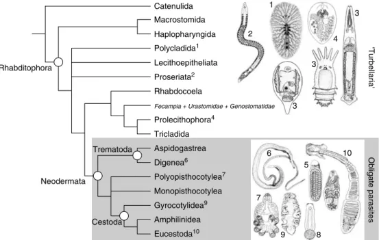
Figure 1.2 provides a consensus view of the major flatworm groups and a hypothesis of their interrelationships based on molecular and mor- phological estimates (Littlewood et al., 1999b);
individual estimates may be found in Brooks (1989), Carranza et al. (1997), Ehlers (1984, 1985), Littlewood et al. (1999b) and Zamparo et al.(2001). The ‘Turbellaria’ is a paraphyletic assemblage and the relationships between its constituent taxa and the Neodermata are yet to be satisfactorily resolved (see section on ‘The Origins of Obligate Parasitism’). Catenulid flat- worms employ a different mitochondrial genetic code than the remaining flatworms, the Rhabditophora (Telford et al., 2000), and appear to have few, if any, convincing morphological synapomorphies with the Rhabditophora.
Nevertheless, molecular data routinely resolve the Catenulida as the most basal members of the Platyhelminthes and they have long been con- sidered flatworms (Ehlers, 1984). Most of the turbellarian groups include marine and fresh- water examples and only some of the triclad turbellarians are terrestrial. The majority of the
‘Turbellaria’ are very small, and of these most are meiofaunal, except the Polycladida, which are often large and strictly marine. A number of turbellarian lineages include symbiotic and some parasitic species (see section on
‘Parasitism in the Platyhelminthes’), but the obli- gate parasites (Neodermata) form a convincing monophyletic group.
Flatworms have no fossil record, beyond one or two trace fossils of uncertain origin or limited utility (Conway Morris, 1981;
Upeniece, 2001; Valentine, 2004), including Quaternary turbellarian eggs (Binford, 1982), a typhloplanoid rhabdocoel in Eocene amber (Poinar, 2003) and shell pitting in marine bivalves caused by digeneans (Ruiz and Lindberg, 1989). This paucity of dateable evi- dence seriously hampers an understanding of the timing of key evolutionary events in the his- tory of flatworm radiation (Littlewood and Donovan, 2003). To unpick the evolutionary history of parasite evolution and radiation objectively is no easy task and we must do it based on our knowledge of extant species, our
Rhabditophora
Catenulida
Tricladida Prolecithophora4 Rhabdocoela Proseriata2 Polycladida1 Lecithoepitheliata Macrostomida
Gyrocotylidea9 Monopisthocotylea Polyopisthocotylea7 Digenea6
Aspidogastrea
Eucestoda10 Amphilinidea Haplopharyngida
Trematoda
Cestoda Neodermata
Obligate parasites
Fecampia + Urastomidae + Genostomatidae
'Turbellaria'
1
2
3 3
3
4
5 6
7
8 9
10
Fig. 1.2. Interrelationships of the major groups of Platyhelminthes based on a consensus of
morphological and molecular estimates. Parasitic flatworms, the Neodermata, form a monophyletic group although their interrelationships are estimated differently by different molecular analyses (see Fig. 1.3).
ability to reconstruct phylogenies and the extent to which we can recognize shared ancestral characters. Considering present day host associations, which provides an insight into the diversity of adaptations exhibited by parasitic flatworms, and to some extent refer- ence to the phylogeny of the various intermedi- ate and definitive hosts may provide an understanding of the evolutionary history shared by the parasites and their hosts. This process depends on parasites having cospeci- ated with their hosts. However, strict cophyly, where parasite speciation events are unequivo- cally mapped on to a fully congruent host tree, requires coevolution to have occurred in the absence of host switching, and we know this to be relatively rare. Evolutionarily ancient line- ages of hosts are not necessarily parasitized by evolutionarily ancient lineages of parasites;
Szidat (1956) postulated that the more ‘primi- tive’ a host the more ‘primitive’ the parasites it harbours and there are many cases where this appears to hold among the parasitic flatworms.
However, it is not a hypothesis that can be used unreservedly or uncritically to infer past host associations. In the absence of reliable phylo- genies, hypotheses such as this have fuelled a storytelling approach to evolutionary parasitol- ogy that has led to erroneous interpretations of comparative data (Brooks and McLennan, 1993). Constructing scenarios that lead from ectoparasitism to endoparasitism or iteratively from one, to two, to three, to four hosts (and never, for example, from one to three, or three to one) may be compelling and persuasive, but in the absence of means by which we can test alternative hypotheses these stories remain of limited value. Although not phylogenetically based, Combes (2001) reviewed the bewilder- ing range of interactions between hosts and parasites in an ecological and evolutionary context; flatworms provided many examples.
Phylogenetic analysis provides an evolu- tionary framework with which we can map the evolution of characters, including those perti- nent to parasitism, that define the diversity and biology of extant flatworms, and from which ancestral conditions may be inferred. Alternative scenarios can be tested in terms of how parsi- moniously they may map on to the evolution- ary hypothesis to hand and through these
means we may begin to reconstruct evolution- ary history. Host phylogenies and the compli- cated field of cophylogeny mapping offer even greater opportunities to infer the nature of his- torical cospeciation and host switching events (Page, 2003), but there are few cases where these data have been suitably sampled among the parasitic flatworms (see also Klassen, 1992). Of course, phylogenies themselves are not without error or bias, depending on the source of data, the method by which it is coded, the models of evolution employed in their analysis, and to varying degrees the indi- vidual user’s predelictions and/or competence.
Furthermore, there are many evolutionary sce- narios that cannot be distinguished unequivo- cally from phylogenies alone; it is the combination of phylogenies and comparative data that offer the best opportunities to differ- entiate between competing possibilities (Brooks and McLennan, 2002).
Presented here are recent estimates of flatworm phylogenies at various taxonomic levels from which one can infer the radiation of parasitic flatworm taxa and the evolution of parasitism through the mapping of comparative data. A phylogenetic approach to inferring the evolution of parasitism within the Platyhelminthes is relatively recent. Most effort has been expended on resolving the phyloge- nies and mapping the characters that are used to build them, rather than collating comparative data and viewing these data in the light of evo- lutionary ecology (Morand and Poulin, 2003).
Phylogenies are hypotheses and remain so until the weight of independent evidence pro- vides overwhelming support for a particular evolutionary scenario to become accepted.
Few platyhelminth phylogenies receive over- whelming support from helminth systematists, and whilst it is surely tiresome for comparative biologists to have to be aware of changing phy- logenies, substantial progress in platyhelminth phylogenetics allows us to interpret recent esti- mates with some confidence. In other words, resolved phylogenies are a worthwhile goal, and biologists would do well to employ them to interpret comparative data in the light of evo- lution. However, first it is important to survey the scope of parasitism as it is found throughout the Platyhelminthes.
Parasitism in the Platyhelminthes Parasitic flatworms, as detailed in this volume, form a monophyletic group known as the Neodermata, i.e. the tapeworms (Cestoda) and flukes (Trematoda) and Monogenea share a common ancestor. These taxa are obligate par- asites, albeit with free-living stages that make up their life cycles, and they derive all their nutrition from their hosts. However, the ances- tor of these worms and its many descendants has not been the only flatworm to take the path to parasitism. Many species within the para- phyletic ‘Turbellaria’ have become intimately associated with other organisms as commen- sals, and others may be classified as obligate parasites; Jennings (1971) provides a compre- hensive summary of the diversity of parasitic and commensal turbellarians, and Rohde (1994) notes that only among the Catenulida, Macrostomida, Haplopharyngida and Lecitho- epitheliata are there no known species that live in symbiosis with other larger Metazoa. The lesser-known parasitic and commensal turbel- larian parasites are worthy of attention, not just from an evolutionary perspective, but because they may hold the key to understand- ing parasitism throughout the phylum, and how it began in the Neodermata. Differentiating between a commensal and a parasite is not only important ecologically, but consider- ing the full gamut of host–flatworm interac- tions may provide a greater understanding of the parasitic forms’ ability to have become so successful.
Feeding directly or indirectly from their hosts, parasites set themselves apart from com- mensals as their growth and/or development is arrested in the absence of a suitable host through lack of nutrition; more usually, para- sitic species in the phase of their life cycle requiring a host will die if one is not available and without a host they are unable to com- plete their life cycle. Commensals, whilst often found in association with their hosts, are not so dependent on them; their nutrition may be facilitated by their hosts but they do not neces- sarily die in the absence of a host. There is obviously a continuum between the terms
‘free-living’ and ‘parasitic’, with ‘commensal’
somewhere between the two; where one draws the line between commensal and
parasite is certainly subjective. This continuum has played a role in the early history of under- standing the evolution of parasitism within the phylum with a number of cases of parasitic or commensal species of turbellarian being pro- posed as the sister group to the Neodermata.
Such ‘missing links’ have the potential to pro- vide useful insight into the evolution of para- sitism as well as to confound an understanding;
it would be wrong to propose evolutionary relationships based on ecological habit.
Comparative studies have highlighted the vari- ous (frequently convergent) adaptations to par- asitism found among the parasitic turbellarians.
Functional genomics (Newmark and Sánchez Alvarado, 2002), including gene knockout through RNAi (Orii et al., 2003), are estab- lished for planarian species and it will be inter- esting to see if similar studies with parasitic flatworms can add anything to our understand- ing of the evolution of parasitism within the phylum.
Notable parasites among the ‘Turbellaria’
include the Fecampiidae, Notenteridae, Genostomatidae and Urastomidae. Many of these species have been shown to produce very large numbers of eggs, presumably to off- set the huge loss among those individuals unable to find a suitable host (Rohde, 1994).
Entirely endoparasitic, fecampiids infect crus- taceans (including crabs, amphipods and bar- nacles) and myzostomids; the one species of notenterid (Notentera ivanovi) infects a poly- chaete annelid; genostomatids infect crus- taceans and fish, and urastomids are found only in bivalve molluscs. As a whole, these families infect the major groups that neoder- matans are found in, and when it was discov- ered that certain features concerning sperm ultrastructure characterized these parasitic turbellarians and the Neodermata, it was sug- gested, not for the first time, that these parasitic turbellarians were the closest living relatives of the Neodermata ( Joffe and Kornakova, 1998;
Kornakova and Joffe, 1999); additional evi- dence linking the Fecampiidae with the Neodermata is also discussed in (Williams, 1988, 1994; Watson et al., 1992; Watson and Rohde, 1993a,b; Rohde, 1994). However, molecular systematic studies have not con- firmed this Revertospermata hypothesis and Rohde (1994) suggested that the morphological
similarities were most likely due to convergent evolution. Molecular phylogenetic estimates have routinely placed the non-neodermatan revertospermatans in a clade with the pro- lecithophoran and triclad turbellarians (Littlewood et al., 1999b; Norèn and Jondelius, 1999; Joffe and Kornakova, 2001; Littlewood and Olson, 2001; Lockyer et al., 2003a); see section on ‘The Origins of Obligate Parasitism’.
Few turbellarian groups do not have one or more commensal or parasitic species; for example, among the dalyellioids (Acholadidae, Graffillidae, Provorticidae, Pterastericolidae and Umagillidae) the lecithophorans (Typhloplanidae), the triclads (Bdellouridae, Procerodidae and Micropharyngidae), the polyclads (Apidioplanidae, Emprosthopharyn- gidae, Hoploplanidae, Latocestidae, Lepto- planidae, Stylochidae and Prosthiostomidae), the prolecithophorans (Cylindrostomatidae, Hypotrichinidae and Plagiostomidae) and all the temnocephalans. Together these taxa have managed to live on or in a bewildering array of other animal groups. Jennings (1971) and Cannon (1986) documented over 200 species belonging to 35 families of turbellarians living in permanent association with another organ- ism. These species occupy various positions in and on their hosts depending on whether they are feeding with assistance from the host or feeding on the host themselves (Jennings, 1997). As Jennings noted, there is ‘a continu- ous spectrum of often overlapping nutritional strategies from ecto- and entozoic predation (generally supplemented by opportunistic commensalism) through full commensalism (in which ingestion of the host’s food may inci- dentally include ingestion of digestive enzymes) to obligate entoparasitism’. If we consider the platyhelminths as a whole, this latter stage is taken to its acme in the Cestoda, which have no gut and rely entirely on the passage of nutri- ents through their modified epidermis.
Amongst the Neodermata this modified synci- tial epidermis (i.e. the neodermis) performs this, including other, critical roles. For the most part, a ciliated, cellular epidermis character- izes turbellarians, but many of the commensal and parasitic forms have developed independ- ently a syncitial epidermis; Genostoma, Kronborgia, and the temnocephalans offer well-documented examples and a review of
epidermal morphology and development among parasitic and commensal turbellarians is provided by Tyler and Tyler (1997).
As Tyler and Hooge (2004) note, the developmental mechanism by which the cili- ated epidermis is replaced by neodermis is not unique to the neodermatan flatworms.
However, the Neodermata have specifically adapted this mechanism as they attack hosts in the life cycle. They speculate that the general phenomenon of epidermal-shedding and replacement with unciliated, syncitial, insunk (i.e. nuclei lie below the body wall muscula- ture) teguments may have pre-adapted the group to parasitism. Clearly, the neodermis plays a critical role in defending the parasite against the host, whether it is from its immune system, or more frequently among enteric groups, its digestive system.
The Origins of Obligate Parasitism – The Appearance of the Neodermata The Neodermata share a number of unique features (apomorphies), of which some have undoubtedly contributed to the successful radiation of the group and the main lineages.
Apomorphies that unite these taxa include:
• multiciliated ectoderm limited to ‘larval’
stages and shed later and replaced by a synci- tial neodermis with sub-epidermal perikarya each separately connected to surface layer;
• protonephridia with a two-cell weir;
• epidermal locomotory cilia with single, cra- nial rootlet;
• epithelial sensory receptors with electron- dense collars;
• complete incorporation of both axonemes in sperm body;
• two long and one short insertions in nuclear small subunit (SSU) ribosomal DNA (rDNA).
Acknowledging the monophyly of the Neodermata as first proposed by Ehlers (1984) allows us to argue that obligate parasitism, as found in the constituent Trematoda, Monogenea and Cestoda, has its origins in a major single evolutionary event. This does not mean it hap- pened quickly, or without intermediate forms, but the lineage that gave rise to the Neodermata
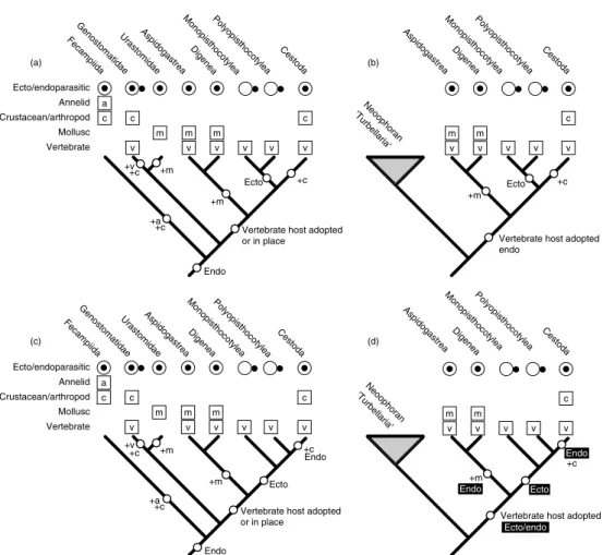
surely began with a single speciation event and continued with a remarkable adaptive radia- tion. Not withstanding the many forays into parasitism made by turbellarian flatworms, it is without doubt that the emergence of the Neodermata that allowed the platyhelminths to engage successfully in a lifestyle that ties them so closely to the lives and evolution of so many other organisms. To infer the plesiomor- phic (‘primitive’) characters of the common neodermatan ancestors we need to determine their interrelationships and to find the sister group to the Neodermata; which lineage of platyhelminths is most closely related to the obligate parasites? There have been a number of phylogenetic estimates from morphological and molecular data yielding a variety of solu- tions (see Fig. 1.3).
One group of turbellarians, which have provided a compelling link to the obligate par- asites, is the Temnocephalida. With various degrees of adaptation towards their ectocom- mensal existence with freshwater crustaceans,
including loss of locomotory cilia and posses- sion of a distinct posterior sucker (Cannon and Joffe, 2001), it is easy to visualize a general- ized transition towards obligate parasitism by placing the temnocephalans as sister group to the Neodermata. Indeed, an early cladistic analysis yielded just this answer (Brooks and McLennan, 1993; Fig. 1.3b). Other candidate lineages have been the Urastomidae and Genostomatidae, forming with the Neodermata the Mediofusata, as part of the Revertospermata hypothesis; Revertospermata = Fecampiida + Mediofusata (Kornakova and Joffe, 1999;
Fig. 1.3c). Ehlers was the first to suggest another revertospermatan group, the Fecampiidae, as a possible sister group to the Neodermata, although he settled on a ‘dalyel- lioid’ clade, which included Temnocephalida, Fecampiidae and Udonellidae (Ehlers, 1985;
Fig. 1.3a). Udonellidae has since been shown to be unequivocally a group of highly modified monopisthocotylean monogeneans (Littlewood et al., 1998), but the morphologists have
(a) Ehlers (1984); morphology (b) Brooks and MacLennan (1993); morphology (c) Kornakova and Joffe (1999); morphology
'Turbellaria' Neodermata 'Turbellaria' Neodermata 'Turbellaria' Neodermata
Mediofusata Revertospermata
'Turbellaria' Neodermata
(d) Zamparo et al. (2003); morphology (e) Lockyer et al. (2003 a,b); complete 18S and 28S rDNA
‘Turbellaria’
‘Turbellaria’:
Temnocephalida Urastomidae
Fecampiida Genostomatidae Prolecithophora
Tricladida
Rhabdocoela Neodermata
Hosts of putative outgroup taxa
Fecampiida : crustaceans, myzostomids, annelids
Genostomatidae : crustaceans, fish Urastomidae : bivalve molluscs Temnocephalida : crustaceans Fecampiidae
Urastomidae
Urastomidae Genostomatidae Fecampiida Temnocephalida
'Dalyellioida'
Fig. 1.3. Four morphological and one molecular estimate of the interrelationships of the Platyhelminthes with emphasis on identifying the sister group to the obligate parasites (Neodermata). Currently, the two main competing hypotheses concern (c) a revertospermatan clade and (e) a large clade of neoophoran turbellarians.
frequently placed parasitic turbellarian groups as close relatives of the Neodermata (Fig. 1.3a–d). Certainly, if one includes the spermatological features in a morphological cladistic analysis, the Revertospermata are resolved (Littlewood et al., 1999b) and a web- based listing of turbellarian taxonomy recog- nizes the integrity of this grouping (http://
devbio.umesci.maine.edu/styler/turbellaria/).
In contrast, molecular systematic estimates of flatworm phylogeny from nuclear ribosomal data are quite different, and consistently recog- nize a large clade of neoophoran turbellarians as the likely sister group to the Neodermata; the parasitic turbellarians along with the triclads and prolecithophorans form a well-supported sister group (Fig. 1.3e).
As with many other taxonomic groups, molecular phylogenetic estimates for the flat- worms have suffered from poor sampling, sin- gle gene sampling, long-branch attraction and inadequate analyses, and although some of these issues have now been addressed, addi- tional data are needed to support or refute the various morphological and molecular esti- mates; summaries of earlier molecular studies can be found in Littlewood et al. (1999a), Littlewood and Olson (2001), Lockyer et al.
(2003a) and Baguñà and Riutort (2004).
A review of the recent literature might suggest that there are too many polarized opinions and entrenched viewpoints on molecules versus morphology or characters being homologous or homoplasious for the evidence to speak for itself. However, if one can argue that molecular data do not come quite as loaded with a priori statements on homology as has been shown or claimed for morphology, as I would, then the two ribosomal genes sampled so far both suggest that a large clade of turbel- larians is the most likely sister group to the Neodermata (Fig. 1.3e). Nevertheless, consen- sus of opinion based on congruence between independent data sets remains an elusive but necessary goal.
The solutions provided by neither molec- ular nor morphological data allow a simple story to be told concerning the stem group neodermatans, whether considered individu- ally or collectively (Fig. 1.4). The sister group according to molecular data is so large that plesiomorphic characters for the clade are all
but non-existent and hold little clue as to the origins of parasitism, except that the ancestral neodermatan was endoparasitic and adopted a vertebrate as its host first (Littlewood et al., 1999a; Fig. 1.4d). Without being able to rec- oncile a parasitic or commensal sister group to the Neodermata, whether intuitive, attractive or indeed correct, the most parsimonious solu- tion presently is that the vertebrates have always been host to neodermatans and so vertebrates must have been the first host. This scenario has generated disquiet amongst those workers who would argue that parasites with complex life cycles most likely parasitized the first interme- diate hosts first, on an evolutionary as well as an ontogenetic scale, and thereafter recruited other hosts, i.e. trematodes first used molluscs and cestodes first used arthropods before they each ultimately involved vertebrates as their definitive hosts. A common argument is that as vertebrates were eating these molluscs and crustaceans, the parasites simply adapted to survive within the guts of the vertebrates. Was this a one-host life cycle with vertebrates, as it is among monogeneans? The argument will fail if it can be shown that the associations between the major parasitic lineages and vertebrates are not homologous. If the Revertospermata hypothesis is correct, and similar sperm mor- phology can be demonstrated to have arisen through common ancestry, rather than conver- gent evolution, then a two-host life cycle with an arthropod and a vertebrate may be the most parsimonious scenario; although the relative position of the Urastomidae could easily make a two-host life cycle with a mollusc and a ver- tebrate plausible. See Cribb et al. (2001a, 2003) for further studies on the origins and radiation of trematode life cycles.
The interrelationships among the Neodermata are just as important in reconcil- ing this conundrum, and yet unfortunately they seem just as confused. In this case the confu- sion arises from alternative solutions provided by molecular data. Morphological analysis consistently resolves the Trematoda (Digenea and Aspidogastrea) as sister taxon to the Cercomeromorpha (Cestoda and Monogenea).
Historically, the Monogenea have been resolved as a monophyletic group by most morpholo- gists offering at least four synapomorphies:
(i) larva with three ciliated zones; (ii) larva and