N N PT P TE EL L W WE EB B C CO OU UR RS SE E – – A AD DV VA AN N CE C ED D C CL LI IN N IC I CA AL L P PR RO OT TE EO OM MI IC CS S
LECTURE-19 MS for PTM analysis
Handout PREAMBLE
Proteins are the most dynamic entities that govern all the activities of a living cell. Any disorder can be tracked down to a change in either the concentration of proteins or the overall structure and function of the proteins. The central dogma focuses from DNA to RNA to protein, with the latter acting as the key players in cellular processes. The DNA counterpart of the central dogma only directs the sequence of amino acids in the proteins. Many proteins undergo chemical modifications at certain amino acid residues following translation. These modifications are essential for normal functioning of the protein and are carried out by one or more enzyme catalyzed reactions. Post- translational modifications like phosphorylation and glycosylation impart important characteristic features to the proteins. Understanding the protein modifications is one of the most challenging fields of proteomics. Studying PTMs is considered very important, because, the unusual modification pattern of some proteins also lead to their loss/gain in function leading to disorders. In this lecture we will focus on various kinds of post- translational modifications that a protein usually undergoes and how to detect them, with a special focus on mass spectrometry based analysis methods.
N N PT P TE EL L W WE EB B C CO OU UR RS SE E – – A AD DV VA AN N CE C ED D C CL LI IN N IC I CA AL L P PR RO OT TE EO OM MI IC CS S
OUTLINE OF LECTURE 1. Introduction
2. Post translational modifications
3. Gel-based method for identifying post-translational modifications 4. Limitations of gel-based method for PTM analysis
5. MS based method for identification of post-translational modifications a) Phospho-proteome analysis
b) Glyco-proteome analysis 6. Conclusion
N N PT P TE EL L W WE EB B C CO OU UR RS SE E – – A AD DV VA AN N CE C ED D C CL LI IN N IC I CA AL L P PR RO OT TE EO OM MI IC CS S
BOX-1: TERMINOLOGY
Post-translational modification (PTM): The chemical modifications that take place at certain amino acid residues after the protein is synthesized by translation are known as post-translational modifications. These are essential for normal functioning of the protein.
Phosphorylation: The process by which a phosphate group is attached to certain amino acid side chains in the protein, most commonly serine, threonine and tyrosine.
Glycosylation: The attachment of sugar moieties to nitrogen or oxygen atoms present in the side chains of amino acids like aspargine, serine or threonine.
Acylation: The process by which an acyl group is linked to the side chain of amino acids like aspargine, glutamine or lysine.
Alkylation: Addition of alkyl groups, most commonly a methyl group to amino acids such as lysine or arginine. Other longer chain alkyl groups may also be attached in some cases.
Hydroxylation: This PTM is most often found on proline and lysine residues which make up the collagen tissue. It enables crosslinking and therefore strengthening of the muscle fibres.
N N PT P TE EL L W WE EB B C CO OU UR RS SE E – – A AD DV VA AN N CE C ED D C CL LI IN N IC I CA AL L P PR RO OT TE EO OM MI IC CS S
1. INTRODUCTION
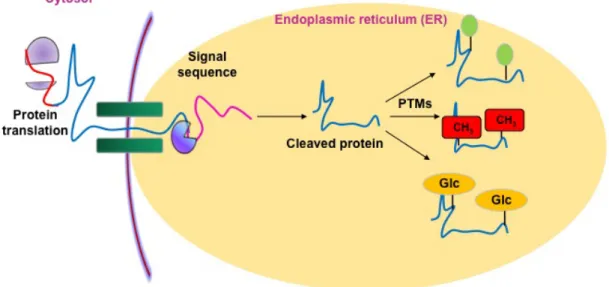
Post-translational modifications (PTMs) are chemical modifications that take place at certain amino acid residues after the protein is synthesized by translation (Fig 1). These post-translational modifications are essential for normal functioning of the protein. Let us consider we have a 10 Kb DNA fragment, which contains five genes. Each gene gives rise to mRNA, which also possesses 5 splice variants each. Hence the entire mRNA pool consists of 25 different kinds of mRNA. Now each mRNA gives rise to three different isoforms of proteins, varying in degrees of post-translational modifications, for example, one is phosphorylated, one is glycosylated and the other is ADP ribosylated.
Therefore, the number of total protein pool will become 75. Therefore, in this hypothetical scenario, from DNA to Protein, five genes give rise to 75 different kinds of proteins all having varying functions.
Fig 1. An overview of post-translational modifications (PTM)
N N PT P TE EL L W WE EB B C CO OU UR RS SE E – – A AD DV VA AN N CE C ED D C CL LI IN N IC I CA AL L P PR RO OT TE EO OM MI IC CS S
Now let’s consider one of the proteins is acting abnormally. On sequencing the entire 10 Kb fragment you found no mutation. Now you wonder if the genetic code hasn’t changed, why the protein is behaving abnormally? Further analysis of the entire proteome reveals that a related kinase has a gain in function mutation. On scanning the proteome of the 10 Kb fragment, now you find that one particular protein is heavily phosphorylated, more than usual. From this example you can understand that changes at post-translational modifications can drastically affect the protein function.
Illustration 1: An overview of PTMs
Once the protein has been synthesized by the ribosome from its corresponding mRNA in the cytosol, many proteins get directed towards the endoplasmic reticulum for further modification. Certain N and C terminal sequences are often cleaved in the ER after which they are modified by various enzymes at specific amino acid residues. These modified proteins then undergo proper folding to give the functional protein.
Illustration 2: Increased complexity of proteome due to PTMs
The final structure of functional proteins most often does not correlate directly with the corresponding gene sequence. This is due to the PTMs that occur at various amino acid residues in the protein, which cause changes in interactions between the amino acid side chains thereby modifying the protein structure. This further increases the complexity of the proteome as compared to the genome.
N N PT P TE EL L W WE EB B C CO OU UR RS SE E – – A AD DV VA AN N CE C ED D C CL LI IN N IC I CA AL L P PR RO OT TE EO OM MI IC CS S
Many cancers result due to hyperactivity of Mitogen Activated Protein Kinases (MAP Kinases). These kinases activate transcription factors, which regulate gene expressions at large. For example, in breast cancer, a correlation between cancer onset and up-regulation of MAP kinase activity is depicted by many studies. Sequence analysis suggests that there is no mutation in the gene encoding these kinases and hence further analysis of the system revealed the abundance of growth factors and peptides and molecules or ligands, which tend to increase the signaling cascade thereby activating MAP kinase. Once activated, MAP kinase activates the downstream molecules, thereby influencing gene expression. Hence it becomes necessary to study the phosphorylation patterns in various diseases so that these kinases can be sought as a potent target for therapy. It is now clear as to why analysis of post-translational modifications is emerging as one of the most promising discipline of proteomics. There are many ways of analyzing post-translational modifications in proteins; most common being dyes specific for the modifications or special fragmentation spectrum in mass spectrometry.
N N PT P TE EL L W WE EB B C CO OU UR RS SE E – – A AD DV VA AN N CE C ED D C CL LI IN N IC I CA AL L P PR RO OT TE EO OM MI IC CS S
2. POST TRANSLATIONAL MODIFICATIONS
Post-translational modification broadly involves any change in the overall chemical nature of the protein that occurs after the protein is translated from the mRNA. The DNA sequence only dictates the amino acid sequence of the protein. The fate of protein post synthesis is governed entirely by the environment, interacting molecules etc, i.e., the cell overall decides the fate of the protein as per its requirement. The most common type of post-translational modification involves phosphorylation, glycosylation and disulphide linkage formation (Fig 2 and 3).
Fig 2. Most common types of PTM
N N PT P TE EL L W WE EB B C CO OU UR RS SE E – – A AD DV VA AN N CE C ED D C CL LI IN N IC I CA AL L P PR RO OT TE EO OM MI IC CS S
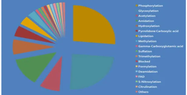
Fig 3. Known PTMs and amino acid residues modified by them
Illustration 3: Increased complexity of proteome due to PTMs
There are several types of post-translational modifications that can take place at different amino acid residues. The most commonly observed PTMs include phosphorylation, glycosylation, methylation as well as hydroxylation and acylation.
Many of these modifications, particularly phosphorylation, serve as regulatory mechanisms for protein action.
N N PT P TE EL L W WE EB B C CO OU UR RS SE E – – A AD DV VA AN N CE C ED D C CL LI IN N IC I CA AL L P PR RO OT TE EO OM MI IC CS S
PHOSPHORYLATION
Phosphorylation is the most common type of post-translational modification.
Phosphorylation refers to addition of phosphate groups to specific residues on proteins;
especially, serine, threonine and tyrosine residues. Protein phosphorylation is brought about by special enzymes, which are known as kinases. The kinases may be divided into broadly three classes based on the residue they phosphorylate. Phosphorylation is brought about by removing the γ phosphate group of ATP and adding it to the OH group of either of the hydroxyl containing amino acids (Fig 4).
Fig 4. A scheme for phosphorylation in general
Protein phosphorylation is an extremely important process, critical for many biological pathways, such as signal transduction. One of the most studied signal transduction pathways involve the RTK (Receptor tyrosine kinase), in which receptor dimerization occurs, i.e. the receptors auto-phosphorylate each other and serve as a platform or
N N PT P TE EL L W WE EB B C CO OU UR RS SE E – – A AD DV VA AN N CE C ED D C CL LI IN N IC I CA AL L P PR RO OT TE EO OM MI IC CS S
adaptor for other proteins to come and bind. Subsequently many other proteins bind and get phosphorylated. The phosphorylation of the protein renders them active and they are able to interact with many downstream molecules as well, which ultimately leads to change in gene expression.
Illustration 4: Phosphorylation reactions
Phosphorylation of amino acid residues is carried out by a class of enzymes known as kinases that most commonly modify side chains of amino acids containing a hydroxyl group. Phosphorylation requires the presence of a phosphate donor molecule such as ATP, GTP or other phoshorylated substrates. Serine is the most commonly phosphorylated residue followed by threonine and tyrosine. Removal of phosphate groups is carried out by the phosphatase enzyme and thus this forms one of the most important mechanisms for regulation of proteins.
Many cancerous pathways involve the activation of MAP kinases, which tend to increase the gene expression of many oncogenes by activating many transcription factors by phosphorylating them. Regulation of phosphorylation is brought about by another set of enzymes known as phosphatases, which remove the γ phosphate group from the amino acid residues thereby rendering them inactive. A detailed study of the phospho-proteome is necessary for elucidating the transcription factors that are elevated during onset of cancer and other diseases.
N N PT P TE EL L W WE EB B C CO OU UR RS SE E – – A AD DV VA AN N CE C ED D C CL LI IN N IC I CA AL L P PR RO OT TE EO OM MI IC CS S
GLYCOSYLATION
Glycosylation refers to the enzymatic process of addition of carbohydrate moieties to the protein. Glycosylation is carried out either in the endoplasmic reticulum or the golgi apparatus and accordingly they are either N-linked or O-linked glycosylation. N-linked glycosylation is so called because the glycan group is attached to the nitrogen atom of asparagine or arginine whereas, in O-linked glycosylation, the glycan group is attached to the oxygen atom of serine, threonine or tyrosine. Glycosylation not only affects protein folding, but also assists in protein trafficking. Glycosylation usually tends to solubilize a protein and also it is via these glycan groups that many proteins interact with each other, especially the extracellular matrix proteins. The blood grouping system in humans are due to the presence of these wide varieties of glycan moieties, which act as antigenic counterparts during blood transfusion.
Illustration 5: Glycosylation reactions
Glycosylation involves the enzymatic addition of saccharide molecules to amino acid side chains. This can be of two types: N-linked glycosylation, which links sugar residues to the amide group of aspargine and O-linked glycosylation, which links the sugar moieties to the hydroxyl groups of serine or threonine. Suitable glycosyl transferase enzymes catalyze these reactions. Sugar residues that are attached most commonly include galactose, mannose, glucose, N-acetylglucosamine, N-acetylgalactosamie as well as fucose.
LIPID ANCHORS
Addition of lipid chains or long chain CoA chains to the proteins is another form of post- translational modification, usually, done to add hydrophobic character into the proteins and also is thus a mode of targeting proteins. Proteins of the membranes contain extreme hydrophobic patches in them to interact with the lipid bilayers of the
N N PT P TE EL L W WE EB B C CO OU UR RS SE E – – A AD DV VA AN N CE C ED D C CL LI IN N IC I CA AL L P PR RO OT TE EO OM MI IC CS S
membranes. N-terminal glycine is often modified by addition of myristic group catalyzed by the enzyme, N-myristoyl transferase. Often this modification adds to the function of the protein, for example, the Net protein of HIV1 binds its own mysristoylated N terminus for activity.
N-TERMINAL AND C-TERMINAL MODIFICATIONS
The most common type of N-terminal modification involves acetylation, where an acetyl group from Acetyl CoA is added to the lysine residue at the N-terminal region of the protein. Acetylation is an important post-translational modification, mainly occurring in the DNA binding protein histones. Histone acetylation/ deacetylation is an important phenomenon governing gene expression. Another important modification is methylation, which can occur at any residue except for the N-terminus. Histone methylation also tends to create local unwinding of the DNA leading to the accessibility of RNA polymerase for the DNA.
Several proteins may contain additional carbon backbones attached to the cysteine residues at the C-terminal region of the proteins. Addition of carbon backbone is brought about by an enzyme known as farnesyl transferase, which transfers a 15- carbon backbone of farnesyl pyrophosphate to the cysteine residue of the protein.
N N PT P TE EL L W WE EB B C CO OU UR RS SE E – – A AD DV VA AN N CE C ED D C CL LI IN N IC I CA AL L P PR RO OT TE EO OM MI IC CS S
UBIQUITINATION
Not even a highly efficient cell can guarantee that all the proteins synthesized at a given time point are properly folded. Misfolded proteins so generated can lead to havoc inside the cell, as they tend to aggregate and inhibit the functions of other proteins.
Inside the cell, the proteins are targeted for destruction by tagging them with a 8 kDa highly conserved protein, Ubiquitin. Upon recognizing the ubiquitin tag, the proteosomal complex digests the proteins into peptides and amino acids released are recycled back.
Ubiquitination is brought about by three enzymes, namely Ubiquitin activating enzyme E1, Ubiquitin conjugating enzyme E2 and Ubiquitin Ligase E3. E1 activates ubiquitin and transfers it to E2. The ubiquitin is then transferred to the target protein on the lysine residue by E3. Ubiquitination is an irreversible process that maintains the normal turnover number of proteins in the cell.
DISULPHIDE BRIDGES
The importance of disulphide bridges in protein structure assembly and functions was first established by famous scientist Anfinsen on Ribonuclease A. When treated with 8 M urea, ribonuclease A completely loses its activity, which regains activity after the urea is dialyzed out.
Disulphide bridges are formed between any two cysteine residues, which can orient themselves in space so that it comes in close proximity and S-S linkage is formed with the elimination of hydrogen. Inside the cells, disulphide linkages are brought about by
N N PT P TE EL L W WE EB B C CO OU UR RS SE E – – A AD DV VA AN N CE C ED D C CL LI IN N IC I CA AL L P PR RO OT TE EO OM MI IC CS S
special chaperones, which fold the proteins in such a way that two distant cysteine come in near contact. Disulphide bridges tend to stabilize the protein structure, making them globular and compact and more soluble. Along with chaperones, the environment of the media also governs the formation of disulphide bridges. The reducing environment of the cytosol usually does not permit formation of disulphide linkages all by themselves and hence the proteins need the assistance of chaperones for linkage formation. In Anfinsen’s experiment, when denaturant like urea, which breaks the 3D structure of the protein, was removed, the protein by virtue of random motion coiled up and as two cysteine residues came in direct contact they formed disulphide bridges, thereby stabilizing the protein and reinforcing the activity.
Chemical reagents like β-mercaptoethanol (BME) or dithiothreitol (DTT) break these disulphide bridges and denature the proteins. However, Arial oxidation tends to reoxidise these bonds and so, to prevent that, iodoacetamide is added so that the sulphide bonds are alkylated and cannot oxidize back.
N N PT P TE EL L W WE EB B C CO OU UR RS SE E – – A AD DV VA AN N CE C ED D C CL LI IN N IC I CA AL L P PR RO OT TE EO OM MI IC CS S
3. GEL-BASED METHODS FOR IDENTIFYING PTM
Gel-based techniques for identifying post-translational modification are effective when radioactive methods are used to detect phosphorylation in proteins. Chiefly, cells are allowed to grow in phosphate labeled media for few generations for accumulation of labeled nucleotide triphosphates. After few generations, the cells are lysed and the proteome is analyzed. Kinases, which accounts for the phosphorylation, use these labeled nucleotide triphosphates to label the proteins. A scintillation counter counts the β-emissions from protein spots in a gel and analyzes the phosphorylated proteins in the gel. However, the radioactive methods are always considered hazardous for human health and β-emissions being moderately penetrative, radioactive methods for gel based phosphorylation detection are discouraged.
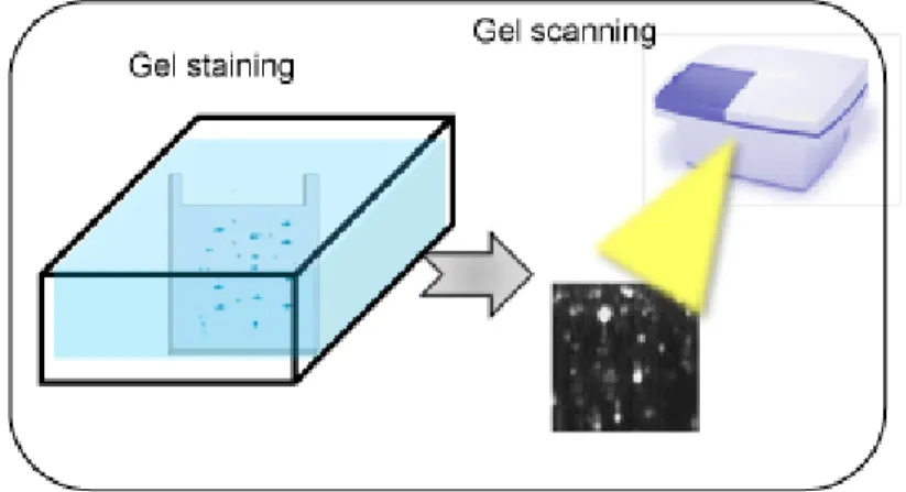
To analyze phosphoproteome, specific dyes are developed, which could interact with the phosphate groups of the proteins and hence differentially stain them (Fig 5).
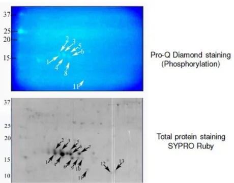
Stains like Pro-Q Diamond is used to differentially stain the phosphoproteome. Dual staining is done in such a case to identify the phosphoproteome from the entire proteome coverage (Fig 6).
Fig 5. A schematic of phosphostaining procedure
N N PT P TE EL L W WE EB B C CO OU UR RS SE E – – A AD DV VA AN N CE C ED D C CL LI IN N IC I CA AL L P PR RO OT TE EO OM MI IC CS S
Fig 6. Dual Staining to identify phosphoproteome and total proteome
Illustration 6: Gel-based detection techniques for PTMs
Protein phosphorylation can be detected using gel-based detection technique. Proteins separated on a 2-DE gel are first placed in a fixing solution containing methanol and acetic acid, which fixes the protein bands on to the gel and minimizes any diffusion.
They are then stained using the Pro-Q-diamond staining solution, which selectively stains only phosphoproteins on the gel. The excess stain is then washed off with a solution of methanol and acetic acid.
The stained gel is then scanned at its excitation wavelength using a gel scanner. The gel image obtained shows the protein bands corresponding to only the phosphoproteins present. This image is saved and the gel is then removed from the scanner for treatment with the second stain, a procedure known as dual staining.
The scanned gel is then removed from the scanner and placed in the SYPRO-Ruby Red fluorescent dye solution. This dye stains all the protein spots present on the gel thereby providing a total protein image with sensitivity down to nanogram level. Excess dye is then washed off using a solution of methanol and acetic acid.
The gel stained with SYPRO-Ruby Red is then scanned in the gel scanner at its
N N PT P TE EL L W WE EB B C CO OU UR RS SE E – – A AD DV VA AN N CE C ED D C CL LI IN N IC I CA AL L P PR RO OT TE EO OM MI IC CS S
proteins present on the gel are detected. This dual staining procedure provides a useful comparative profile of the phosphoproteins and the total proteins on the gel, thereby enabling detection of the phosphorylated proteins.
Protein mixture containing phosphorylated as well as other unmodified proteins can be separated by a suitable electrophoresis technique. SDS-PAGE and two dimensional gel electrophoresis are most commonly used for protein separation. These separated proteins on the gel are used for further analysis.
The separated protein bands are then blotted onto a nitrocellulose membrane. These membranes are then probed either by means of specific anti-phospho-amino acid antibodies or more recently, by motif antibodies that specifically bind to proteins having phosphorylation at a particular amino acid residue. This binding interaction can then be detected by means of suitably labeled secondary antibodies or by autoradiography using a radioactive probe. Thus, the use of immunoblotting technique has been shown to be extremely effective for detection of PTMs.
Stains like Pro-Q Emerald and Sypro Ruby can differentially stain glycoproteins and hence the entire glycoproteome can also be detected using these simpler techniques.
For more sophisticated and sensitive versions of staining, antibodies specific to the phosphotyrosine residues or glycan moeites are developed, which are conjugated to a fluorophore. However, the biggest disadvantage of gel-based methods for detection of post-translational modifications remained in variability, sensitivity and proteome coverage. As a result MS-based approaches are widely used.
N N PT P TE EL L W WE EB B C CO OU UR RS SE E – – A AD DV VA AN N CE C ED D C CL LI IN N IC I CA AL L P PR RO OT TE EO OM MI IC CS S
4. LIMITATIONS OF GEL-BASED METHODS OF PTM ANALYSIS
Despite its simplicity, gel based methods of post-translational modification analysis, could not make big impact because of the variability and reproducibility issues in gels, lack of comprehensive proteome coverage and sensitivity issues. Phosphoproteome accounts for a tiny percentage of the global proteome at any instant and phospho-stain dyes are not sensitive enough to detect the entire phosphoproteome. Also, the issue of diffusion of dyes led to inaccurate measurements in the quantification.
Integral membrane proteins, which are highly hydrophobic in nature are usually under-represented in the gels and hence there is a finite chance of losing them during detection. The membrane proteome accounts for a large number of phosphorylated and lipolyted modifications, all of which are undetected in gel-based approach. The requirement of dual staining also takes away majority of time for the detection and analysis, which is why techniques like microarray and mass spectrometry are more commonly used over gel-based approaches for the PTM analysis.
N N PT P TE EL L W WE EB B C CO OU UR RS SE E – – A AD DV VA AN N CE C ED D C CL LI IN N IC I CA AL L P PR RO OT TE EO OM MI IC CS S
5. MS BASED METHODS FOR IDENTIFICATION OF PTMs
MS-based methods for identifying post-translational modification gradually took over other proteomic approaches because of its sensitivity of detection, low run time and reduced biochemical biasness (this is in reference to specific dyes used to stain specific modifications). The use of mass spectrometry based PTM analysis goes way back to the time when scientists characterized the spectrum of hemoglobin from sickle cell anemia. Sickle cell anemia results from a change in one nucleotide of a codon resulting in the change in one amino acid, from glutamic acid to valine. This change in the hemoglobin is evident not only on the phenotype but also on the mass spectrum as well.
The fragmentation pattern of both normal and sickle cell hemoglobin are the same.
However, one of the ion peaks shifts showed change in mass of 30 Da in case of sickle cell anemia.
Every post-translational modification adds a specific mass to the protein, depending on the number of modifications. For example, a single phosphorylation in the protein would increase the overall protein mass by a magnitude of 80 Da. Identifying the amount of change in the spectrum shift is thus a direct measure of the post-translational modification, in terms of both the number of modifications as well as the site of modification (using tandem MS/MS splitting and sequence determination). However, like every other techniques, MS based approach also faces several limitations, which would be discussed.
N N PT P TE EL L W WE EB B C CO OU UR RS SE E – – A AD DV VA AN N CE C ED D C CL LI IN N IC I CA AL L P PR RO OT TE EO OM MI IC CS S
Illustration 7: MALDI-TOF analysis
Post-translational modifications can be detected by means of mass spectrometry due to the unique fragmentation patterns of phosphorylated seine and threonine residues. The modified protein of interest is digested into smaller peptide fragments using a suitable enzyme like trypsin. This digest is then mixed with a suitable organic matrix such as a- cyano-4-hydroxycinnamic acid, sinapinic acid etc. and then spotted on to a MALDI plate.
The target plate containing the spotted matrix and analyte is placed in a vacuum chamber with high voltage and short laser pulses are applied. The laser energy gets absorbed by the matrix and is transferred to the analyte molecules, which undergo rapid sublimation resulting in gas phase ions. These ions are accelerated and travel through the flight tube at different rates. The lighter ions move rapidly and reach the detector first while the heavier ions migrate slowly. The ions are resolved and detected on the basis of their m/z ratios and a mass spectrum is generated.
Identification of PTMs by MS largely lies in the interpretation of results. Comparison of the list of observed peptide masses from the spectrum generated with the expected peptide masses enables identification of those peptide fragments that contain any PTM due to the added mass of a modifying group. In this hypothetical example, two peptide fragments are found to have different m/z values, differing by 80 daltons and 160 daltons. It is known that the added mass of a phosphate group causes an increase in m/z of 80 daltons. Therefore, this principle of mass difference enables detection of modified fragments.
Illustration 8: Methodology for MALDI analysis
Let us take a test sample which is phosporylated. Let user take the sample as apomyoglobin as known standard and spot it on the MALDI plate. User must play around the spot region to find sweet spot, where the peaks are more in number with high intensity. After 100 profiles user can save the data. In between firing user have option for abort, resume, suspend and clear data. User can select these options depending on the profile data obtained. In most cases the default parameters for peak processing are best suited. Once the PMF data is ready, data in the excel format can be exported, and saved. The mass can be calculated from any two peaks by taking the difference and applying the formulas. For the PTM identification, user need to have the information of observed mass from standard peaks, and even the mass value for each amino acids. The difference in mass between observed and theoretical mass, determines the phosphate group addition i.e. PTM has taken place on this particular amino acid. The 42Da difference corresponds to acetylation, 43Da for trimethylation and 617.6Da for Heme. In similar way, the PTM can be identified if user has a basic knowledge of the amino acid mass. The difference between two adjacent peak mass helps to identify the PTM.
N N PT P TE EL L W WE EB B C CO OU UR RS SE E – – A AD DV VA AN N CE C ED D C CL LI IN N IC I CA AL L P PR RO OT TE EO OM MI IC CS S
5.1. PHOSPHO-PROTEOME ANALYSIS
The phospho-proteome analysis itself is a very challenging task because of the following reasons:
(a) Phospho-proteins account for a minute fraction of the entire proteome at any given instant. The reversible action of phosphorylation, carried out by phosphatases, makes it extremely challenging to detect the phosphorylating moieties. When a ligand binds to the cell surface receptor, the signal transduced into the cell is only transient and not persistent. As a result, the time span during which a protein remains in its phosphorylated form is extremely small.
(b) Phosphorylation sites are heterogenous in proteins. The key residues of phosphorylation in proteins are tyrosine, serine and threonine. One cannot guarantee at a particular moment, which residue is going to be phosphorylated. Hence, developing antibodies against a particular residue for detecting phosphorylation is challenging.
(c) Also, the abundance of these signaling molecules is so small, that a highly sensitive detection technique is necessary for detection of these proteins.
All of these challenges are addressed by MS-based approach for detecting phosphorylation. However, in MS, since the high voltage and particle bombardment leads to fragmentation of the peptides, there is a finite chance of loss of the phosphorylation during fragmentation. Thus, a serious issue that needs to be addressed is the stability of the phosphorylating groups, for example, a phosphorylated histidine is extremely labile, whereas a methylated lysine is extremely stable. At the same time, phosphorylated tyrosine is extremely stable whereas, phosphorylated serine and
N N PT P TE EL L W WE EB B C CO OU UR RS SE E – – A AD DV VA AN N CE C ED D C CL LI IN N IC I CA AL L P PR RO OT TE EO OM MI IC CS S
threonine are extremely labile and lose the phosphate group by a phenomenon of beta elimination. The beta elimination complicates the MS spectrum by creating anomalous fragments and hence it becomes difficult to identify the residues by standard database search. However, this problem has been addressed by the availability of precursor ion scanning whereby prior to particle bombardment, the precursor ion is scanned and recorded and then allowed to fragment further.
Enrichment of proteome for target phosphoproteome is essential for its analysis.
Usually, three different techniques are employed for this purpose:
Immobilized Metal Affinity Chromatography (IMAC): This approach is based on the principle of affinity of phosphates with metal cations like zinc, gallium and iron. Passing the proteins extracted overall through this column selectively adsorbs the phosphoproteins.
Chemical derivatization: Beta elimination under normal condition results in dehydroalanine and dehydroamino butyric acid residues. These can be detected after chemical modification with ethane thiol.
Immunoprecipitation: Phospho-specific antibodies can be used to selectively precipitate phosphoproteins and thus separating them from entire proteome.
Once the phosphoproteome is collected, they are trypsinized and the peptides so generated are passed though Titanium oxide columns for enrichment of the phosphopepetides. Usually, this chromatographic technique is coupled with MS analysis. The addition of one phosphate group to a peptide increases the mass of the
N N PT P TE EL L W WE EB B C CO OU UR RS SE E – – A AD DV VA AN N CE C ED D C CL LI IN N IC I CA AL L P PR RO OT TE EO OM MI IC CS S
peptide by 80 Da. The purpose of MS in this respect is to identify both the protein by virtue of the peptide and also identify the key phosphorylated residues.
Illustration 9: LC-MS/MS based analysis
Liquid chromatography coupled with mass spectrometry serves as a useful technique for enrichment and identification of proteins having a particular type of PTM from a complex mixture. The complex protein sample is loaded onto a miniaturized affinity column, which will interact specifically with proteins having the PTM of interest. Here, use of immobilized metal affinity chromatography columns containing ions such as Ga3+, Zn2+, Fe3+ or TiO2 which have been found to specifically chelate the phosphorylated proteins are depicted. Unwanted proteins are removed by washing the column with a suitable buffer solution after which the phosphorylated protein of interest is eluted out by modifying the buffer solution.
The protein purified by liquid chromatography is then subjected to typtic digestion followed by analysis using tandem mass spectrometry. Here use of MALDI-TOF-TOF- MS for resolution of the generated ion fragments is demonstrated. Separation is based on the flight time of the ions and greater resolution is achieved due to the presence of two mass analyzers. The peptide ion spectrum generated is analyzed by comparing it with the expected spectrum, thereby allowing determination of modified peptides having different m/z values.
N N PT P TE EL L W WE EB B C CO OU UR RS SE E – – A AD DV VA AN N CE C ED D C CL LI IN N IC I CA AL L P PR RO OT TE EO OM MI IC CS S
GLYCOPROTEOME ANALYSIS
Glycan groups attached to the amino acid residues are relatively more stable than phosphate moieties and hence can be easily detected by MS based approach, provided the glycan chain is not too big and only contributes to a size of 20-40 Da.
Like phosphoproteome analysis, the first task for glycoproteome analysis is to selectively enrich the pool of glycoproteome. This can be done by passing the proteome through a lectin column, whereby glycoproteins would selectively adsorb to the beads by affinity towards lectin. Another approach for selective enrichment of glycoproteome was described by Liu et al, using hydrazide chemistry. Hydrazide beads were mixed with the crude high abundant protein depleted serum and the hydrazide beads selectively adsorbed the glycoproteins. These proteins are then trypsinized and chemically treated to detach them from the beads and then analyzed by MS/MS for both protein identification and site of glycosylation. Ideally, the glycopeptides are chemically or enzymatically treated such that they release the glycan chains either fully or partially, and all these fragments are then analyzed.
N N PT P TE EL L W WE EB B C CO OU UR RS SE E – – A AD DV VA AN N CE C ED D C CL LI IN N IC I CA AL L P PR RO OT TE EO OM MI IC CS S
6. CONCLUSION
MS-based approach for post-translational modification has greatly revolutionized this proteomics field. As discussed in this lecture, post-translational modification is extremely important for studying the dynamic functionalities of the proteins subjected to various conditions. These post-translational modifications are key to develop many diagnostic tools and drugs for treatment. Hence a complete analysis of PTMs needs to be performed before hand and with advancement in mass spectrometry, it is now easily possible.
N N PT P TE EL L W WE EB B C CO OU UR RS SE E – – A AD DV VA AN N CE C ED D C CL LI IN N IC I CA AL L P PR RO OT TE EO OM MI IC CS S
REFERENCES
Proteomics- Introduction to Methods and Applications. Ed. Agniezka kraj and jerzy silberring. Wiley publication, 2012.
Leitner, A., M. Sturm, and W. Lindner. "Tools for analyzing the phosphoproteome and other phosphorylated biomolecules: a review." Anal.Chim.Acta 703.1 (2011): 19- 30.
Liu, T., et al. "Human plasma N-glycoproteome analysis by immunoaffinity subtraction, hydrazide chemistry, and mass spectrometry." J.Proteome.Res. 4.6 (2005):
2070-80.
Meevissen, M. H., et al. "Targeted glycoproteomic analysis reveals that kappa-5 is a major, uniquely glycosylated component of Schistosoma mansoni egg antigens."
Mol.Cell Proteomics. 10.5 (2011): M110.
Zhang, Y., et al. "Combining various strategies to increase the coverage of the plant cell wall glycoproteome." Phytochemistry 72.10 (2011): 1109-23.
Berggren, K. N. et al. An improved formulation of SYPRO Ruby protein gel stain:
Comparison with the original formulation and with a ruthenium II tris
(bathophenanthroline disulfonate) formulation. Proteomics 2002, 2, 486-498.
Reinders, J. & Sickmann, A. Modificomics: Posttranslational modifications beyond protein phosphorylation and glycosylation. Biomolecular Engineering 2007, 24:169-177.
N N PT P TE EL L W WE EB B C CO OU UR RS SE E – – A AD DV VA AN N CE C ED D C CL LI IN N IC I CA AL L P PR RO OT TE EO OM MI IC CS S
Zhang, H. et al., Phosphoprotein Analysis Using Antibodies Broadly Reactive against Phosphorylated Motifs. J Biol. Chem. 2002, 277(42):39379-39387.
Delom, F. & Chevet, E. Phosphoprotein analysis: from proteins to proteomes.
Proteome Science 2006, 4:!5.
Andersson L, Porath J: Isolation of phosphoproteins by immobilized metal (Fe3+) affinity chromatography. Anal Biochem1986, 154:250-254.
Posewitz MC, Tempst P: Immobilized gallium(III) affinity chromatography of phosphopeptides. Anal Chem 1999, 71:2883-2892.
Neville DC, Rozanas CR, Price EM, Gruis DB, Verkman AS, Townsend RR: Evidence for phosphorylation of serine 753 in CFTR using a novel metal-ion affinity resin and matrix assisted laser desorption mass spectrometry. Protein Sci 1997, 6:2436-2445.
Larsen MR, Thingholm TE, Jensen ON, Roepstorff P, Jorgensen TJ: Highly selective enrichment of phosphorylated peptides from peptide mixtures using titanium dioxide microcolumns. Mol Cell Proteomics 2005, 4:873-886.
Oda Y, Nagasu T, Chait BT: Enrichment analysis of phosphorylated proteins as a tool for probing the phosphoproteome. Nat Biotechnol 2001, 19:379-382.
Janek K, Wenschuh H, Bienert M, Krause E: Phosphopeptide analysis by positive and negative ion matrix-assisted laser desorption/ionization mass spectrometry.
Rapid Commun Mass Spectrom 2001, 15:1593-1599.
Wilm M, Mann M: Analytical properties of the nanoelectrospray ion source. Anal Chem 1996, 68:1-8.
N N PT P TE EL L W WE EB B C CO OU UR RS SE E – – A AD DV VA AN N CE C ED D C CL LI IN N IC I CA AL L P PR RO OT TE EO OM MI IC CS S
Erdjan Salih, Phosphoproteomics by mass spectrometry and classical protein chemistry approaches. Mass Spectrometry Reviews 2005, 24:828-846.
Morelle, W. et al., The use of mass spectrometry for the proteomic analysis of glycosylation. Proteomics 2006, 6:3993-4015.
Ptacek, J. et al., Global analysis of protein phosphorylation in yeast. Nature Letters 2005, 438:679-684.
Zhu, H. et al., Global Analysis of Protein Activities Using Proteome Chips. Science 2001, 293:2101-2105.