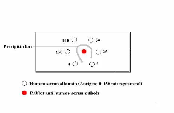
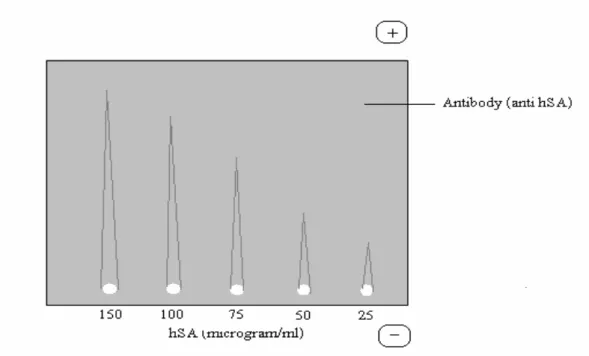
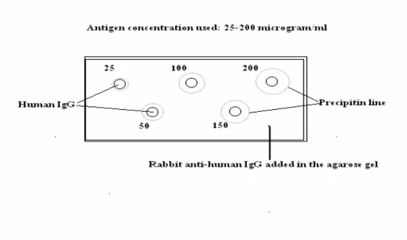
Freund's adjuvant should be used when a small amount of the immunogen is available for immunization. Each of the reactants develops a concentration gradient with the highest concentration near the periphery of the well. The peripheral wells are filled with different concentrations of the same Ag (0-150 micrograms/ml human serum albumin), and the Ab (rabbit anti-human albumin antibody) is added in the central well.
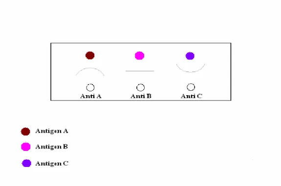
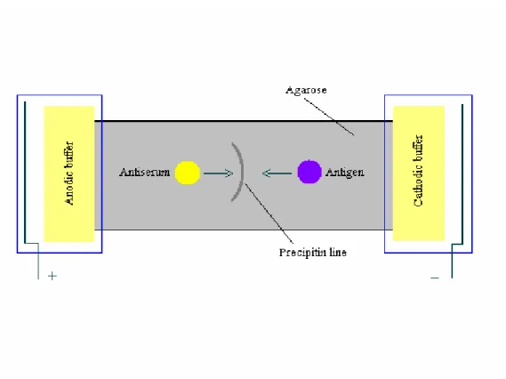
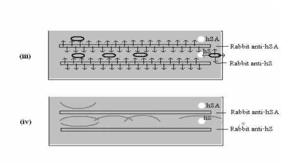
The shape of a precipitin line provides a rough estimate of the relative molecular mass of a globular protein antigen. The principle of immunoelectrophoresis is the formation of precipitate lines (insoluble immune complexes) at the equivalence point (equivalence zone) of the antigen and the corresponding antibody. As with the above method, the Abs are not charged due to the choice of buffer.
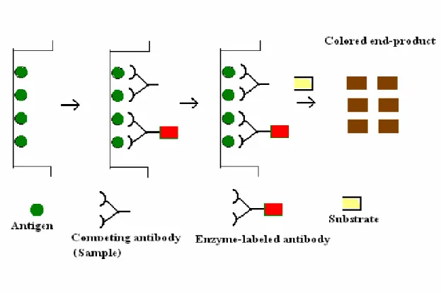
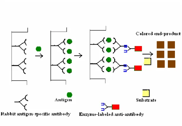
The result is that the rocket-shaped precipitin lines are formed (Fig. 7); the closed areas are proportional to the concentration of Ag in the sample. Higher absorbance more should be the concentration of the antigen or antibody being detected. This type of ELISA is used to detect a pre-exposure to Hepatitis C or Hepatitis E virus by measuring the level of virus-specific IgG antibody in the serum of the suspected patient.
For example, if the 1st antibody is raised in rabbit against an antigen 'X', the 2nd antibody in a mouse (say a monoclonal antibody) is produced using the same antigen ('X'). Immunoblotting can be used to determine a number of important characteristics of protein antigens, the presence and amount of an antigen, the relative molecular mass of the polypeptide chain and the efficiency of extraction of the antigen. Transfer of proteins from the gel to nitrocellulose membrane can be accomplished in one of two ways.
Buffer is pulled through the gel by a heavy weight on top of the membrane sheet. The current is directed at right angles to the gel, causing the electrophoretically resolved proteins to migrate out of it. The proteins in the gel under the influence of an applied electric field migrate out of the gel and are electroblotted on the membrane below within 60-90 minutes.
Larger proteins take longer to transfer from the gel than smaller ones. The gel electrophoresis technique involves denaturing the antigen sample so that only antibodies that recognize denaturation-resistant epitopes will bind the antigen. Partial or incomplete transfer of electrophoretically resolved proteins from the gel to the membrane also limits the detection of antigens by the probe antibody.
The major limitation in using monoclonal antibodies for immunoblotting is that many will not recognize epitopes that are destroyed by the denaturing reagents used to prepare the sample.
No. Property Polyclonal antibodies Monoclonal antibodies
Typhoid remains one of the leading causes of morbidity and mortality in India. The patient's serum is tested in two-fold serial dilutions against each of the different salmonella antigen suspensions. The agglutination test is performed for each of the selected Salmonella antigens in small serum tubes (7 X 1 cm); six of these are used for six serum dilutions (1:2 to 1:128), and in the 7th tube saline is used instead of human serum (non-serum) as a negative control.
The titer of the patient's serum for each salmonella antigenic suspension is read as the highest dilution of serum that gives visible agglutination, e.g. The strength of the reaction for the infecting serotype progressively increases to a maximum titer by the end of the 3rd week. Thus, the antibiotic(s) sensitivity tests are performed to determine (i) the degree of sensitivity or resistance of the pathogen isolated from the patient.
Strains of most pathogenic species differ from each other in their sensitivity, so it is necessary that the sensitivity pattern of the specific strain isolated from the patient be determined by sensitivity tests performed in the laboratory. The MIC is that minimum concentration of a given antibiotic at which no visible growth of the pathogenic microorganism occurs in vitro. Such a report implies that the organism found in the patient's blood sample is responsible for infection in the patient.
This organism could be killed in vivo by administration of four times the MIC of the selected antibiotic(s) to which the pathogenic organism is sensitive. The MIC of a particular drug can be determined by any of the following three methods. The MIC is generally considered to be the lowest concentration of a given antibiotic that prevents the development of visible growth of the test organism.
A comparison of the size of the zone with that produced in a parallel test with a control strain provides a measure of the MIC. However, knowing these limitations, the results of the antibiotic susceptibility test are of great importance for the successful management of the patient with the administration of the selected dose of the selected drugs and in the evaluation of new antibiotics (Phillips 1986). The discs will be placed in the seed medium within a short and standard time of planting the plates.
A predetermined, continuous and stable gradient of drug concentrations is created directly under the strip. The MIC is read directly from the scale printed on the E test strip at the point where the ellipse of growth inhibition crosses the strip.