Citation:Ghosh, A.; Singh, V.K.;
Singh, V.; Basu, S.; Pati, F. Recent Advancements in Molecular Therapeutics for Corneal Scar Treatment.Cells2022,11, 3310.
https://doi.org/10.3390/
cells11203310
Academic Editor: Dimitrios Karamichos
Received: 14 July 2022 Accepted: 12 October 2022 Published: 21 October 2022 Publisher’s Note:MDPI stays neutral with regard to jurisdictional claims in published maps and institutional affil- iations.
Copyright: © 2022 by the authors.
Licensee MDPI, Basel, Switzerland.
This article is an open access article distributed under the terms and conditions of the Creative Commons Attribution (CC BY) license (https://
creativecommons.org/licenses/by/
4.0/).
cells
Review
Recent Advancements in Molecular Therapeutics for Corneal Scar Treatment
Anwesha Ghosh1, Vijay K. Singh2,3, Vivek Singh2,3 , Sayan Basu2,3 and Falguni Pati1,*
1 Department of Biomedical Engineering, Indian Institute of Technology Hyderabad, Kandi, Sangareddy 502284, Telangana, India
2 SSR-Stem Cell Biology Laboratory, Center for Regenerative Ophthalmology, L V Prasad Eye Institute, Hyderabad 500034, Telangana, India
3 Centre for Ocular Regeneration (CORE), L.V. Prasad Eye Institute, Hyderabad 500034, Telangana, India
* Correspondence: [email protected]
Abstract:The process of corneal wound healing is complex and induces scar formation. Corneal scar- ring is a leading cause of blindness worldwide. The fibrotic healing of a major ocular wound disrupts the highly organized fibrillar collagen arrangement of the corneal stroma, rendering it opaque. The process of regaining this organized extracellular matrix (ECM) arrangement of the stromal layer to restore corneal transparency is complicated. The surface retention capacity of ocular drugs is poor, and there is a large gap between suitable corneal donors and clinical requirements. Therefore, a more efficient way of treating corneal scarring is needed. The eight major classes of interventions targeted as therapeutic tools for healing scarred corneas include those based on exosomes, targeted gene therapy, microRNAs, recombinant viral vectors, histone deacetylase inhibitors, bioactive molecules, growth factors, and nanotechnology. This review highlights the recent advancements in molecular therapeutics to restore a cornea without scarring. It also provides a scope to overcome the limitations of present studies and perform robust clinical research using these strategies.
Keywords:corneal scar; molecular therapeutics; exosome; si/microRNAs; recombinant viral vectors;
bioactive molecules; nanotechnology
1. Introduction
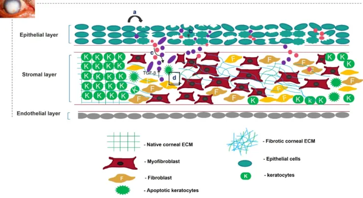
Unregulated tissue fibrosis is generally associated with the development of chronic diseases. Ocular insults to the cornea, such as alkali burns, injuries, and inflammation, trigger the complex process of wound healing to maintain its structural integrity, functional integrity, and transparency [1]. This is mainly characterized by the migration and prolif- eration of corneal epithelial cells, followed by the differentiation of stromal keratocytes into myofibroblasts, the excess deposition of extracellular matrix (ECM) components, an increase in alpha-smooth muscle actin (α-SMA) expression, and ECM remodeling, result- ing in corneal opacity [2,3]. The accumulation of these events leads to a scarred cornea (Figure1). Corneal scarring is the fourth most common cause of blindness, affecting approx- imately five million people globally each year [4]. A scarred cornea is classified according to its degree of opacity. Nebular opacity is caused by superficial scars on the ocular surface.
As the severity of corneal scarring increases, the degree of opacity progresses from nebular to macular and leucoma [5]. Superficial scars heal over a period of three months, restoring vision; however, deep scars generally worsen, leading to blindness [6].
Finding an appropriate therapeutic technique to heal a scarred cornea efficiently has proven difficult. Corneal transplantation is an unsuitable therapy for treating scarring because of the lack of appropriate corneal donors [7]. The use of the amniotic membrane as an ocular bandage because of its anti-angiogenic and antifibrotic properties is a popular treatment method. However, its use is limited because of ethical problems related to tissue donation, potential risks of the transmission of diseases, inter-amnion variability, and
Cells2022,11, 3310. https://doi.org/10.3390/cells11203310 https://www.mdpi.com/journal/cells
Cells2022,11, 3310 2 of 31
reproducibility [8]. Topical ocular drops are a simple way of treating a scarred cornea.
However, their low bioavailability and rapid drainage from the ocular surface limit their popularity [9]. Therefore, there is a need for a therapeutic method that can effectively harness the body’s signaling cascade to halt fibrosis and heal the corneal wound.
Cells 2022, 11, x. https://doi.org/10.3390/xxxxx www.mdpi.com/journal/cells
because of the lack of appropriate corneal donors [7]. The use of the amniotic membrane as an ocular bandage because of its anti-angiogenic and antifibrotic properties is a popular treatment method. However, its use is limited because of ethical problems related to tissue donation, potential risks of the transmission of diseases, inter-amnion variability, and re- producibility [8]. Topical ocular drops are a simple way of treating a scarred cornea. How- ever, their low bioavailability and rapid drainage from the ocular surface limit their pop- ularity [9]. Therefore, there is a need for a therapeutic method that can effectively harness the body’s signaling cascade to halt fibrosis and heal the corneal wound.
This review highlights the recent advancements in therapeutic approaches to treating a scarred cornea at the molecular level. The following eight classes of interventions have been targeted as therapeutic tools for treating scarred cornea: exosomes, microRNAs (miRNAs), recombinant viral vectors, bioactive molecules, growth factors, histone deacetylase inhibitors, and nanotechnology. Exosomes can be used as delivery vehicles for transporting therapeutic cargo, such as antifibrotic miRNAs or proteins from stem cells to the target cells. Small interfering RNAs (siRNAs) or miRNAs can be used to silence the expression of specific fibrotic genes and treat corneal scarring. A scarred cornea can also be treated by modulating the levels of growth factors, such as insulin-like growth factor 1 (IGF-1) and transforming growth factor beta (TGF-β), which are critical for fibrosis. Epi- genetic modulators, such as histone deacetylase inhibitors, or clinically accepted biomol- ecules, such as chitosan, glucosamine, Lycium barbarum polysaccharide, decorin, TPCA-1, or acetylcholine, are recent medicaments that can heal a scarred cornea via their respective biological mechanisms. This review also highlights the advances in nanotechnology for corneal treatment. Understanding the limitations of the existing corneal scarring reme- dies might provide insights for developing new therapeutic modules to restore vision in scarred-cornea patients.
Figure 1. Overview of corneal wound-healing process and scar formation. After an ocular wound, specifically epithelial wounding, (a) the epithelial cells undergo apoptosis, (b) and epithelial cell markers are lost because of corneal epithelial injury, triggering their migration and proliferation. A deep injury disrupts Bowman’s membrane, allowing the invasion of neutrophils (pink) and macro- phages (purple) into the stromal layer and releasing TGF-β. This increases the concentration of TGF- Figure 1.Overview of corneal wound-healing process and scar formation. After an ocular wound, specifically epithelial wounding, (a) the epithelial cells undergo apoptosis, (b) and epithelial cell markers are lost because of corneal epithelial injury, triggering their migration and proliferation.
A deep injury disrupts Bowman’s membrane, allowing the invasion of neutrophils (pink) and macrophages (purple) into the stromal layer and releasing TGF-β. This increases the concentration of TGF-βin the stromal layer (c). Therefore, the keratocytes undergo apoptosis, differentiate into myofibroblasts, and disrupt the arrangement of the corneal ECM (d).
This review highlights the recent advancements in therapeutic approaches to treat- ing a scarred cornea at the molecular level. The following eight classes of interventions have been targeted as therapeutic tools for treating scarred cornea: exosomes, microR- NAs (miRNAs), recombinant viral vectors, bioactive molecules, growth factors, histone deacetylase inhibitors, and nanotechnology. Exosomes can be used as delivery vehicles for transporting therapeutic cargo, such as antifibrotic miRNAs or proteins from stem cells to the target cells. Small interfering RNAs (siRNAs) or miRNAs can be used to silence the expression of specific fibrotic genes and treat corneal scarring. A scarred cornea can also be treated by modulating the levels of growth factors, such as insulin-like growth factor 1 (IGF-1) and transforming growth factor beta (TGF-β), which are critical for fibro- sis. Epigenetic modulators, such as histone deacetylase inhibitors, or clinically accepted biomolecules, such as chitosan, glucosamine,Lycium barbarumpolysaccharide, decorin, TPCA-1, or acetylcholine, are recent medicaments that can heal a scarred cornea via their respective biological mechanisms. This review also highlights the advances in nanotechnol- ogy for corneal treatment. Understanding the limitations of the existing corneal scarring remedies might provide insights for developing new therapeutic modules to restore vision in scarred-cornea patients.
Cells2022,11, 3310 3 of 31
2. Scarring of Cornea
The cornea is a structurally and functionally integrated avascular organ with three cellular layers interrupted by two acellular layers [10]. It is resistant to minor wounds;
however, major ocular trauma can compromise its transparency [11]. A significant abrasion might disrupt the basement and Bowman’s membranes, leading to interactions between the epithelial and stromal layers of the cornea. This interaction initiates the complex wound- healing process [12,13]. Various growth factors, such as fibroblast growth factor (FGF), epidermal growth factor (EGF), and IGF-1, cross the disrupted Bowman’s membrane from the tear film to reach the stromal layer of the cornea during the initial wound-healing pro- cess [14,15]. In the epithelial layer, wounded epithelial cells undergo apoptosis, migration, and proliferation. The concentration of interleukin-1 (IL-1) increases in the epithelial layer upon injury. Therefore, IL-1 crosses the disrupted Bowman’s membrane into the stromal layer and becomes a major factor in killing keratocytes [16].
In the stromal layer, the corneal injury increases the expression of keratocyte membrane- boundαVβ/αβintegrins [17] and various metalloproteases, such as matrix metalloprotease 2 (MMP2) or MMP9 [18]. This cumulatively activates TGF-βfrom latency [19,20]. Neu- trophils, monocytes, and macrophages infiltrate the wounded area during the healing process. They either become incorporated into fibrous DNA-like structures known as neu- trophil extracellular traps of ECM or secrete TGF-β, increasing the concentration of TGF-β in the stromal layer [21–24]. Therefore, TGF-βtakes over the control of the corneal wound- healing process. Quiescent keratocytes transdifferentiate into myofibroblasts, enlarged spindle-shaped cells characterized by the neo-expression ofα-SMA in the presence of TGF- β. Incorporatingα-SMA into the corneal ECM generates a contractile force that disrupts the orthogonal arrangement of its collagen fibrils, altering its normal curvature [25,26].
The active form of TGF-βfirst binds to the keratocyte plasma-membrane-bound TGF- βtype I/II receptor (TBRI/II) and switches on the downstream SMAD-dependent signaling cascade [27]. This increases the expression of various fibrotic genes, such as MMPs and connective tissue growth factor (CTGF) [28]. In addition to TGF-β, other neuropeptides, such as calcitonin gene-related peptide, vasoactive intestinal peptide, and substance P (SP), are released from the axonal nerve endings of corneal epithelial layers. These neu- ropeptides cross the disrupted Bowman’s membrane to reach the stromal layer of the cornea. Substance P binds to the neurokinin 1 receptor (NK1R) of keratocytes and activates three different signaling pathways [29]. The SP/NK1R complex activates the RhoA/ROCK pathway to phosphorylate LIM kinase (LIMK). This inactivates cofilin (an actin-cleaving enzyme) [30], preventing actin degradation and promoting actin polymerization. Actin mostly remains bound to myocardin-related transcription factors A/B (MRTFA/B); how- ever, the increased actin polymerization allows myocardin-related transcription factor A/B (MRTFA/B) to bind to serum response factor, activating profibrotic genes, such as MMPs [31,32]. The SP/NK-IR complex activates phospholipase C-beta and adenylyl cyclase, increasing the intracellular calcium level and cyclic adenosine monophosphate (cAMP) concentrations. This switches on protein kinase A and activates the extracellular signal-regulated kinase/mitogen-activated protein kinase (ERK/MAPK) pathway, increas- ing the proliferation of keratocytes and fibroblast cells [33,34]. The increased Ca2+ions bind to calmodulin and cohere with the Kca3.1 ion channel of keratocytes to hyperpolarize the cells. This causes the cells to skip the G1 phase of the cell cycle, promoting fibrotic cell proliferation [35]. The increase in cellular stress increases the intracellular reactive oxygen species (ROS) levels, which share a positive feedback loop with the secretion of various growth factors, such as TGF-β, proinflammatory cytokines, etc. [36] Interleukin 10 (IL-10), a proinflammatory cytokine, binds to its cognate receptor and activates the nuclear factor kappa B (NF-κβ) pathway, increasing stress and other inflammatory genes’
expression [37]. TGF-βalso increases the expression of bromodomain-containing protein 4 (BRD4), increasing the expression of kelch-like ECH-associated protein 1 (keap1). Keap1 might bind to the antioxidant gene nuclear factor erythroid 2–related factor 2 (Nrf-2) and undergo proteasomal degradation [38,39]. Cellular stress also increases the expression of
Cells2022,11, 3310 4 of 31
the ubiquitin-specific peptidase-10 (USP10) gene. The USP10 gene stabilizes p53, increasing apoptosis of wounded keratocytes. However, it also deubiquitinates integrins, contributing to the activation of TGF-βand fibrosis [40]. The apoptotic cells also release TGF-βand other inflammatory cytokines, completing the loop of fibrosis [41,42] (Figure2).
Cells 2022, 11, x. https://doi.org/10.3390/xxxxx www.mdpi.com/journal/cells
[37]. TGF-β also increases the expression of bromodomain-containing protein 4 (BRD4), increasing the expression of kelch-like ECH-associated protein 1 (keap1). Keap1 might bind to the antioxidant gene nuclear factor erythroid 2–related factor 2 (Nrf-2) and un- dergo proteasomal degradation [38,39]. Cellular stress also increases the expression of the ubiquitin-specific peptidase-10 (USP10) gene. The USP10 gene stabilizes p53, increasing apoptosis of wounded keratocytes. However, it also deubiquitinates integrins, contrib- uting to the activation of TGF-β and fibrosis [40]. The apoptotic cells also release TGF-β and other inflammatory cytokines, completing the loop of fibrosis [41,42] (Figure 2).
Figure 2. Mechanism of corneal scarring. This figure represents the keratocytes of the stromal layer, the epithelial layer, the disrupted Bowman’s membrane, and the stromal layer of the human cornea.
The mechanism of corneal scarring is depicted in a single keratocyte cell for simplicity; (a) activation of transforming growth factor-β (TGF-β) from its latent state; (b) SMAD-dependent TGF-β signaling pathway; (c) infiltration of neutrophils and macrophages into the stromal layer and their secretion of active TGF-β; (d) CTGF (connective tissue growth factor) signaling pathway; (e) substance-P- mediated activation of the Rho/ROCK signaling cascade, leading to actin polymerization and MMP gene expression; (f) increase in intracellular Ca level by Substance-P-mediated PCL-β activation; (g) Kca3.1 ion-channel-mediated hyperpolarization of the keratocyte plasma membrane and subse- quent influx of Ca ions; (h) Nrf2 (nuclear factor erythroid 2–related factor 2)-mediated antioxidant gene production; however, BRD4 (bromodomain-containing protein 4) increases keap1 expression, leading to the formation of the kelch-like ECH-associated protein 1 (keap1)/Nrf2 complex and their subsequent degradation; (i) deubiquitylation of integrins by the USP10/G3BP complex.
The complex signaling cascade signifies the corneal wound healing and scarring pro- cess. The corneal scarring process continues even after the wound has healed, making the condition worse. Keratocytes return to their quiescent state after wound healing, and the immune cells disappear. However, the disrupted Bowman’s membrane does not regener- ate because of its low regenerative capacity. This causes extracellular vesicles and persist- ing fibroblast cells to continuously invade the stromal layer, accumulating TGF-β and re- peating corneal ECM remodeling. Therefore, a scarred cornea is an immediate outcome of the multiplexed corneal wound-healing process [43].
Good vision requires a healthy corneal stromal ECM. The wound-healing process replaces the clear transparent stromal tissues with fibrotic tissues containing a disor- ganized collagen fibril arrangement and different proteoglycan content. There is an Figure 2.Mechanism of corneal scarring. This figure represents the keratocytes of the stromal layer, the epithelial layer, the disrupted Bowman’s membrane, and the stromal layer of the human cornea.
The mechanism of corneal scarring is depicted in a single keratocyte cell for simplicity; (a) activation of transforming growth factor-β(TGF-β) from its latent state; (b) SMAD-dependent TGF-βsignaling pathway; (c) infiltration of neutrophils and macrophages into the stromal layer and their secretion of active TGF-β; (d) CTGF (connective tissue growth factor) signaling pathway; (e) substance-P- mediated activation of the Rho/ROCK signaling cascade, leading to actin polymerization and MMP gene expression; (f) increase in intracellular Ca level by Substance-P-mediated PCL-βactivation;
(g) Kca3.1 ion-channel-mediated hyperpolarization of the keratocyte plasma membrane and subse- quent influx of Ca ions; (h) Nrf2 (nuclear factor erythroid 2–related factor 2)-mediated antioxidant gene production; however, BRD4 (bromodomain-containing protein 4) increases keap1 expression, leading to the formation of the kelch-like ECH-associated protein 1 (keap1)/Nrf2 complex and their subsequent degradation; (i) deubiquitylation of integrins by the USP10/G3BP complex.
The complex signaling cascade signifies the corneal wound healing and scarring process. The corneal scarring process continues even after the wound has healed, making the condition worse. Keratocytes return to their quiescent state after wound healing, and the immune cells disappear. However, the disrupted Bowman’s membrane does not regenerate because of its low regenerative capacity. This causes extracellular vesicles and persisting fibroblast cells to continuously invade the stromal layer, accumulating TGF-βand repeating corneal ECM remodeling. Therefore, a scarred cornea is an immediate outcome of the multiplexed corneal wound-healing process [43].
Good vision requires a healthy corneal stromal ECM. The wound-healing process replaces the clear transparent stromal tissues with fibrotic tissues containing a disorga- nized collagen fibril arrangement and different proteoglycan content. There is an increase in the synthesis of chondroitin sulfate, hyaluronan, and biglycan and a decrease in the synthesis of keratin sulfate and lumican, which are required for maintaining corneal transparency [44,45]. This disordered, newly remodeled corneal ECM also lacks normal curvature and possesses low mechanical strength and elasticity. It refracts the incident
Cells2022,11, 3310 5 of 31
light in all directions; therefore, the light is not focused on the retina, leading to vision impairment [46]. Externally stimulating the body’s response in attenuating the signaling cascade of corneal fibrosis might be a better approach to recovering a scarless cornea.
3. Regeneration of Scarless Cornea
Restoring useful vision is the primary concern of patients with corneal scarring. This can be achieved by preventing the excessive migration of corneal ECM components during wound closure. TGF-β, the growth factor responsible for disrupting the corneal ECM, must remain inactive during the wound-healing process. This review focuses on various thera- peutic tools, such as exosomes, small oligo-nucleotide sequences, and bioactive molecules that can either directly interrupt the TGF-βsignaling pathway, prevent the secretion of active TGF-β, or modulate a secondary molecule that disrupts the TGF-βsignaling cas- cade. Detailed mechanistic knowledge of these therapeutic tools is required to understand their true potential and overcome current limitations. Successfully implementing these techniques might eliminate the need for corneal transplantation.
3.1. Exosomes as A Therapeutic Tool
Exosomes are membrane-bound vesicles released by cells into their extracellular space.
They have emerged as a novel therapeutic approach over cell-based therapy for treat- ing corneal scarring [47]. Most stem-cell-based therapies face several challenges, such as immunogenicity, the high cost of production, ethical concerns, stability, and survival.
However, exosomes have less stringent safety and regulatory requirements than other stem-cell-based therapies. These microvesicles act as biological vehicles that deliver parent cell cargo to the target cells. The exosomes are internalized by the target cells by either receptor-mediated endocytosis or simple membrane fusion. Upon reaching the cytoplasm of the target cell, the exosomes release their contents, activating various signaling cas- cades [48]. Exosomes can be isolated from stem cells using simple techniques, such as ultracentrifugation, ultrafiltration, size exclusion chromatography, and immunity capture.
These vesicles can deliver antifibrotic proteins and miRNAs from stem cells to the ocular surface to modulate the therapeutic signaling cascade of the scarred cornea.
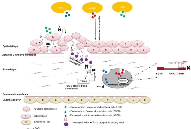
An increase in the concentration of chemokines and cytokines at the injury site is the hallmark of wound healing. Interleukin 8 (IL-8), a proinflammatory cytokine, binds to glycosaminoglycans (GAGs). In this GAG-bound form, IL-8 interacts with the C-X-C motif chemokine receptor 1 (CXCR1) and CXCR2 of neutrophils that infiltrate at the site of injury, leading to oligomerization and a haptotactic gradient. This haptotactic gradient directs the migration of neutrophils to the wounded area and seeds the process of corneal fibrosis [49].
Corneal stromal stem cells (CSSCs) express tumor necrosis factor (TNF)-stimulated gene 6 (TSG-6), a hyaluronan-binding protein, during inflammation [50]. This 35 kDa protein interacts with IL-8 via its link module domain and disrupts the binding of IL-8 and GAGs, preventing neutrophil migration [51,52]. Shojaati et al. [53] isolated extracellular vesicles (EVs) from CSSCs, mixed them with fibrin gel, and administered the EV–fibrin gel to the surface of an eye with corneal debridement. After 2 weeks of debridement, the EV-treated cornea showed minor corneal scarring compared to CSSC-treated and untreated controls.
EVs were more effective in preventing neutrophil infiltration, reducing fibrotic marker expression, and restoring corneal morphology.
Cultured CSSCs treated with exosomes isolated from adipose-derived stem cells (AD- SCs) showed optimal proliferation, reduced apoptosis, increased aldehyde dehydrogenase 1 (ALDH1), decreased MMP1, MMP3, and MMP9 expression, and increased collagen I, II, III, IV, and V expression compared to untreated CSC [54]. Therefore, ADSC-derived exosomes might revive the plasticity of transformed corneal keratocytes or stromal cells by increasing keratocyte marker expression and ECM production. The therapeutic effects of ADSCs and ADSC-derived exosomes could be attributed to microRNA-19a, which binds the 30UTR of homeodomain-containing serine/threonine kinase 2 (HIPK2). MicroRNA- 19a post-transcriptionally suppresses homeodomain-interacting protein kinase 2 (HIPK2),
preventing the activation of the Jun N-terminal kinase (JNK) pathway and fibrosis [55].
Corneal keratocytes, when co-cultured with ADSC-derived exosomes, reduce HIPK2, phos- phorylated SMAD3, and p53 expression, preventing the transformation of keratocytes into myofibroblasts and apoptosis of the nearby injured cells. This condition was reversed by the lentivirus-mediated overexpression of HIPK2 in keratocytes, confirming the role of miRNA-19a in corneal scar healing [56].
In a study, human corneal epithelial cells (HCEs) were treated with recombinant throm- bospondin 1 (TSP1) to evaluate the effectiveness of corneal epithelial-derived exosomes in corneal wound healing. They were subjected to the same hypoxic stress encountered in corneal wound healing. Exosomes with TSP-1 as a cargo protein decrease paraptosis (apop- tosis caused by hypoxic conditions) [57]. EVs derived from human placenta mesenchymal stem cells were used to treat alkali-burned mouse corneas. The EVs decreased vascular en- dothelial growth factor (VEGF) expression after 48 h, and corneal restoration was observed 14 d post-treatment. The HCEs treated with EVs showed a reduction in the expression of profibrotic genes (IL-10, IL-1β, TNF-α, and NF-κβ) and the apoptosis gene cas8, revealing their anti-inflammatory and anti-apoptotic potential [58]. The therapeutic effect of this exosome is due to the reduction in inflammation and apoptosis. Increased apoptosis in nearby wounded cells attracts inflammatory molecules and neutrophils. These neutrophils are entrapped in fibrous DNA-containing structures and release neutrophil esterase (neu- trophil esterase trap), enhancing the transformation of keratocytes into myofibroblasts, mediated by TGF-β[59,60].
The ECM also possesses factors that can reduce proinflammatory cytokines upon receiving an inflammatory insult and attract macrophages, dendritic cells, and lympho- cytes to the injured site. Yin et al. [61] processed the ECM from lymph, cartilage, and corneal tissues into micro-sized particles and compared their efficiency in corneal wound healing. The treatment of IL-1β-pretreated rabbit corneal keratocytes with these ECM microparticles reduced proinflammatory cytokine expression and prevented keratocytes from transforming into myofibroblasts. However, the keratocytes were more elongated than native keratocytes and ECM microparticles in the untreated control group. They also addressed the efficacy of ECM microparticles in modulating the gene expression of the tear film or lacrimal gland. Inflammation decreases tear production; increases tear film osmolarity and osmotic stress; and activates MAP kinase and NF-κβsignaling cascades, causing various inflammatory mediators to aggravate the ocular surface damage [62]. The rabbit cornea was injured by performing a superior lamellar keratectomy. Treatment with fibrin gel (FG)-encapsulated lymph node extracellular matrix microparticles increased Mucin 5AC (Muc5AC) and lactoferrin expression, causing infiltration by neutrophils, a reduction in corneal haze, a reduction in TGF-β,α-SMA, and COL-1 expression, and an increase in the thickness of the mucin layer. Therefore, tissue-derived microparticles could be potent therapeutics for ocular surface homeostasis and prevent corneal fibrosis.
Exosome-based therapy is still in its nascency and requires preclinical research to be used for treating scars. Exosomes isolated from corneal stromal stem cells effectively prevent corneal scarring and require more preclinical safety and toxicity studies. Similarly, ADSC-derived exosomes reduce apoptosis and restore keratocyte marker expression in corneal stromal cells. Further characterization of these experiments is required to check their preclinical efficacy and gain better molecular insight to understand their therapeutic benefits. Exosomes from HCEs can hasten the corneal wound-healing process and prevent corneal scarring (Figure3). The status of exosome-based therapy for corneal scar treatment is summarized in Table1. Moreover, the parent cells could be engineered to overexpress specific antifibrotic proteins. Exosomes consisting of this protein can be used in combination with tissue-derived microparticles to increase the effectiveness of exosome-based therapy for a scarred cornea.
Cells2022,11, 3310 7 of 31
Cells 2022, 11, x FOR PEER REVIEW 9 of 33
Cells 2022, 11, x. https://doi.org/10.3390/xxxxx www.mdpi.com/journal/cells
Figure 3. Role of exosomes in the treatment of scars. Exosomes from different cell types (ADSCs, CSSCs, and HECs) are loaded within the fibrin gel or encapsulated in the hydrogel and applied on the ocular surface; (a) exosomes from HECs taken up by the epithelial cells prevent paraptosis (apoptosis caused by hypoxic conditions); (b) epithelial cells treated with exosomes do not undergo apoptosis; (c) exosomes from ADSCs contain miR-19a, which post-transcriptionally silences HIPK2 and attenuates the JNK pathway; (d) exosomes from CSSCs contain TSG-6 protein, and once they are taken up by keratocytes, the TSG-6 proteins are secreted within them; (e) TSG-6 protein binds to GAG via its LINK domain and prevents the binding of IL8 to the GAG of corneal stroma; (f) the unbound form of IL-8 cannot be presented to the CXC1/2 receptor of neutrophils. It attenuates the infiltration of neutrophils to the wounded area of the cornea.
3.2. Targeted Gene Silencing for the Generation of A Scarless Cornea
The idea of directly targeting the upregulated genes of the corneal scarring signaling cascade to heal a scarred cornea has attracted researchers over the past few years. Sema- phorin 3A (SEMA-3A), ubiquitin-specific protease-10 (USP 10), and calmodulin/calcium- activated K+ channel 3.1 (Kca3.1) are upregulated during the corneal wound-healing pro- cess. The increased expression of these genes contributes to corneal fibrosis. Therefore, targeting these genes using siRNA can mitigate the scarred corneal environment. The complete knockout of a targeted gene is undesirable because genetically modifying a mammalian cell is difficult, time-consuming, and expensive; moreover, knocking out a gene could have lethal consequences. Therefore, the in vivo delivery of small oligonucle- otide sequences, designed to target certain mRNAs, using a suitable delivery vehicle to silence a target gene might be an effective way to prevent corneal fibrosis.
3.2.1. Targeting Semaphorin 3A in Scarless Cornea Regeneration
The epithelial cells around the wounded area secrete various growth factors in re- sponse to an injury. Epidermal growth factor-1 (EGF-1) is one such factor that binds to
Figure 3. Role of exosomes in the treatment of scars. Exosomes from different cell types (ADSCs, CSSCs, and HECs) are loaded within the fibrin gel or encapsulated in the hydrogel and applied on the ocular surface; (a) exosomes from HECs taken up by the epithelial cells prevent paraptosis (apoptosis caused by hypoxic conditions); (b) epithelial cells treated with exosomes do not undergo apoptosis; (c) exosomes from ADSCs contain miR-19a, which post-transcriptionally silences HIPK2 and attenuates the JNK pathway; (d) exosomes from CSSCs contain TSG-6 protein, and once they are taken up by keratocytes, the TSG-6 proteins are secreted within them; (e) TSG-6 protein binds to GAG via its LINK domain and prevents the binding of IL8 to the GAG of corneal stroma; (f) the unbound form of IL-8 cannot be presented to the CXC1/2 receptor of neutrophils. It attenuates the infiltration of neutrophils to the wounded area of the cornea.
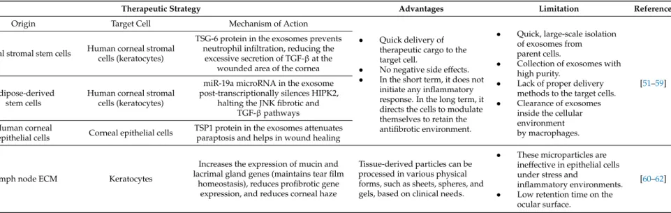
Table 1.Exosomes as therapeutic tools in reviving a cornea without scarring.
Therapeutic Method Therapeutic Strategy Advantages Limitation References
Exosomes
Origin Target Cell Mechanism of Action
• Quick delivery of therapeutic cargo to the target cell.
• No negative side effects.
• In the short term, it does not initiate any inflammatory response. In the long term, it directs the cells to modulate themselves to retain the antifibrotic environment.
• Quick, large-scale isolation of exosomes from parent cells.
• Collection of exosomes with high purity.
• Lack of proper delivery methods to the target cells.
• Clearance of exosomes inside the cellular environment by macrophages.
[51–59]
Corneal stromal stem cells Human corneal stromal cells (keratocytes)
TSG-6 protein in the exosomes prevents neutrophil infiltration, reducing the
excessive secretion of TGF-βat the wounded area of the cornea
Adipose-derived stem cells
Human corneal stromal cells (keratocytes)
miR-19a microRNA in the exosome post-transcriptionally silences HIPK2,
halting the JNK fibrotic and TGF-βpathways Human corneal
epithelial cells Corneal epithelial cells TSP1 protein in the exosomes attenuates paraptosis and helps in wound healing
Tissue-derived
microparticles Lymph node ECM Keratocytes
Increases the expression of mucin and lacrimal gland genes (maintains tear film
homeostasis), reduces profibrotic gene expression, and reduces corneal haze
Tissue-derived particles can be processed in various physical forms, such as sheets, spheres, and gels, based on clinical needs.
• These microparticles are ineffective in epithelial cells under stress and
inflammatory environments.
• Low retention time on the ocular surface.
[60–62]
Cells2022,11, 3310 9 of 31
3.2. Targeted Gene Silencing for the Generation of A Scarless Cornea
The idea of directly targeting the upregulated genes of the corneal scarring signaling cascade to heal a scarred cornea has attracted researchers over the past few years. Semaphorin 3A (SEMA-3A), ubiquitin-specific protease-10 (USP 10), and calmodulin/calcium-activated K+ channel 3.1 (Kca3.1) are upregulated during the corneal wound-healing process. The increased expression of these genes contributes to corneal fibrosis. Therefore, targeting these genes using siRNA can mitigate the scarred corneal environment. The complete knockout of a targeted gene is undesirable because genetically modifying a mammalian cell is difficult, time-consuming, and expensive; moreover, knocking out a gene could have lethal consequences. Therefore, the in vivo delivery of small oligonucleotide sequences, designed to target certain mRNAs, using a suitable delivery vehicle to silence a target gene might be an effective way to prevent corneal fibrosis.
3.2.1. Targeting Semaphorin 3A in Scarless Cornea Regeneration
The epithelial cells around the wounded area secrete various growth factors in re- sponse to an injury. Epidermal growth factor-1 (EGF-1) is one such factor that binds to EGF-1 receptors (expressed in large numbers) on the surface of fibroblasts [63]. The binding of EGF-1 to its cognate receptor on the fibroblast of the epithelial layer or the stromal layer of the cornea increases the expression of Semaphorin 3A (SEMA3A). The semaphorin family consists of a large group of proteins that can be either secreted or cell surface-bound [64].
Jeon et al. [65] showed the increased expression of SEMA3A in corneal epithelial and stro- mal keratocyte cells, indicating its overexpression during wound healing. Similar SEMA3A expression was observed in cultured corneal fibroblasts. Increased expression was observed in fibroblasts pretreated with TGF-β. SEMA3A, in combination with TGF-β, leads to a significant increase in profibrotic marker gene expression, indicating that SEMA3A only supports the TGF-β-mediated expression of fibrotic genes.
Morishige et al. [66] also showed a similar increase in SEMA3A expression in epithelial cells during wound healing. Native epithelial cells were transfected with siRNA targeting SEMA3A; however, its expression was unaltered. In fibroblasts transfected with similar siRNA, there was a reduction in all of the isoforms of SEMA3A, confirming that wound healing increases the level of SEMA3A in fibroblasts. The underlying mechanism behind this role of SEMA3A is still unclear. Yamazaki et al. [67] showed that SM-345431 (vinaxan- thone), a SEMA-3A inhibitor, promoted neural regeneration in a murine dry-eye model.
This inhibitor also maintained corneal epithelial integrity and nerve density in the dry-eye model, providing a molecular basis for targeting SEMA-3A.
3.2.2. Silencing USP10, A Deubiquitinase, Can Prevent Corneal Scarring
The cellular stress associated with corneal wound healing increases the local lev- els of USP10, a deubiquitinase with dual function. The elevated USP10 is translocated into the nucleus immediately after the wound. It binds to p53 to prevent cellular ubiq- uitylation [68] and increases apoptosis of the wounded and surrounding keratocytes or epithelial cells, allowing neutrophil and macrophage infiltration. In the later stages of wound healing, when acute cellular stress is relieved, USP10 accumulates in the cyto- plasm and either binds to Ras GTPase-activating protein-binding protein (G3BP), a Ras- GTPase-activating protein-binding protein, or the USP10 modulator, which deubiquitinates integrins [69]. This increases the integrin levels on the cell surface and TGF-β-mediated fi- brosis. Lipofectamine-mediated transfection of USP10-siRNA in wounded porcine corneas improved the corneal stromal ECM arrangement. It also decreased fibrotic marker (fi- bronectin andα-SMA) expression andαVβ1,5 integrin expression on the surface of corneal fibroblasts [70]. Bowmil et al. [71] used a self-deliverable siRNA (siRNA) targeting USP10 conjugated to cys3 and cholesterol to treat wounded rabbit corneas. These siRNAs could penetrate to a depth of 324µm in the corneal stromal layer within 24 h of transfection and reduce the expression of collagen III and fibrotic markers. They also reduced immune cell
infiltration and apoptosis in the cells surrounding the wound. This finding supports that molecular therapeutic tools should target USP10 for scarless corneal wound healing.
3.2.3. Knockout of Kca3.1 Ion Channel for Preventing Corneal Scarring
Ca2+signaling is integral to corneal fibrosis. The axonal nerve endings of the cornea secrete neuropeptides such as substance P (SP) in response to injury. Substance P (SP) binds to NK1Ron on keratocytes and activates phospholipase C-β, which cleaves phospholipid 2,3-bisphosphate to inositol-3-phosphate (IP3) and diacylglycerol. IP3 induces intracel- lular calcium by opening the IP3-gated Ca2+ion channels of the endoplasmic reticulum.
Calmodulin is a Ca2+-binding protein that binds to Ca2+ions and activates the Kca3.1 ion channel [72]. The activated K+ ion channels cause an efflux of K+ions and an influx of Ca2+ions, resulting in the hyperpolarization of keratocytes. Hyperpolarization activates the transformation of keratocytes into fibroblasts and enables their evasion of the G1/S cell cycle checkpoint, which increases fibroblast proliferation. This increased fibroblast proliferation expedites corneal wound healing, leading to TGF-β-mediated myofibroblast formation, which results in corneal fibrosis [73,74]. Integrin on the cell surface is activated upon binding to specific ligands [75]. It forms macromolecular complexes with ion channels and helps in its localization to the plasma membrane of cells [76]. The increased integrin expression observed on the keratocyte cell membrane during corneal fibrosis indicates an increased accumulation of Kca3.1 on the plasma membrane. The accumulated Kca3.1 hyperpolarizes the keratocytes, contributing to fibrosis. Kca3.1 knockout mice showed reduced haze formation because of reduced fibrotic gene expression and lowα-SMA [77].
Human corneal fibroblasts (HCFs) pretreated with TGF-βand incubated with TRAM-34 showed a decrease in profibrotic gene expression compared to only-TGF-β-activated HCFs.
Therefore, it is likely that Kca3.1 inhibition prevents the differentiation of keratocytes into myofibroblasts and corneal fibrosis [78]. The targeted silencing of genes such as SEMA3A, USP10, and Kca3.1 can prevent the differentiation of keratocytes into myofibroblasts in the presence of TGF-β, preventing corneal scar formation (Table2).
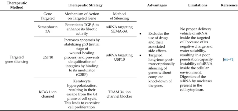
Table 2.Targeted gene knockdown and protein overexpression as therapeutic tools for preventing corneal scarring.
Therapeutic
Method Therapeutic Strategy Advantages Limitations References
Targeted gene silencing
Gene Targeted
Mechanism of Action on Targeted Gene
Method of Silencing
• Excludes the use of drugs and their associated side effects.
• Targeted long-term post- transcriptionally silencing of genes without complete knockdown of the gene.
No proper delivery vehicle of siRNA inside the targeted cell because of its negative charge and water solubility, resulting in a poor penetration capacity.
Instability of siRNA inside the cellular environment.
Digestion of the siRNA by nucleases present in the cell cytoplasm.
[64–71]
Semaphorin 3A
Potentiates TGF-βto enhance its fibrotic
activity
siRNA targeting SEMA-3A
USP10
Increases apoptosis by stabilizing p53 (initial
stage of wound-healing process) and prevents
ubiquitination of integrins by binding
to its modulator (G3BP)
siRNA targeting USP10
KCa3.1 ion channel
Keratocyte hyperpolarization,
resulting in their escape from the G1
phase of cell cycle.
This leads to excessive cell proliferation.
TRAM 34, ion channel blocker
Cells2022,11, 3310 11 of 31
Table 2.Cont.
Therapeutic
Method Therapeutic Strategy Advantages Limitations References
Targeted gene overexpression
Gene Targeted
Mechanism of Action on Targeted Gene
Method of Overexpression
• This strategy can be applied to modify both wild and mutant cell types.
• Non-drug method for treating scarring.
Extra stress on the cell, as cellular resources are wasted in translating and exporting the specific protein.
Instability of plasmid DNA expression vectors containing the gene of interest or misincorporation of the gene of interest in the case of
homologous recombi- nation methods.
[78–85]
KLF4 Suppresses EMT Lentiviral vector
Id3
Sequestering bHLH transcription factors and preventing the downregulation of epithelial cell markers
to hinder EMT.
pcDNA3- mCherry LIC
mammalian expression vector construct
SMAD7
Prevents nuclear localization of SMAD2/3 and attenuates the TGF-β
pathway by preventing the phosphorylation
of SMAD3.
Recombinant adeno-associated
viral vector
Abbreviations: USP10: ubiquitin-specific protease-1; Kca3.1 ion channel: K+ channel 3.1 ion channel; KLF4:
Krüppel-like factor 4; Id3: inhibitor of differentiation 3; SMAD7: mothers against decapentaplegic homolog 7.
3.3. Protein Overexpression in Preventing Corneal Scarring
The cornea is an ideal tissue for gene therapy, as the ocular surface can be an excellent platform for the topical application of various viral/non-viral gene delivery vectors. The overexpression of specific genes in corneal epithelial or stromal cells that block the TGF-β signaling cascade can prevent scar formation.
3.3.1. Overexpression of KLF4 in Preventing Scar Formation
Epithelial cells express Krüppel-like factor 4 (KLF4), which coordinates the apical and basal polarity of epithelial cells. It suppresses EMT to maintain the plane of epithelial cell division and epithelial cell homeostasis. Corneal injuries lead to the loss of native epithelial markers, such as E-cadherin 1 (CDH1) and zona occludens, and the transfor- mation of epithelial cells to fibroblast-like cells that secrete TGF-β. Fujimoto et al. [79]
reported low KLF4 expression in epithelial cells that migrate towards the wound. Human corneal epithelial cells (HCEs) transfected with siRNA targeting KLF4 showed decreased epithelial markers, increased mesenchymal markers, and increased profibrotic gene ex- pression. The lentiviral-vector-mediated overexpression of KLF4 in HCEs showed the increased expression of corneal epithelial cell markers. However, KLF4 overexpression did not affect mesenchymal gene expression, such as fibronectin 1 and N-cadherin 2.
KLF4-overexpressing HCEs and the HCE control were pretreated with TGF-βfor 30 min.
KLF4-overexpressing HCEs showed 20% less SMAD2/3 phosphorylation compared to the HCE control. However, KLF4-overexpressing HCEs incubated with TGF-βfor four hours showed SMAD2/3 phosphorylation levels similar to the TGF-βpretreatment levels.
Therefore, KLF4 might contribute to scar prevention due to EMT suppression and a reduc- tion in SMAD2/3 phosphorylation, limiting its nuclear localization and TGF-β-mediated corneal fibrosis.
3.3.2. Overexpression of Id3 for Reviving Cornea without Scars
Human corneal stromal fibroblasts overexpressing inhibitor of differentiation 3 (Id3) in the presence of TGF-βprevent myofibroblast formation. Id3 overexpression also reduces the expression of fibrotic genes, such as fibronectin,α-SMA, collagen I, and collagen IV [80].
After TGF-βtreatment, the Id3-overexpressing cells showed: TGF-βbinding to its cognate receptors (TGF-βRII and TGF-βRI); the phosphorylation of SMAD2/3 and its translocation to the nucleus; and the activation of transcription factors, such as the ZEB, SNAIL, and basic helix-loop-helix (bHLH) families [81]. The bHLH transcription factor consists of
two alpha helixes separated by a loop and a specific DNA-binding domain (DBD). The DBD binds to the E-box DNA consensus sequence and promotes EMT [82]. The Id3 gene, belonging to a class V group of the bHLH family, consists of an HLH domain. However, it lacks the DNA-binding domain and forms dimers with other bHLH transcription factors.
The sequestration of the bHLH transcription factor prevents it from binding to the E-Box and thereby suppresses EMT. Therefore, Id3 acts as a dominant-negative regulator of this transcription factor [83]. Id3 overexpression prevents the TGF-β-mediated profibrotic cascade in HCF. However, an in vivo animal-based study is needed to establish its efficacy in reducing corneal scarring.
3.3.3. Overexpression of SMAD7 for Regenerating Cornea without Scars
SMAD7 is a TGF-βsignaling pathway inhibitor that binds to TGF-βR1 via its MH2 domain. This binding prevents SMAD3 phosphorylation and its interaction with SMAD2 and SMAD4, inhibiting the nuclear localization of the SMAD complex and TGF-β-mediated fibrosis [84,85]. Gupta S et al. [86] reported an increase in theα-SMA-positive cell count in HCFs transfected with siRNA targeting SMAD after TGF-βtreatment. Theα-SMA-positive cell count was reduced in TGF-β-treated HCFs after transfection with the recombinant AAV5-SMAD7 viral vector. Rabbit corneal wounds treated with recombinant AVV5-SMAD7 resulted in reduced corneal haze and profibrotic gene expression after four weeks. More- over, there were no signs of infection, intraocular inflammation, redness, or ocular dis- charge, establishing the safety and efficacy of this formulation. Therefore, recombinant AVV5-SMAD7-mediated gene delivery is safe and therapeutically efficient in preventing corneal scarring.
An overview of antifibrotic genes with the potential to heal corneal scarring is sum- marized in Table2. Further validation of their effectiveness and safety in preclinical and clinical studies is required. Establishing this antifibrotic environment in corneal cells using a gene therapy approach is restricted by the lack of long-term stability, high costs, and low efficiency. Therefore, various bioactive compounds, such as histone deacetylase (HDAC) inhibitors, glucosamine, and chitosan, with the potential to induce similar antifibrotic conditions must be considered.
3.4. MicroRNA Therapies in Regenerating Cornea without Scars
MicroRNAs are 21-nucleotide-long, non-coding RNAs that can post-transcriptionally silence the expression of their target genes. These microRNAs form an RNA-induced silencing complex (RISC) with Argonaute proteins. These proteins guide them towards the target mRNA, where they pair with the target mRNA and silence its expression [87].
MicroRNAs in the cornea affect various cellular processes, such as cell migration, differentiation, proliferation, and metabolism. Any dysregulation in miRNA levels during corneal injury can lead to corneal neovascularization and scar formation. An J et al. [88]
observed a sharp decrease in the expression of miR-204 in a murine corneal wound model.
MicroRNA-204 is abundant in the cornea. It inhibits corneal cell proliferation by G1 phase arrest. Therefore, low miR-204 levels during corneal wounds account for the excessive proliferation of transformed epithelial cells, myofibroblasts, ECM remodeling, and fibrosis during quick wound healing. Wang Y et al. [89] compared lens epithelial cells from healthy donors and patients with posterior capsular opacification (PCO). They showed increasedα- SMA and vimentin and decreased E-cadherin in the diseased cells, indicating opacification.
An in vitro PCO model was developed and transfected with miR-204. The lens epithelium cells showed increased E-cadherin and decreasedα-SMA compared to non-transfected con- trols and miR-204-5p-inhibitor-transfected cells. MicroRNA-204 prevents TGF-β-mediated EMT and fibrosis by targeting SMAD4, disrupting the SMAD2/3-SMAD4 complex. It also decreases the expression of the Hey/HMGA transcription factors, which induce EMT through CDH1 repression and SNAIL activation [90,91]. A subconjunctival injection of a recombinant adeno-associated vector (rAAV-miR-204) prevented neovascularization in murine alkali-burned corneas, indicating the anti-angiogenic role of miR-204 [92].
Cells2022,11, 3310 13 of 31
Ratuszny et al. [93] showed the significant upregulation of miR-145- and TGF-β- induced myofibroblasts in scarred human corneas compared to normal corneal and un- treated corneal fibroblasts. The TGF-β-mediated upregulation of miR-145 in the myofi- broblast inducesα-SMA expression by downregulating KLF4. miR-145 mediates the post-transcriptional silencing of KLF4 by targeting its 3’UTR [94]. miR-145 silencing using anti-miR-145 resulted in decreasedα-SMA and increased KLF4 expression. The lipofectamine-mediated transfection of a miR-145 inhibitor caused the stimulated cells to reduce collagen gel contraction in TGF-β-pretreated corneal fibroblast cells. This miR-145 inhibitor decreased the migratory capacity of myofibroblasts by 50%. Sun et al. [95] re- ported that miR-145 can directly target the 30UTR of KLF4, mediating post-transcriptional gene silencing. KLF4 is an EMT suppressor that interacts directly with the promoter of SMAD7 and upregulates its expression. The increased expression of SMAD7 prevents SMAD2/3 phosphorylation and localization, disrupting the TGF-βsignaling pathway [96].
Therefore, miR-145 is a promising target for further therapeutic exploration to prevent corneal scar formation.
Micro-RNA133b modulates connective tissue-like growth factor (CTGF), a down- stream molecule of the TGF-βsignaling pathway [97]. CTGF is a member of the CNN family of proteins. It has an N-terminal insulin-like growth factor-binding protein domain that binds to IGF receptors, accelerating cellular differentiation and ECM secretion. Their C-terminal domain increases cellular proliferation and DNA synthesis in the presence of epidermal growth factor (EGF) and its receptor [98]. Zhao et al. [99] showed that miR- 133B conjugated to gold nanoparticles (AuNPs) reduced the expression of myofibroblast- inducing genes (α-SMA and type 1 collagen) in corneal stromal cells. The transplantation of miR-133B/AuNPs with collagen in the rabbit cornea after lamellar keratoplasty inhibited corneal haze and scarring.
The downregulation of miR-145 and the upregulation of miR-204 and miR-133b can prevent TGF-β-mediated corneal fibrosis (Table3). MicroRNA-based gene targeting is a promising therapeutic approach to prevent and heal corneal scarring (Figure4).
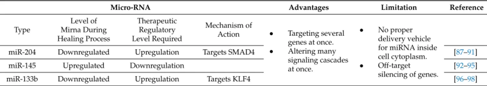
Table 3.MicroRNAs as therapeutic tools in reviving a cornea without scarring.
Micro-RNA Advantages Limitation Reference
Type
Level of Mirna During Healing Process
Therapeutic Regulatory Level Required
Mechanism of
Action • Targeting several genes at once.
• Altering many signaling cascades at once.
• No proper delivery vehicle for miRNA inside cell cytoplasm.
• Off-target silencing of genes.
miR-204 Downregulated Upregulation Targets SMAD4 [87–91]
miR-145 Upregulated Downregulation [92–95]
miR-133b Downregulated Upregulation Targets KLF4 [96–98]
Abbreviations: SMAD4: mothers against decapentaplegic homolog 4; KLF4: Krüppel-like factor 4.
Cells 2022, 11, x FOR PEER REVIEW 14 of 33
Cells 2022, 11, x. https://doi.org/10.3390/xxxxx www.mdpi.com/journal/cells
Micro-RNA133b modulates connective tissue-like growth factor (CTGF), a down- stream molecule of the TGF-β signaling pathway [97]. CTGF is a member of the CNN family of proteins. It has an N-terminal insulin-like growth factor-binding protein domain that binds to IGF receptors, accelerating cellular differentiation and ECM secretion. Their C-terminal domain increases cellular proliferation and DNA synthesis in the presence of epidermal growth factor (EGF) and its receptor [98]. Zhao et al. [99]showed that miR-133B conjugated to gold nanoparticles (AuNPs) reduced the expression of myofibroblast-in- ducing genes (α-SMA and type 1 collagen) in corneal stromal cells. The transplantation of miR-133B/AuNPs with collagen in the rabbit cornea after lamellar keratoplasty inhibited corneal haze and scarring.
The downregulation of miR-145 and the upregulation of miR-204 and miR-133b can prevent TGF-β-mediated corneal fibrosis (Table 3). MicroRNA-based gene targeting is a promising therapeutic approach to prevent and heal corneal scarring (Figure 4).
Table 3. MicroRNAs as therapeutic tools in reviving a cornea without scarring.
Micro-RNA Advantages Limitation Reference Type Level of Mirna During
Healing Process
Therapeutic Regulatory Level Required
Mechanism
of Action • Target- ing several genes at once.
• Altering many signaling cascades at once.
• No proper delivery vehicle for miRNA inside cell cytoplasm.
• Off-target silencing of genes.
miR-204 Downregulated Upregulation Targets
SMAD4 [87–91]
miR-145 Upregulated Downregula-
tion [92–95]
miR-133b Downregulated Upregulation Targets
KLF4 [96–98]
Abbreviations: SMAD4: mothers against decapentaplegic homolog 4; KLF4: Krüppel-like factor 4.
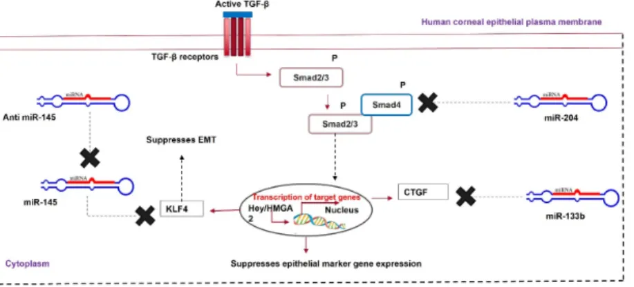
Figure 4. MicroRNA therapy for corneal scarring. Upregulation of miR-204 and miR-133b and downregulation of miR-145 help in healing a scarred cornea via modulating Krüppel-like factor 4 (KLF4), Smad2/3, and CTGF.
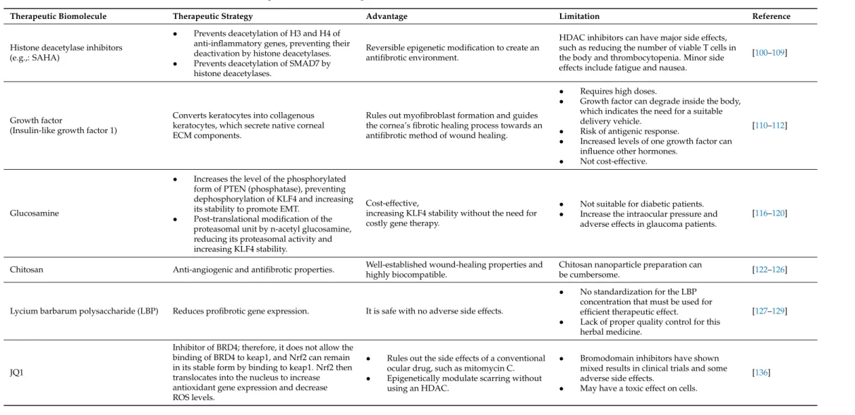
3.5. Bioactive Molecules as HDAC Inhibitors in the Regeneration of Scarless Cornea The wound-healing process of an injured cornea involves three overlapping phases:
the inflammatory phase, the proliferative or fibrotic phase, and the final remodeling phase
Figure 4. MicroRNA therapy for corneal scarring. Upregulation of miR-204 and miR-133b and downregulation of miR-145 help in healing a scarred cornea via modulating Krüppel-like factor 4 (KLF4), Smad2/3, and CTGF.
3.5. Bioactive Molecules as HDAC Inhibitors in the Regeneration of Scarless Cornea
The wound-healing process of an injured cornea involves three overlapping phases:
the inflammatory phase, the proliferative or fibrotic phase, and the final remodeling phase [100]. These events change the native morphology of the corneal stromal ECM, rendering it opaque. During the initial inflammatory phase of the healing process, corneal cells exhibit high levels of proinflammatory cytokines. During the initial phase of wound healing, the infiltrated neutrophils, monocytes, or activated macrophages secrete TGF-β, increasing its local concentration around the wounded area. TGF-βguides the transition of the wound from the inflammatory phase to the fibrotic phase by decreasing the local proinflammatory cytokine level. The timely termination of the initial inflammatory phase is required for the cells to enter the cell proliferation and ECM synthesis phase, healing the wound [101]. A lesser-known pathway that TGF-βuses for this transition is by mod- ulating histone acetylation. TGF-βrecruits histone deacetylases (HDACs), removing the acetyl group from the lysine residues of histones H3 and H4 of anti-inflammatory genes.
Histone acetylation is associated with anti-inflammatory gene activation; therefore, HDAC is recruited in the presence of TGF-β, deactivating various anti-inflammatory genes and enabling the transition to the fibrotic phase [102–106]. Trichostatin A (TSA), a deacetylase inhibitor, prevents the deacetylation of histone H3 or H4 of anti-inflammatory genes. TSA prevented the TGF-β-mediated transformation of corneal fibroblasts to myofibroblasts in vitro and also reduced the corneal opacity in a rabbit model of corneal haze [107,108].
A possible mechanism of action for HDAC inhibitors is increasing the local concen- tration of TGF-βduring wounding, leading to HDAC recruitment, SMAD7 deacetylation, SMAD7 ubiquitinylation, and proteasomal degradation. This paves the way for TGF-β- mediated corneal scarring. Similarly, suberoylanilide hydroxamic acid (SAHA), an HDAC inhibitor, reducedα-SMA and MMP9 expression in equine corneal fibroblasts treated with TGF-β. This shows that SAHA can prevent TGF-β-mediated corneal fibrosis [109]. Epige- netic modulators, such as TSA and SAHA, might be useful bioactive molecules for treating corneal scars (Table4).
3.6. Guided Wound Healing to Prevent Scarring
Corneal wound healing is accompanied by the secretion of various growth factors.
During wound healing, these growth factors interact with their cognate receptors present on the surface of the cell to regulate the synthesis of collagen, proteoglycan, and other ECM components’ deposition. Various growth factors, such as IGF-1, platelet-derived growth factor (PDGF), TGF-β, and fibroblast growth factor 1 (FGF-1), either reach the wound site from the tear film or are secreted by the surrounding wounded or apoptotic epithelial cells.
Etheredge et al. [110] reported that FGF-1 stimulates keratocyte proliferation; however, it inhibits the synthesis of type 1 procollagen and keratan sulfate. Type 1 procollagen is an essential component of the stromal ECM, and keratan sulfate directs collagen assembly to maintain corneal transparency. FGF-1 induces the formation of hypercellular keratocytes with densely packed cells in the sparse matrix. IGF-1 turns these hypercellular keratocytes into collagenous keratocytes that secrete ECM components, similar to native keratocytes.
Therefore, the IGF-1/IGF-receptor signaling pathway prevents the transformation of ker- atocytes into myofibroblasts and ECM remodeling (Figure5). However, this is not a predominant signaling pathway during wound healing because the local concentration of TGF-βin the wound microenvironment is high. Therefore, the TGF-β/TGF-βRI path- way takes charge, and TGF-βbinds to its receptor on keratocytes to synthesize biglycan, fibronectin containing extra domain A, and hyaluronan. This proteoglycan disrupts the spacing between collagen fibrils, reducing the corneal transparency. It also guides the differentiation of keratocytes into myofibroblasts, characterized by the neo-expression of α-SMA.α-SMA incorporates actin into collagen fibrils and exerts a contractile force on the ECM, disrupting the normal corneal architecture.
Therefore, an increase in the local concentration of IGF-1 during corneal wounding can provide insights into this pathway because this pathway directs the synthesis of fibrillar
Cells2022,11, 3310 15 of 31
collagen similarly to the native one. This process of guided wound healing avoids scar formation (Figure4). Sarenac et al. [111] reported that keratocytes treated with TGF-βand IGF1 showed significantly lower SMAD3 nuclear localization and higher SMAD7 compared to TGF-β-treated keratocytes. IGF-1 also increased keratocyte markers, such as keratocan or ALDH3, when TGF-β-pretreated keratocytes were incubated with IGF-1 alone or together with SAHA. The TGF-β-pretreated keratocytes incubated with IGF-1 and halofuginone showed high proliferation and no myofibroblast trans-differentiation. Ghiasi et al. [112]
used hyaluronic acid as a vehicle to deliver substance P and IGF-1 to the surface of a photo-ablated rabbit cornea. IGF-1 and substance P synergistically promoted epithelial wound healing after a week of treatment, with a marked decrease in the wounded areas.
TGF-βheals corneal wounds and is a hallmark of the process of scarring. In contrast, IGF-1 guides the process of wound healing and circumvents scarring (Table4). So far, IGF-1 has been studied as an external growth factor, either alone or together with a bioactive compound, such as SAHA or substance P. Bioengineering keratocytes to overexpress IGF-1 can increase its local concentration and allow it to control the process of wound healing.
Cells 2022, 11, x FOR PEER REVIEW 18 of 33
Cells 2022, 11, x. https://doi.org/10.3390/xxxxx www.mdpi.com/journal/cells
Figure 5. Role of growth factors in scar prevention. Various growth factors, such as fibroblast growth factor (FGF) or insulin-like growth factor (IGF-1), are secreted from the tear film. These growth factors cross the disrupted Bowman’s membrane to reach the corneal stromal layer. FGF converts keratocytes to hypercellular keratocytes. IGF-1 converts hypercellular keratocytes to colla- genous keratocytes, which secrete ECM components similarly to the native cornea. Meanwhile, in the epithelial layer, after wounding, epithelial cells undergo apoptosis, and the apoptotic epithelial cells release TGF-β. Neutrophils, macrophages, and monocytes, which migrate from the epithelial layer to the stromal layer, secrete TGF-β, thereby increasing its local concentration. Keratocytes in the presence of TGF-β are converted to myofibroblasts, which disrupt the highly organized fibrillar arrangement of the corneal ECM.
3.7. Clinical Therapy for Scar Prevention
A gold-standard technique to prevent corneal scarring is still under research. Various biomolecules, such as decorin, TPCA-1, glucosamine, acetylcholine, and chitosan, have the potential to heal a scarred cornea via their respective biological pathways. Chen et al.
[113] reported the significance of glucosamine in corneal scar treatment. This amino sugar finds clinical application in osteoarthritis management [114]. However, it significantly en- hances intraocular pressure [115]. Park et al. [116] noted that glucosamine attenuated renal fibrosis by downregulating SMAD2 phosphorylation; therefore, a study of glucosamine’s effects on corneal fibrosis can provide new insights into its clinical applications. Chen’s group observed an increase in the expression, stability, and nuclear localization of KLF4 and a decrease in fibrotic gene expression in HCFs treated with glucosamine and TGF-β.
The mechanism underlying the glucosamine-mediated increase in KLF4 expression is still under research. However, a study by Wang DF et al. [117] reported that glucosamine in- creases the expression of the phosphorylated form of PTEN (a phosphatase), leading to an increase in the phosphorylated form of KLF4. This increases the P300 histone acetylase activity, mediating H3 histone acetylation [118,119].
Glucosamine can form O-linked N-acetylglucosamine via glucosamine transferase activity and the post-translational modification of the proteasome complex to prevent KLF4 degradation[120]. Moreover, KLF4 interacts with TGF-β control elements and pre- vents a proinflammatory environment[121]. Therefore, glucosamine might find clinical applications in healing corneal scarring (Figure 6).
Figure 5.Role of growth factors in scar prevention. Various growth factors, such as fibroblast growth factor (FGF) or insulin-like growth factor (IGF-1), are secreted from the tear film. These growth factors cross the disrupted Bowman’s membrane to reach the corneal stromal layer. FGF converts keratocytes to hypercellular keratocytes. IGF-1 converts hypercellular keratocytes to collagenous keratocytes, which secrete ECM components similarly to the native cornea. Meanwhile, in the epithelial layer, after wounding, epithelial cells undergo apoptosis, and the apoptotic epithelial cells release TGF-β.
Neutrophils, macrophages, and monocytes, which migrate from the epithelial layer to the stromal layer, secrete TGF-β, thereby increasing its local concentration. Keratocytes in the presence of TGF-β are converted to myofibroblasts, which disrupt the highly organized fibrillar arrangement of the corneal ECM.
3.7. Clinical Therapy for Scar Prevention
A gold-standard technique to prevent corneal scarring is still under research. Various biomolecules, such as decorin, TPCA-1, glucosamine, acetylcholine, and chitosan, have the potential to heal a scarred cornea via their respective biological pathways. Chen et al. [113]
reported the significance of glucosamine in corneal scar treatment. This amino sugar finds clinical application in osteoarthritis management [114]. However, it significantly enhances intraocular pressure [115]. Park et al. [116] noted that glucosamine attenuated renal fibrosis by downregulating SMAD2 phosphorylation; therefore, a study of glucosamine’s effects on corneal fibrosis can provide new insights into its clinical applications. Chen’s group observed an increase in the expression, stability, and nuclear localization of KLF4 and a decrease in fibrotic gene expression in HCFs treated with glucosamine and TGF-β. The mechanism underlying the glucosamine-mediated increase in KLF4 expression is still under