Siddhartha Sankar Ghosh, Department of Life Sciences and Bioengineering, Indian Institute of Technology Guwahati, India for the award of the degree of Doctor of Philosophy. This is to confirm that the thesis entitled "Recombinant sFRPs in Wnt/β-Catenin Signaling Targeted Cancer Therapy" is submitted to the Indian Institute of Technology Guwahati by Archita Ghoshal for the award of the degree of Doctor of Philosophy in the Department of Biosciences and Bioengineering, is a bonafide record of research work done by her. Siddhartha Sankar Ghosh, who has been a guide to me in every sense of the term.
More specifically, the role of sFRPs in regulating cell growth by modulating the Wnt pathway has been considered. Further, the therapeutic implications of GST-tagged sFRP1, alone and in combination with conventional chemotherapeutic drugs, ie. Studies with Western blotting and semi-quantitative PCR-based expression of essential molecules from the Wnt pathway validated the molecular mechanisms.
The therapeutic efficacy of the proteins on mammalian cancer cells was then determined, both alone and in the co-therapy module. Furthermore, the functionality of the proteins was evaluated based on their role in inhibiting the Wnt/β-catenin signaling pathway and induction of apoptosis.
Introduction and Review of Literature 1
- Recombinant proteins 6
- Role of Secreted Frizzled-related Proteins (sFRPs) in Wnt signaling10
- Polymeric nanoparticles 15
- Fluorescent nanoclusters 16
- Nanoparticles and Recombinant proteins: an interplay 16
- Key questions and scope of research 17
- Objectives of the thesis 18
- Salient features of this work 18
The approval of insulin in 1982 by the US FDA (Food and Drug Administration) ushered in the era of recombinant therapeutic proteins. Since then, over 100 such recombinant proteins have been approved for clinical use, and many more are in the pipeline. A major challenge in curing cancer lies in the fact that non-malignant cells must be preserved while cancer cells are targeted.
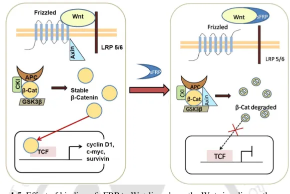
In absence of these sFRPs, β-catenin, a downstream molecule of the Wnt pathway, escapes proteasome degradation and accumulates in the cytoplasm. Post-translational modifications in the form of N-linked glycosylations have been found in the sFRPs, a number of putative sites are in the NLD [48]. Although a few potential sites are also present in the CRD, glycosylation is not essential for binding to Wnt ligand [ 49 ].
However, in the last decade, efforts have been made to encapsulate these drugs in nanocarriers, to obtain better pharmacokinetics, targeted delivery and improved therapeutic benefits [ 54 ]. The common underlying factor linking diagnosis to therapy is that both require accumulation in the diseased site of the body, for example the tumor tissue.
Expression, Purification, and Therapeutic Implications
Material and Methods 22
- Cell culture 22
- Cloning of sFRP1 23
- Expression of GST-sFRP1 in E.coli BL21 (DE3) 23
- Solubilization, Purification and Refolding of GST-sFRP1 protein 24
- Homology modeling and docking studies 25
- Western blotting 25
- Secondary structure characterization with circular dichroism 26
- MALDI TOF-TOF analysis 26
- Cell viability assay 26
- Combination therapy 27
- Cell cycle analysis 27
- Statistical tests 28
Cells were lysed in 10 mM phosphate-buffered saline (PBS) and 1 mM phenylmethylsulfonyl fluoride (PMSF) by sonication. However, when the cells were lysed by sonication and centrifuged, the expressed protein was recovered in an insoluble fraction, possibly as inclusion bodies. Cells were seeded in a 96-well plate at a density of 7000 cells per well in DMEM containing 10% FBS.
Both HeLa and MCF-7 cells were tested for the above combinations with an MTT assay following the same protocol. Both HeLa and MCF-7 cells were treated with 12 nM protein alone and in combination with cisplatin (2 µg/ml and 5 µg/ml, respectively) or doxorubicin (0.4 µg/ml and 0.2 µg/ml, respectively). ml). Cells were seeded at a density of approximately 10 cells per well, in a six-well tissue culture plate.
The dose of protein, which exerts the maximum effect on cell viability, was chosen; for the drugs, a concentration was selected at which approximately 80% of the cells were viable. Next, cells were centrifuged, washed with cold PBS and incubated with 0.2 mg/ml RNase A (Amresco) at 37 °C for 1 h.
Results and Discussion 28
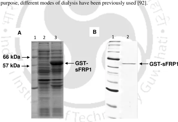
- Cloning, expression and purification of GST-sFRP1 28
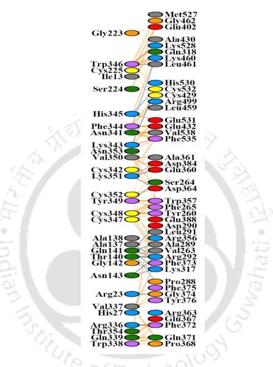
- Homology modeling and docking studies 32
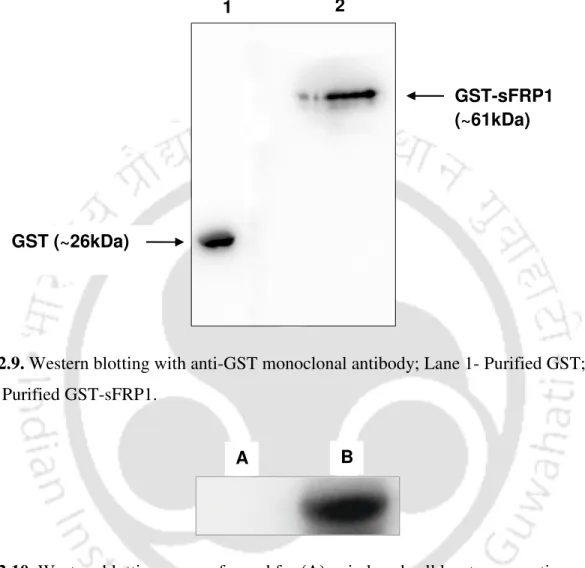
- Western blot analysis 39
- Secondary structure characterization with circular dichroism 40
- Analysis of MALDI TOF-TOF data 41
- Cell viability assay 42
- Combination therapy 44
- Cell cycle analysis 47
- Effect of GST-SFRP1 on non-cancerous cell line HEK-293 50
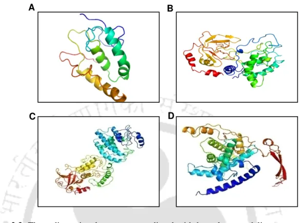
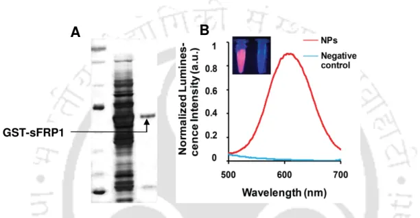
A single band of GST-sFRP1 corresponding to a molecular weight of 61 kDa was obtained after purification at the desired position in a 12% SDS-PAGE (Figure 2.2B). The structural integrity of the binding domain in the predicted whole molecular structure of sFRP1 as well as in GST-sFRP1 was confirmed using the same method (Figure 2.4C and Figure 2.4D, respectively). Also, the crystal structure of GST obtained from PDB (ID: 1UA5) was aligned with the predicted GST-sFRP1 to confirm the reliability of the predicted model (Figure 2.4E).
Here, MALDI together with time-of-flight analysis was used for the characterization of the recombinant sFRP1; the mass spectrometry data is shown in Figure 2.12. Cell viability was unchanged when purified GST alone (to a concentration of 40 nM) was added to cells (Figure 2.14). The optimized ratio obtained via MTT assays in the case of HeLa and MCF-7, when treated with GST-sFRP1 together with cisplatin (Figure 2.15A and Figure 2.15B respectively) and doxorubicin (Figure 2.16A and Figure 2.16B respectively).
Microscopic images of control and treated HeLa and MCF-7 cells are illustrated in Figure 2.17 and Figure 2.18, respectively. FACS-based analysis was performed to determine the impact of GST-sFRP1 (alone and in combination with chemotherapeutic agents) on cell cycle of HeLa (Figure 2.19A and Figure 2.19B) and MCF-7 (Figure 2.19C and Figure 2.19D) beacon. cells.
Conclusions 51
Targeting Wnt Canonical Signaling by Recombinant sFRP1 Bound
Material and Methods 57
- Synthesis of Chitosan-Au NC-Alginate NPs (Chi-Au NC-Alg NPs) 57
- Expression, purification of GST tagged human sFRP1 58
- Characterization of Chi-Au NC-Alg NPs 58
- Imaging of Chi-Au NC-Alg NPs with TEM and fluorescence
- Release studies of protein 59
- Mammalian cell culture 59
- Stability studies of NPs and tracking the release of protein 59
- Assessment of cell viability 60
- Tracking NPs with high-end deconvolution microscopy 60
- Preparation of whole cell protein lysate and western blotting 60
- Acridine orange (AO) / Ethidium bromide (EB) dual staining 61
- Expression profiling for downstream genes 62
- Combination therapy with cisplatin 62
- Cell cycle analysis 62
- Detection of apoptosis by fluorescein isothiocyanate (FITC)
After checking the luminescence of the Au NCs in UV transilluminator, 2 mL of the synthesized chitosan-Au NC was taken into a separate tube. GST-sFRP1 was purified to near homogeneity by affinity chromatography, using glutathione-agarose beads, which bind to GST. [27] The purified GST-tagged human SFRP1 protein was dialyzed against 10 mM Tris buffer at pH 7.5 for 7 hours. Cells were maintained in DMEM containing 10% FBS, 100 U/ml penicillin, 100 µg/ml streptomycin in a humidified atmosphere at 37°C in an incubator with 5% carbon dioxide.
For this purpose, HeLa cells were seeded in a six-well plate at a density of 10 cells per well and allowed to attach for 8 h. After allowing the cells to adhere for 8 h, they were treated with Chi-Au NC-Alg NPs, GST-sFRP1, and sFRP1-NPs for 48 h, respectively. Monitoring the localization of NPs was essential to confirm whether they remained in the media or were taken up by the cells.
With this intention, cells were treated with both for 24 h in 60 mm culture Petri dishes containing serum-free medium. To distinguish between healthy and membrane-compromised cells, the cells were simultaneously stained with AO and EB. At the end of the 48 h treatment period, the medium was discarded, the cells were washed thoroughly with 10 mM PBS, and AO and EB were added at concentrations of 2 µg/ml and 10 µg/ml, respectively.
After incubation for 5 min in the dark, cells were washed with fresh PBS and visualized under a fluorescence microscope (Nikon ECLIPSE TS100). The effect of protein-conjugated NPs on the cell cycle pattern of HeLa cells was evaluated by FACS using PI. Cells were seeded at a density of 105 cells per well in a six-well plate and allowed to attach overnight.
Then the cells were treated separately with recombinant sFRP1 and sFRP1-NPs, alone and in combination with cisplatin. Samples were treated for 48 hours and at the end of that, cells were harvested by trypsinization and fixed with 70% ethanol in ice for 1 hour. Cells were collected by trypsinization, washed with PBS and stained with Annexin V-FITC and PI, according to the manufacturer's protocol provided with Annexin V-FITC apoptosis detection kit (BD Biosciences).
Results and Discussion 63
- Recombinant GST-sFRP1 bound to Chi-Au NC-Alg NPs 63
- Binding and release studies of recombinant protein 69
- Stability of Au NCs in cell culture media studied by probing
- Tracking NPs for anti-cancer function 73
- Targeting the Wnt signaling pathway 76
GST-sFRP1 binding studies were performed by probing the luminescence of Au NCs. The sustained release profile obtained in this case is ideal for sustained protein activity. In this effort, the luminescence of the NPs was quenched by 20% in the initial hours, likely due to the instability of the NPs (in the absence of protein) in the medium (Figure 3.9B).
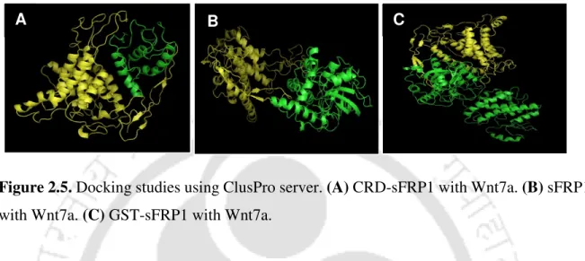
The effect was enhanced by sFRP1-NP treatment, where a significant population of cells showed AO and EB staining (Figure 3.14). However, the significantly increased efficacy of the co-therapy can be attributed to the blockade of the Wnt pathway by sFRP1-NP. The structure of the binding domain (CRD or cysteine-rich domain) of sFRP4 was predicted using the PHYRE2 Protein Fold Recognition Server, together with those of sFRP4 and GST-sFRP4 [ 83 ].
A weak GST band was also observed at 26 kDa, which may be due to cleavage of the recombinant protein. Docking studies were performed for GST-sFRP4 with one of the Wnt ligands known to bind to sFRP4 [139]. These data provided evidence of β-catenin-dependent regulation of the canonical Wnt pathway after treatment with GST-sFRP4.
Herein, it was established that recombinant sFRP4 suppressed the expression of key members of the Wnt pathway involved in aberrant proliferation of cancer cells. Notably, the effect of sFRP4 in blocking the Wnt signal was not impaired even during the course of the combination module. To confirm whether the recombinant sFRP4 was functional and effectively modulated the Wnt pathway as intended, the expression of a central molecule of the canonical Wnt signal was examined by Western blotting.
After double coating the sample with a gold film, the cells were imaged with FESEM (Sigma, Zeiss). Zeta potential studies (Figure 5.2A) showed that the net surface charge of the synthesized NPs was -27. This image shows the presence of NPs around and on the cell surface.
Western blotting experiments were performed to probe the expression of β-catenin, a central molecule of the Wnt signaling cascade (Figure 5.8A). These results provide categorical evidence in favor of a role for GST-sFRP4 in antagonizing the Wnt/β-catenin pathway. Furthermore, the low toxicity of Ag NCs [189] ensures that they can be used for in vivo therapy, unlike many other toxic nanoclusters [190] .
The effect of this action was manifested in the form of a G2/M arrest of the cell cycle.
![Figure 1.8. Application of theranostic nanoparticles in targeting and therapy; idea of the image conceptualized from Reference [52]](https://thumb-ap.123doks.com/thumbv2/azpdfnet/10518227.0/40.918.180.778.127.533/figure-application-theranostic-nanoparticles-targeting-therapy-conceptualized-reference.webp)