I would like to thank the funding agencies that gave me the financial support to carry out my research. Last but not least; I would like to thank my family for supporting me throughout this journey.
CHAPTER 2: Fabrication and characterization of silk-based multilayered angle-ply construct for damaged
CHAPTER 4: Design and fabrication of 3D-printed biomimetic construct to recapitulate form and function of
Development of an in situ formulation of silk hydrogel for nucleus pulposus (NP) tissue
LIST OF TABLES
The stress-strain curve of cyclic mechanical testing up to 50 cycles for horizontal and vertical fiber compression directions. H" represents hydrogel in the device (II) digital images showing the adhesion of hydrogel, (a-b) for native NP gel and (cd) fabricated hydrogel (SH with 0.5% dWJECM), (III) the stress-strain curve of the peel test, and (IV) Young's modulus derived from the stress-strain curve of the peel test, (B) assessment of the injectability: (I) the diagram and digital image represent the test procedure, (II) a typical stress-strain curve of the injectability test with a cross head speed of 1 mm.min-1, (II) 30 mm.min-1, and (III) 100 mm.min-1, and (C) limited mechanical pressure test: (I) diagram showing the limited and unconstrained compression tests, (II) the digital images showing the device set up for the closed compression tests, (a) open and (b) compressed state.
Introduction and Literature Review
Introduction and Literature Review
Introduction
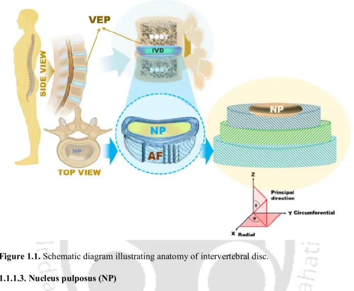
- Anatomy, physiology and function of intervertebral disc (IVD)
- Annulus fibrosus (AF)
- Transition zone (TZ)
- Nucleus pulposus (NP)
- Vertebral end plate (VEP)
- Degeneration of IVD or degenerative disc disease (IDD)
- Imbalance in matrix turnover
- Inflammatory pathways
- Oxidative stress
- Altered disc nutrition and cell death
- Genetic predisposition
- Current diagnostic tools
- Grading of IDD
- Current clinical solutions for IDD
- Conservative management 1. Physical exercises
- Surgical options
- Biological therapies - the regenerative strategies for treatment of IDD
It is reported that the prevalence of LBP is higher (7.5%) in rural areas than in urban areas (5.5%). Most patients (up to 80%) with acute LBP benefit from this conservative strategy over several weeks[91].
Review of literature
- Tissue engineering approaches for IVD regeneration
- Biomaterials for tissue engineering
- Tissue engineering approaches for annulus fibrosus (AF) regeneration 1. Electrospun mats
- Tissue engineering approaches for nucleus pulposus (NP) regeneration
Wilda et al., investigated the effectiveness of poly(d,l-lactide) (PDLLA)/bioglass composite foam to repair AF tissue of degenerated IVD[237]. Cabraja et al., developed a PGA and hyaluronan (HA) based resorbable scaffold for AF tissue regeneration and repair [238]. Pirvu et al., investigated the potential of a poly(trimethylene carbonate) (PTMC)-based scaffold seeded with human bone marrow-derived mesenchymal stromal cells for AF rupture repair in a bovine organ culture annulotomy model under 14 days of dynamic loading[244].
Similarly, Whatley et al. polyurethane (PU) based biomimetic elastic vertebral disc using a specially designed computer-aided 3D fabrication device[280]. Similarly, Moriguchi et al. tissue-engineered collagen (AF)/alginate (NP) total disc which, when implanted into the canine vertebral column, integrated into the host tissue, maintained disc height and other biochemical properties after 16 weeks of implantation[ 254 ].
MOTIVATION AND OBJECTIVE OF THE PRESENT INVESTIGATION
MOTIVATION AND OBJECTIVES OF THE PRESENT INVESTIGATION
SF can be isolated from both mulberry (i.e., Bombyx mori, BM SF) and non-mulberry (e.g., Antheraea assamensis, AA SF and Philosamia ricini, PR SF) sources. 3D printing technology can be used to summarize the complex tissue architecture with the highest accuracy and precision in a high throughput manner. Silk hydrogel can be manufactured in a variety of ways, including using chemicals or applying physical stimuli.
This self-gelling property may be a potential platform for NP regeneration therapy via in situ gelation. Taking advantage of all these properties of WJ, a silk protein composite hydrogel can be designed, providing a minimally invasive injectable hydrogel for NP tissue replacement.
Fabrication and Characterization of Silk Based Multilayered Angle-ply Construct for Damaged
Annulus Fibrosus (AF) Tissue Replacement
ABSTRACT
Introduction
However, these surgical interventions are only effective in symptomatic pain relief without restoring the biomechanical functions of the IVD, which may result in disintegration of adjacent segments. Replication of AF anatomical shapes using different biomaterials has included both natural and synthetic polymers, but few studies have focused on recapitulating tissue structural features. Most importantly, the introduction of lamellar frameworks in AF tissue engineering may be critical to this field due to the direct link between hierarchical features and tissue mechanical functions [239].
The multi-scale, hierarchical, collagen fiber-reinforced composite structure of AF is responsible for shock absorption and flexibility of the spine. This type of organization creates an angular layer structure that is essential for proper biochemical and biomechanical function of the AF.
Materials and methods
- Isolation of silk fibroin protein
- Preparation of biological lamellar replicates
- Scanning electron microscopy (SEM)
- Wide angle x-ray diffraction (WAXD)
- Fourier transform infrared spectroscopy (FTIR)
- Mechanical properties
- Biological assessments
- Isolation and culture of porcine annulus fibrosus (AF) cells and bone marrow derived human mesenchymal stem cells (hMSCs)
- Cell seeding and maintenance within constructs
- Imaging of cellular alignment in lamellar constructs
- Cell proliferation
- Histology of the cell seeded constructs
- Biochemical assay for secreted extracellular matrix (total collagen and sulfated glycosaminoglycan; sGAG)
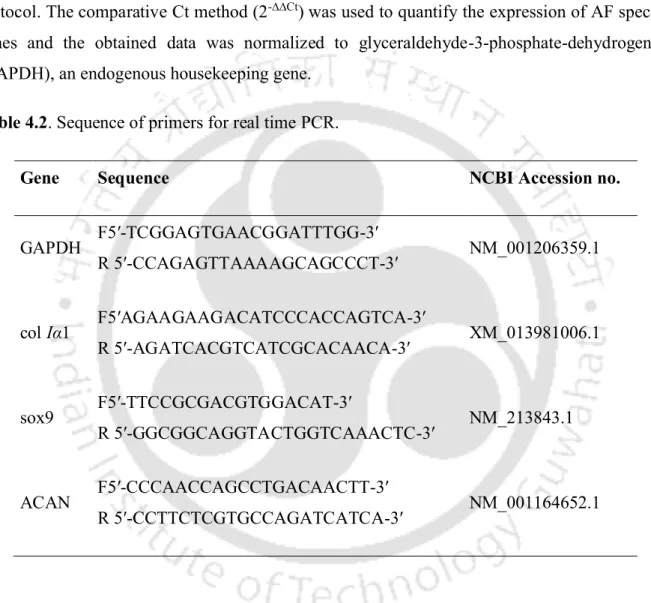
- Real time PCR analysis
- In vivo response to lamellar constructs
- Statistical analysis
The lamellar scaffolds seeded with primary porcine AF cells and hMSCs were washed in PBS followed by fixation in 10% (v/v) NBF (neutral buffered formalin) for 24 h before histological analysis. For this experiment, sections were deparaffinized, hydrated and permeabilized, followed by incubation with 1% bovine serum albumin (BSA) at 37 °C for 30 min. Then the sections were washed and incubated with universal secondary antibodies (Vectastain ABC kit, CA, U.S.A.) followed by development of .
The samples were then fixed in formalin and paraffinized followed by sectioning for hematoxylin and eosin (H&E, Sigma-Aldrich, U.S.A.) staining. For immunofluorescence, the treated sections were blocked with 1% (w/v) bovine serum albumin (BSA, Sigma, U.S.A.) for 30 min at room temperature (RT, 25 °C) followed by incubation with primary antibody against CD 68 (diluted) 1: 200, Abcam, U.K.) for 1 hour at RT.
Results
- Scaffold features
- Mechanical properties of constructs
- Cell survival, proliferation and alignment study
- Histology and immunohistochemistry analysis
- Quantitative analysis of ECM deposition
- Real time PCR analysis
- In vivo assessments
The results showed that cells (in both cases; porcine AF cells and differentiated hMSCs) were homogeneously distributed throughout the constructs and arranged in a lamellar fashion, adhering to the lamellar walls after 2 weeks of culture (Figure 2.4 , A and D). Alcian blue staining revealed even deposition of sGAG within the entire lamellar constructs for both cases, but with more intense blue staining for porcine AF cell-seeded constructs (Figure 2.4, B and E). Similarly, from immunohistochemistry, type I collagen was abundantly secreted by both cells, ie, porcine AF cells and differentiated hMSCs, after 2 weeks of culture in chondrogenic medium (Figure 2.4, C and F).
For porcine AF cells, the mRNA expression level of the three genes increased with time (days 7 and 14), and the maximum expression was observed for col Iα 1 when maintained in chondrogenic medium (Figure 2.5C). After one week, the retrieved constructs showed immune cells (mainly macrophages, confirmed by immunofluorescence for CD68) around the constructs (Figure 2.6B, II).
Discussion
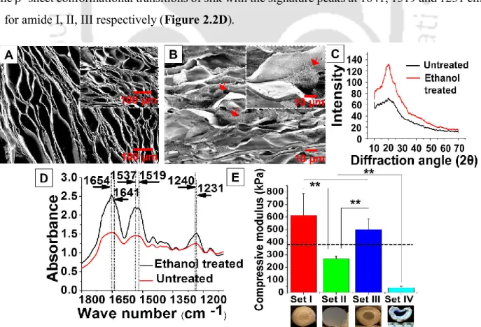
SEM and fluorescent-based image analysis revealed the transversely aligned and lamellar features of the constructs (Figure 2.1C, I-II and Figure 2.2A). Electron microscopy of the cross-sections of the scaffolds revealed circular openings (44 to 78 µm) of the lamellar channels. In this study, the cellular compatibility of the constructs was assessed with two cell types; primary porcine AF cells and bone marrow-derived hMSCs.
H&E staining of the constructs revealed cellular infiltration and extension in the lamellae (Figure 2.4, A and D). In the present study, recapitulation of the internal architecture of the AF has been achieved using the tissue engineering approach, but it is necessary to address other daunting challenges for the clinical application of existing technologies.
Significant findings
Fabrication and Characterization of a Seamless Full Thickness Disc-like Angle-ply Construct with Tailored
Mechanical Properties Modulating Extracellular Matrix Secretion by Annulus Fibrosus (AF) cells
Introduction
Static tension of various magnitudes (i.e., physico-mechanical cues of the substrate) acts as an important modulator for AF cell behavior[399]. It is very important to choose suitable mechanical properties of the substrate for AF tissue engineering. Silk as a biomaterial has been used in tissue engineering for decades due to its widespread versatility, eg, biocompatibility, least immunogenicity, tunable biodegradability, and mechanical properties[191].
Furthermore, through this study, we thoroughly investigated the effects of structural organization and mechanical properties on extracellular matrix secretion and specific gene expression using our developed silk AF model that provided differential mechanical properties. The effect of substrate morphology and its bulk mechanical properties on growth, expression and biochemical properties of AF cells was also investigated.
Materials and methods
- Isolation of mulberry silk fibroin
- Isolation of non-mulberry silk fibroin
- Preparation of BM/AA and BM/PR SF blends
- Fabrication of disc-like angle-ply constructs
- Field emission scanning electron microscopy (FESEM) study
- Fourier transform infrared spectroscopy (FTIR)
- Porosity measurement of lamellar scaffolds
- Swelling property of lamellar scaffolds
- In vitro enzymatic degradation
- Mechanical properties
- Cell culture on lamellar SF scaffolds
- Isolation and culture of annulus fibrosus (AF) cells
- Seeding and culture of AF cells within lamellar constructs
- Cell proliferation assay
- Assessment of viability and alignment of seeded cells on lamellar constructs
- Histology of lamellar constructs
- Biochemical assays for DNA, sGAG and collagen content
- Real time PCR analysis
- In vitro inflammatory response study
- Statistical analysis
SF lamellar constructs were placed in 3 ml of phosphate buffered saline (PBS, pH 7.4) containing 2 U.mg-1 enzyme. The viability and extent of AF cells seeded in the lamellar constructs were visualized using the live/dead assay kit (Invitrogen, Life Technologies, U.S.A.). Cell-seeded constructs were then stained with the live/dead assay kit according to the manufacture's protocol.
The constructs were then thoroughly washed and incubated with Hoechst 33342 (Invitrogen, Life Technology, USA) for 30 min in the dark. For histological analysis, the cell-seeded lamellar constructs were fixed overnight in 10% (v/v) NBF (neutral buffered formalin).
Results
- Construct features
- Porosity measurement of lamellar scaffolds
- Swelling property of lamellar constructs
- In vitro enzymatic degradation
- Mechanical properties
- In vitro biological studies
- Cells survival and proliferation on lamellar scaffolds
- Histology and immunohistochemistry analysis
- Biochemical quantification of ECM components
- Real time PCR analysis
- In vitro inflammatory response study
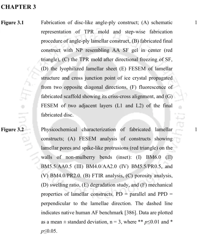
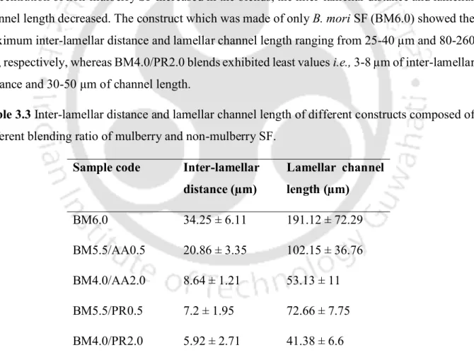
It was also found that the lamellar walls of each mix exhibited spike-like bumps whose distribution and density increased as the non-mulberry SF concentration increased in the mixes, while smooth wall boundaries were observed in BM6.0 ( figure 3.2A, II -V). FESEM images showed cell distribution in all cases after three weeks of culture (Figure 3.3E). Compressive strength was studied in two ways; parallel (PD) and perpendicular (PPD) to the direction of the lamellae (Figure 3.2F).
Cells were observed to be well distributed in a lamellar manner within all constructs, which was also observed in the FESEM study (Figure 3.3E, I-V). By immunohistochemistry, unlike sGAG, it was observed that maximal type I collagen was deposited in BM4.0/AA2.0 and sufficient amount of deposition was also found in BM6.0 and BM5.5/AA0.5, but significantly decreases in both BM4.0/PR2.0 and BM5.5/PR0.5 (Figure 3.4C, I-V).
Discussion
This increasing order of compressive modulus may be related to the physicochemical properties of the manufactured constructs. This may be due to the increased gross hydrophobicity of constructs that recalled water molecule to interact. This may be due to the reduced pore size/porosity in mixtures which restricts the enzyme molecules from passing through the constructs.
This may be due to the preferential selection and response of a specific cell population to a particular matrix stiffness. We also investigated the mechanosensing behavior of AF cells responding to the variations in matrix stiffness.
Significant findings
Design and Fabrication of 3D-printed Biomimetic Construct to Recapitulate Form and Function of Intervertebral Disc
IVD)
Introduction
The principle of 3D printing can be described as the development of spatially defined structures with biomaterials using 3D printer technologies. 3D printing technology has been used to print various tissues, including skin, liver, heart, blood vessels, bone, cartilage, as well as for IVDs using various biomaterials. One of the limitations that hinders the production of biomimetic artificial organs using 3D printing technology is the selection of an appropriate biomaterial-based ink (biomaterial ink).
Researchers have optimized silk fibroin-based biomaterial inks for 3D printing technology [442] by enzymatic crosslinking [443] in conjunction with polyethylene glycol (PEG), polyvinylpyrrolidone (PVP in methacrylate photo-crosslinkable form [446]), mixed with gelatin and enzymatic cross-linking [447] and in non-cross-linking form, with a mixture of gelatin, for 3D printing of various tissues [448] This is the first time we have reported the fabrication of a native structural prototype of AF using 3D printing technology.
Materials and methods
- Isolation of Bombyx mori silk fibroin
- Preparation of carrageenan solution
- Formulation of silk-carrageenan based biomaterial ink
- Characterization of the silk-carrageenan based biomaterial ink 1. Rheological studies
- Computer-aided design (CAD) model of annulus fibrosus (AF)
- Characterization of the 3D-printed constructs
- Field emission scanning electron microscopy (FESEM)
- Fourier transform infrared (FTIR) spectroscopy
- Swelling behavior of the 3D printed silk based scaffolds
- In vitro enzymatic degradation
- Mechanical properties
- Biological response study of the 3D-printed constructs 1. Isolation of primary porcine AF cell
- Isolation of adipose derived stem cells from porcine subcutaneous adipose tissue Adipose derived stem cells (ADSCs) were isolated from porcine subcutaneous fat tissue following
- Cell seeding and proliferation assay within 3D-printed constructs
- Cell viability study
- Histological analysis
- Biochemical analysis
- Real time PCR analysis
- In vitro inflammatory response study
- In vivo response to biomaterial ink
- Statistical analysis
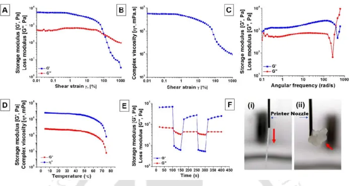
A rheometer (MCR 302, Anton Paar, Austria) with a 25 mm diameter standard parallel steel plate was used to measure the rheological parameters of the formulated biomaterial paint. Analysis of the printed filament dimension and pore size of the 3D printed cellular constructs were done using FESEM (Zeiss, Germany) at an operating voltage of 2–3 kV. This assay was used to determine the biodegradability of the 3D printed constructs under physiological conditions.
The compressive modulus was determined in both directions of the 3D printed constructions of the angular mesh (10 x 10 x 10 mm); horizontal plane and vertical plane. Trypan blue dye (Sigma-Aldrich, USA) was used to check the viability of isolated AF cells.
Results
- Rheological characterization of the biomaterial ink
- Silk-carrageenan biomaterial ink based 3D printing of AF architecture
- Physiochemical characterization of the 3D printed scaffolds 1. Field emission electron microscopy (FESEM) analysis
- Furrier transform infrared spectra (FTIR) analysis
- Swelling behavior
- In vitro enzymatic degradation
- Mechanical properties
- Biological response to 3D printed constructs 1. Cell viability and proliferation study
The 3D CAD model of the AF region of the IVD was designed based on the native structure (Figure 4.2, A-B). 3D CAD model of AF structure; (A) CAD model fed to the 3D bioprinter: (i) model representing the angled lattice structure (i-ii) design showing the top and side view of the CAD model, (iii) showing G-code for a small part of construction, (B) (i) prototype of the 3D multi-layer CAD model representing the structure after flipping by 90° that would mimic the native AF architecture, (ii) multi-layer CAD model of the filaments arranged at an angle of ± 30° but alternating direction in each successive layer, and (iii) G-code for the full structure. The fiber diameter and spacing between fibers in the printed constructs were maintained as designed in the CAD model.
The pore size of the 3D printed scaffolds and their interconnectedness were evaluated by FESEM. A significant difference was observed in the degradation rate of the 3D printed constructs treated with enzyme and the control group (p≤0.001).