COMPARATIVE SKELETAL ANATOMY
COMPARATIVE
SKELETAL ANATOMY
A PHOTOGRAPHIC ATLAS FOR MEDICAL EXAMINERS, CORONERS, FORENSIC
ANTHROPOLOGISTS, AND ARCHAEOLOGISTS
By
BRADLEy].}loAMS, PhD
Office ofChiefMedical Examiner, New York, NY
PAMELA]. CRABTREE, PhD
Department ofAnthropology, New York University, New York, NY
Photographs by
GINA SANTUCCI
Authors
Bradley1. Adams, PhD
Office of Chief Medical Examiner 520 1st Avenue
NewYork, NY 10016 [email protected]
ISBN: 978-1-58829-844-7
PamelaJ.Crabtree, PhD DepartmentAnthropology New York University 25 Waverly Place New York, NY 10003 [email protected]
e-ISBN: 978-1-59745-132-1
Library of Congress Control Number: 2008921061
©2008 Humana Press, a part of Springer Science-Business Media, LLC
All rights reserved. This work may not be translated or copied in whole or in part without the written permission of the publisher (Humana Press, 999 Riverview Drive, Suite 208, Totowa, NJ 07512 USA), except for brief excerpts in connection with reviews or scholarly analysis. Use in connection with any form of information storage and retrieval , electronic adaptation, computer software , or by similar or dissimilar methodology now known or hereafter developed is forbidden .
The use in this publication of trade names, trademarks, service marks, and similar terms, even if they are not identified as such, is not to be taken as an expression of opinion as to whether or not they are subject to proprietary rights . While the advice and information in this book are believed to be true and accurate at the date of going to press, neither the authors nor the editors nor the publisher can accept any legal responsibility for any errors or omissions that may be made. The publisher makes no warranty, express or implied, with respect to the material contained herein.
Cover illustration: bear skull showing upper and lower dentition (see discussion in Chapier 4).
Printed on acid-free paper 9 8 7 6 5 4 3 2 I springer.com
PREFACE
Bones are frequently encountered in both archaeological and forensic contexts. In either situation it is critical that human remains are differentiated from non-human remains. In the realm of forensic investigations, this is usually the final determination. In the archaeo- logical context, greater precision in identification may be warranted in order to draw con- clusions about ancient diets, animal husbandry and hunting practices, and environmental reconstructions. This photographic atlas is designed to assist the archaeologist or forensic scientist (primarily zooarchaeologists and forensic anthropologists) in the recognition of various species that are commonly encountered in both contexts. Obviously the ability to differentiate between the bones of various species (let alone simply human vs non-human bones) is dependent upon the training of the analyst, but good reference material is also essential. While there are books dedicated to human osteology and books that focus on animal osteology, there is really nothing that brings the two together. It is our intent to fill this void with the compilation of photographs presented in this atlas. Greater attention is given to the postcranial remains, which are presented in standard anatomical orientations.
In addition, "non-traditional" photographs of the various non-human species are also included in an attempt to bring together both anatomical and artistic images.
For this atlas, the large, non-human mammals include : horse (Equus cabal/us) , cow (Bos taurus), black bear (Ursus americanus), white-tail deer (Odocoileus virginianus), pig (Sus scrofa), goat (Capra hircus), sheep (Ovis aries) , and dog (Canis familiaris).
All of these are compared to a modem adult male human skeleton.
The smaller non-human animals include: raccoon (Procyon lotor) , opossum (Didelphis virginiana), cat (Felis eatus), rabbit (Oryctolagus cuniculus and Sylvilagus floridanus) , turkey (Meleagris gallopavo), duck (Anas platyrhynchos), chicken (Gallus gallus), rat (Rattus norvegicus) , red fox (Vulpes vulpes), and snapping turtle (Chelydra serpentina). All of these are compared to a modem newborn human skeleton.
The first part of this book consists of a brief introduction followed by detailed black and white photographs of the key postcranial elements from the animals listed above.
In order to show size and shape variations between the human and the non-human species selected for this atlas, scaled skeletal elements are pictured side-by-side. For example, a cow humerus and a human humerus are placed side-by-side in order for the reader to observe how they differ. Anterior (i.e., front or cranial in animals) and poste- rior (i.e., back or caudal in animals) views of each bone are presented. In some cases, medial or lateral views are also included.
The second part of the book consists of an overview of common butchering tech- niques used in traditional and commercial meat processing. This is followed by photo- graphs of representative butchered bones. We have included a range of different butchery marks, including both prehistoric cut marks made with stone tools and his- toric cut marks made with cleavers and saws. We have also included examples of sawn human bones from a forensic case associated with intentional body dismemberment.
Since bone was a common raw material throughout antiquity and up until the early 20th century, we have also illustrated a number of example s of worked bone artifacts.
v
VI Preface Overall, we hope that this book will fill a void in the forensic science and archaeo- logical literature, presenting comparisons between human and non-human bones that are useful to the archaeologist and forensic scientist.It is our goal that this book is frequently consulted as a laboratory and field reference guide ... one that gets worn and discolored over the years from continued use and not a book that sits idle on a book shelf.
BradleyJ.Adams Pamela J.Crabtree
CONTENTS
Preface v
About the Author .ix
1 Introduction 1
2 Human vs Horse 9
3 Human vs Cow 29
4 Human vs Bear 45
5 Human vs Deer 75
6 Human vs Pig 97
7 Human vs Goat 117
8 Human vs Sheep 133
9 Human vs Dog 153
10 Human vs Raccoon 177
11 Human vs Opossum 195
12 Human vs Cat 217
13 Human vs Rabbit 235
14 Human vs Turkey 251
15 Human vs Duck 269
16 Human vs Chicken 289
17 Miscellaneous 307
18 Traces of Butchery and Bone Working 323
19 References 347
Vll
ABOUT THE AUTHORS
Bradley
J.
AdamsBradley1.Adams received his BA from the University of Kansas and his MA and PhD degrees from the University of Tennessee. He is currently the Director of the Forensic Anthropology Unit for the Office of ChiefMedical Examiner (OCME) in NewYork City.
He is also affiliated with numerous universities in the NewYork City area. In his present position with the OCME, Dr. Adams and his team are responsible for all forensic anthro- pology casework in the five boroughs of NewYork City (Manhattan, Brooklyn, Queens, the Bronx, and Staten Island). Priorto accepting the position in NewYork City,Dr.Adams was a forensic anthropologist and laboratory manager at the Central Identification Laboratory in Honolulu, Hawaii.
Pamela
J.
CrabtreePamela1.Crabtree is an associate professor of anthropology at NewYork University, where she has taught since 1990. Her area of specialization is zooarchaeology, and she has analyzed a wide variety of faunal collections from late prehistoric and early medieval Europe, the Middle East, and historic North America. Dr. Crabtree is co-author of Exploring Prehistory: How Archaeology Reveals Our Past (2006) and she is co-editor of AncientEurope: Encyclopedia of the Barbarian World 8000Be-AD1000. She is currently a member of the archaeological team that is surveying the IronAge site of Dun Ailinne in Ireland.
ix
1 Introduction
Regardle ss of the context (forensic or archaeological), the correct identification of human and non-human remains is a very serious issue in osteological analyses. While the difference between various species is often very striking, it can also be quite subtle (Figure 1-0 1). Case studies and text books have highlighted similarities between some species, for example the hand and foot bone s (metacarpals and metatarsals) of the human hand and the bear paw in the forensic realm (Byers 2005; Owsley and Mann 1990; Stewart 1979; Ubelaker 1989). These compari sons between the human and bear are also presented in Chapter 4 of this book. Sometimes the morphological similarity between species is quite unusual and counterintuitive. For example , there is a remark- able correspondence between an adult human clavicle and an adult alligator femur (Figure 1-02).
The goal of this book is to create a comprehensive photographic guide for use by experienced archaeologists and forensic scientists to distinguish human remains from a range of common animal species. The atlas illustrates the larger mammal species in comparison to adult human bones, while the smaller mammal , bird, and reptile species are compared to an infant human skeleton. We have chosen to photograph the Old World dome sticates-cattle (Bos tauru s ) ,sheep (Ovis ari es ) , goat (Capra hircus) ,horse (Equus caballus) ,and pig(Sus scroJa)- since the se animal s are frequently found on historic archaeological sites in North America, and they are commonly recovered from Neolithic and later sites in the Eastern Hemisphere. Furthermore , they are also common in modem contexts and could easily end up being submitted as a forensic case.
The atlas include s three domestic bird species; two of them, chicken (Gallus gallus ) and duck (Anas platyrhynchos),were initially domesticated in the Eastern Hemisphere, while the third, turkey (Me leagris gallopavo), was first domesticated by Native Americans. We have also chosen to illustrate a range of North American wild mam- mals, including many that were frequently hunted by Native Americans in pre- Columbian and colonial times. These include black bear(Ursus americanus) ,white-tail deer (Odocoileus virginianus) ,raccoon (Procyon lotor ),and opossum (Didelphis vir- giniana).We have also included two species of rabbits. The smaller rabbit is the native wild rabbit or cotton-tail (Sylvilagus floridanus), while the larger rabbit is a domestic rabbit(Oryctolagus cuniculus) which is originally of European origin. Commensal species are frequently found in historic-period archaeological sites, and we have illus- trated two of the most common , dog (Canisfamiliarisy and cat(Felis catus).We have also included a chapter of miscellaneous photographs (Chapter 17). In this chapter var- ious views are presented of infant and adult human skeletons, selected comparisons between human and red fox (Vulpes vulp es), bobcat (Felis rufus),rat (Rattus norvegi- cus),and snapping turtle (Chelydra serpentina) .The snapping turtle is the only reptile that is included as many of the bones are distinctive is shape and they are commonl y recovered from North American archaeological sites.
1
2 Adams andCrabtree
Fig. 1-01. Compari son from left to right of infant human, adult chicken , and adult cat right femora (anterior views).
Most archaeological faunal remains are the leftovers from prehistoric and historic meals.
Many animal bones showtraces of butchery that reveal the ways in which the carcass was dismembered. Furthermore, it is not unusual for food refuse to be mistaken for human remains and end up in the medical examiner or coronersystem. In this atlas we have illus- trated a rangeof different butchery marks and techniques (Chapter 18), including both pre- historic cut marks made with stone tools and historic cut marks made with cleavers and saws. We have also included examples of sawn and butchered faunal bones and have included schematic diagrams of modern, commercial butchery patterns. Since bone was a commonraw material throughout antiquity and up untilthe early20thcentury, we have also illustrated a numberof examples of worked boneartifacts. Finally, knifecuts and saw marks in bone are not unique to non-human remains. Thereare numerous caseseach year of inten- tional body mutilation using knifes and/or saws. In cases of human dismemberment (usu- ally implying sawing through bones) or disarticulation (usually implying separation between joints)it is quitepossible that a badlydecomposed or skeletonized human bodyportion may
Introduction 3
Fig. 1-02. Comparison of an adult human clavicle with alligator and crocodile femora; note the sim- ilar morphology between the human and nonhuman elements . Top is a left human clavicle, middle is a right Crocodylus acutus femur, bottom is a right Alligator mississippiensis femur.
appear non-human to the untrained eye. A forensic example of postmortem human dismem- berment is also presented in Chapter 18 to show the similarity of tool mark evidence.
The ability to differentiate between complete or fragmentary human and non-human bones is dependent on the training of the analyst and the available reference and/or comparative material. It is truly a skill that requires years of training and experience and is not something that can be gleaned entirely from books. There is no substitute for coursework and training in osteology with actual skeletal material in order to appreci- ate the range of variation within all animal species. An experienced osteologist should always be consulted for confirmation of element type and species if there is any doubt.
ARCHAEOLOGICAL CONTEXT
Animal bones have played critical roles in archaeological interpretation for more than one hundred and fifty years of scientific endeavors . The discovery of the bones of
4 Adams and Crabtree extinct animals in association with simple chipped stone tools in sites in France and Britain helped to establish the antiquity of the human presence in Europe and to over- throw the traditional 6000-year biblical chronology for human life on earth . Faunal remains have also played a crucial role in the reconstruction of early human subsis- tence practices, in the study of animal domestication in both the Eastern Hemisphere and the Americas , and in the analysis of the ways in which historic cities were provi- sioned with food. Large numbers of animal bones are often recovered from archaeological sites , and these bones can be used to study past hunting practices, animal husbandry patterns , and diet. In order to use animal bones in archaeological interpretation , zooar- chaeologists (archaeologists who specialize in the study of faunal remains) must be able to identify the bones, determine sex and age at death when possible, and examine the bones for evidence of butchery marks and traces of bone working.
While archaeologists expect to find human remains in cemeteries, human bones are often found in other contexts. For example, two adult human burials and the remains of several infants were unexpectedly recovered from the habitation area of the early Anglo-Saxon village site of West Stow in eastern England (West 1985: 58-59). This was the case even though the settlement site was associated with a nearby contempo- rary cemetery. In another example, at the late Neolithic site of Hougang near Anyang in China , burials of infants in pits or urns were associated with house construction activi- ties (Chang 1986: 270). In short, zooarchaeologists and physical anthropologists must be able to confidently identify both animal bones and human remains in order to accu- rately interpret past cultures.
The first step in the analysis of animal bones recovered from archaeological sites is the careful identification of both body part and animal species . Precise identification requires a good comparative collection of modern specimens whose species, sex, and age are well-documented. However, a comparative collection must be supplemented by identification guides and atlases that can help the researcher distinguish between differ- ent species. Most zooarchaeological identification guides focus solely on non-human species, (e.g., Brown and Gustafson 1979; Cornwall 1956; Gilbert 1990; Gilbert, et al.
1981; Olsen 1964, 1968) even though human remains are commonly found in archaeo- logical sites. One exception to this is Schmid (1972) who does illustrate human bones, but there is no comparison with subadult human bones.
FORENSIC CONTEXT
Itis equally important for forensic scientists working with human skeletal remains to be able to differentiate between human and non-human bones. In the modern foren- sic context , it is quite common for non-human bones to be mistaken for human remains and end up in the medical examiner or coroner system. It is of obvious importance that they are correctly identified as such, or the consequences could be substantial. It is usu- ally the role of a forensic anthropologist to make this assessment of "human vs. non- human" and generate the appropriate report. In most forensic scenarios, once a determination of non-human is made it is seldom of investigative significance to cor- rectly identify the species. There are numerous skeletal anatomy books dedicated to human osteology (e.g., Bass 2005; Brothwelll981 ; Scheuer and Black 2000; Steele and Bramblett 1988; White 2000; White and Folkens 2(05). Some guides and textbooks on human osteology and forensic anthropology do include sections on differentiating
Introduction 5 between human and non-human remains (e.g., Bass 2005; Byers 2005; Ubelaker 1989) but these are more cursory discussions.
When attempting to differentiate between human and non-human skeletal remains, fragmentation only compounds the problem. If fragmentation is so extreme that gross identification of human versus non-human bone is not possible, microscopic (i.e., histological) techniques can be employed (e.g., Mulhern and Ubelaker 2001) . Under magnification, the shape of the bone cells may be indicative of non-human bone, but this technique is not "fool proof" as some non-human animals (e.g., large dogs, bovines, and non-human primates) are nearly identical to humans microscopically. Our atlas will only focus on the gross assessment of bones.
BOOK TERMINOLOGY AND ORGANIZATION
In constructing this atlas, we have chosen to illustrate examples of both adult and juvenile animal bones, in addition to adult and infant human skeletons. Other guides to the identification of birds and mammals from archaeological sites illustrate only adult bones. However, many animal bones recovered from archaeological sites and within the forensic context are the remains of juvenile animals . Farmers who keep cattle for milk, for example, often slaughter excess male calves during their first year of life. In a meat-oriented economy, farmers frequently choose to slaughter adolescent animals, since these animals are nearly full-grown, and continuing to feed animals beyond ado- lescence results in only limited increases in meat output. We have included illustrations of both adult and juvenile pigs, and we have illustrated both an adult sheep and an immature goat. We have also photographed examples of immature chickens, since most chickens consumed today are quite young.
In general, the animals in this atlas are presented in the order of their size, progress - ing from largest to smallest. The corresponding human and non-human elements are presented alongside each other in order to fully appreciate the variation in size and shape between them. In order to add a scaled perspective, a metric ruler (centimeters) is present in each photograph along with a U.S. penny.
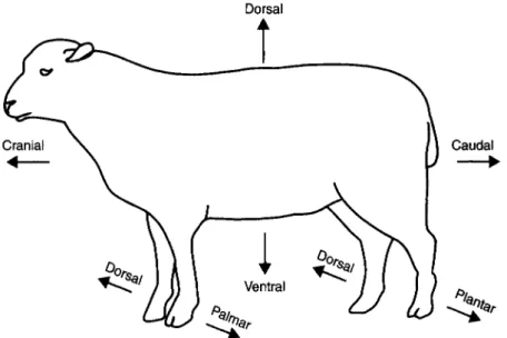
Bipedalism, upright walking on two legs, is one of the most important developments in all of human evolution. However, as a result of bipedalism, many human bones are oriented in somewhat different ways than comparable bones are in other mammals. In addition, the directional terms used to describe parts of the body differ somewhat between humans and other mammals (Figures 1-03 and 1-04). For example, in human osteology the term anterior is used to describe the front portion of a bone, while in quadrupeds the term cranial is used . Similarly, the back portion of the femur is described as posterior in humans, but it is described as caudal in other mammals.
Different terms are also used for the lower portions of mammal limbs. For example, the surface of the forelimb (distal to the radius and ulna) that faces the ground is described as palmar (or volar), while the comparable surface in the hindlimb is described as plantar. The opposite surfaces of the bone are described as dorsal. The terms proximal, distal, medial, and lateral are used to describe surfaces in both human and non-human bones. For humans, we have used the directional terms as described in Bass (2005). For other mammals, they have used the terms as defined in Evans and de Lahunta (1980) and Getty (1975). In describing bird bones. we have followed the terminology used by Cohen and Sergeantson (1996).
6
••••••••••••• Supe rior •••••••••••••••••
.
/ .. /
Dista l ..:
-,-, ...
-,
Adams and Crabtree
Fig. 1-03. Schematic diagram of human skeleton in standard anatomical position (i.e., standing with arms at the side and palmsforward so that no bonesare crossing) labeled with anatomical terminology.
BACKGROUND OF THE SPECIMENS INCLUDED IN THIS BOOK Most of the non-human skeletons that are illustrated in this atlas come from the col- lections of the zooarchaeology laboratory in the Anthropology Department of New York University. The bear skeleton was borrowed from the Department of Mammology of the American Museum of Natural History. Most of the horse bones that are illus- trated here are from a horse skeleton that was borrowed from the Museum Applied Science Center for Archaeology (MASCA) at the University of Pennsylvania Museum.
Introduction 7
Cranial
+---
Dorsal
i
Ventral
Caudal
~
Fig . 1-04. Schematic Diagram of animal skeleton labeled with anatomical terminology.
The raccoon skeleton was borrowed from Susan Ant¢n. The alligator and crocodile femora were provided by the Herpetology Department at the American Museum of Natural History and were photographed by Ilana Solomon and Tam Nguyen. The orig- inal photograph of the turkey skull was provided courtesy of the National Wild Turkey Federation, while Gina Santucci performed the artistic modifications to the photograph . Seth Brewington provided the photograph of the antler comb from Iceland. The horse metacarpus and metatarsus were borrowed from the Zooarchaeology Laboratory in the Anthropology Department at Hunter College. Jeannette Fridie was a great help with many facets of this book. The human remains are from unidentified individuals that were analyzed at the Office of Chief Medical Examiner in New York City. We are grate- ful to everyone who loaned us specimens and assisted in this project.
2 Human vs Horse
Fig. 2-00.A lateral view of the horse's cranium. The horse's dental formula is 3/3.0-1/0-1.3/3.3/3.
Canines are usually seen only in males.
9
10 Adams and Crabtree
Fig. 2-01.A human right humerus (anterior view) is compared to a horse 's right humerus (cranial view). The shaft of the horse's humerus has a large deltoid tuberosity. The proximal end of the horse's humerus includes an intermediate tubercle, which is not seen on the human humerus.
Human vs Horse 11
Fig. 2-02.A humanright humerus (posterior view) is comparedto a horse's right humerus (caudal view).
12 Adams and Crabtree
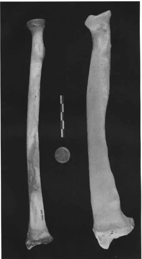
Fig. 2-03.A human right radius and ulna (anterior views) are compared to a horse's radius and ulna (cranial view). Note the large olecranon process on the horse's ulna.
Human vs Horse 13
Fig. 2-04.A human right radius and ulna (posterior views) are compared to a horse 's radius and ulna (caudal view). Note that the horse's ulna tapers to a point about two-thirds of the way down the shaft of the radius.
14 Adams and Crabtree
Fig. 2-05. A human right radius and ulna (lateral views) are compared to a horse 's radius and ulna (lateral view). The horse's ulna is partially fused to the radius in adults.
Human vs Horse 15
Fig. 2-06.A human right femur (anterior view) is compared to a horse 's right femur (cranial view).
The horse's femur shows a well developed third trochanter.
16 Adams andCrabtree
Fig. 2-07. A human right femur (posterior view) compared to a horse 's right femur (caudal view).
Human vs Horse 17
Fig. 2-08.A human right tibia (anterior view) is compared to a horse's right tibia (cranial view). The horse distal tibia includes both a medial and a lateral malleolus . The lateral malleolus is the evolu- tionary remnant of the distal fibula.
18 Adams andCrabtree
Fig. 2-09. A human right tibia (posterior view) is compared to a horse's right tibia (caudal view).
Human vs Horse 19
Fig. 2-10.A human right tibia (lateral view) is compared to a horse' s right tibia (lateral view).
20 Adams and Crabtree
Fig. 2-11. A humanright fibula (medial view) is compared to a horse's right fibula (lateral view). The horse'sfibula is greatly reduced. The rounded headis transversely flattened, andthe shaft tapers to a point.
N
-
Fig.2-12.Ahumanrightscapula(anteriorview)iscomparedtoahorse'srightscapula(medialview).Bothscapulaeareorientedastheywould beinahuman.N N
Fig.2-13.Ahumanrightscapula(posteriorview)iscomparedtoahorse'srightscapula(lateralview).Notethatthespineofthehorse'sscapula risesfromthescapularneck.
Human vs Horse 23
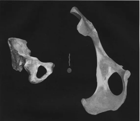
Fig. 2-14.A human right innominate (lateral view) is compared to a horse's right innominate (lateral view). The articular surface on the horse's acetabulum is crescent-shaped.
24 Adams and Crabtree
Fig.2-15.A human rightinnominate (medial view) is comparedtoa horse's right innominate (dorsal view).
Human vs Horse 25
Fig. 2-16.A human axis (C2) is compared to a horse's axis (C2). Both views are lateral. The cervical vertebrae generally reflect the length of the animal 's neck. Note how much longer the horse's axis is when compared to the human axis.
Fig. 2-17.The human sternum (anterior view) is compared to one of the horse's sternabrae .
26 Adams and Crabtree
Fig. 2-18 . A horse's right metacarpus and metatarsus (dorsal views) are shown on the left. The horse 's right metacarpus (volar view) and right metatarsus (plantar view) are shown on the right.
The horse has a single main metacarpus (3rd metacarpal) and metatarsus (3rd metatarsal). The remnants of the 2nd and 4th metacarpals and 2nd and 4th metatarsals can be seen volar/plantar views (shown on the right) . These "splint bones" (lateral metapodia) taper to a point about half way down the shaft of the main metapodial.
Human vs Horse 27
Fig. 2-19. Three caudal, or tail, vertebrae of a horse (dorsal views). While the numbers may vary, most horses have about 18 caudal vertebrae. These bones can sometime be confused with human phalanges.
3 Human vs Cow
Fig. 3-00. Cow skull (basal view). The cow has no upper incisors or canines. The tooth cow's maxil- lary tooth row includes three premolars and three molars on each side of the maxilla. The mandibular dental formula is 3.1.3.3.
29
30 Adams and Crabtree
Fig. 3-01. Human left humerus (anterior view) compared to cow's left humerus (cranial view). The proximal epiphysis of the cow's humerus is shown on the right. On the proximal end, the greater and less tubercles are far more well-developed in the cow than they are in the human. On this distal end, the cow has a barrel-shaped trochlea.
Human vs Cow 31
Fig. 3-02.Human left humerus (posterior view) compared to cow's left humerus (caudal view). The cow's unfused proximal epiphysis is shown on the right. Note that the cow humerus has a particularly deep olecranon fossa when compared to the human example .
32 Adams and Crabtree
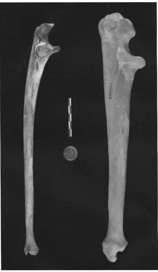
Fig. 3-03. Human left radius and ulna (anterior views) compared to a cow's left radius and ulna (cranial view). The human radius and ulna are roughly equal in size. With the exception of the large olecranon process, the cow's ulna is greatly reduced. While the human radius has a distinct head, the cow's radius has a broad and slightly concave articular surface.
Human vs Cow 33
Fig. 3-04. Human left radius and ulna (posterior views) compared to the cow's left radius and ulna (caudal view). Note that the cow's radius is fused to the lateral side of the radius shaft.
34 Adams and Crabtree
Fig. 3-05. Human left radius and ulna (lateral views) compared to the cow's left radius and ulna (lat- eral views). The shaft of the cow's radius is quite slender.Inadult animals, it is usually fused to the caudal surface of the radius.
Human vs Cow 35
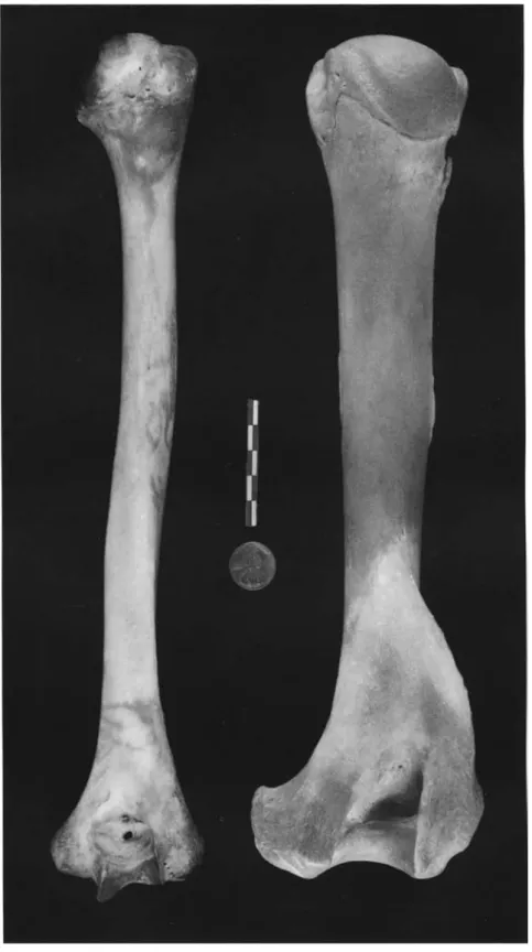
Fig. 3-06.Human left femur (anterior view) compared to cow's left femur (cranial view). The distal epiphysis of the cow's femur is shown separately to the right. The cow skeleton illustrated here is a 6-7 year old ox, or male castrate. While the distal epiphysis usually fuses by about 3.5-4 years of age (Silver 1969) castration has delayed epiphyseal fusion in this specimen . The greatest length of the human femur extends from the head to the distal condyle s; in the cow the greatest length extends from the greater trochanter to the condyles.
36 Adams and Crabtree
Fig. 3-07. Human left femur (posterior view) compared to cow's left femur (caudal view). The cow's unfu sed distal epiphysis is shown on the right. The human femur includes a linea aspera which appears as a projecting ridge for the muscle attachment. This is a unique human feature that is not seen in other mammals as it is an area of attachment for the muscles used in bipedalism. The cow's femur includes a supercondular fossa that can be seen on the lateral portion of the shaft.
Human vs Cow 37
Fig. 3-08.Human left tibia (anterior view) compared to a cow's left tibia (cranial view). The unfused proximal epiphysis of the cow's tibia is shown at right. The shaft of the cow's tibia is significantly more robust. The distal end of the cow's tibia includes two parallel articular facets for articulation with the astragalus.
38 Adams and Crabtree
Fig. 3-09. Human left tibia (posterior view) compared to the cow's tibia (caudal view). The unfused proximal epiphysis of the cow's tibia is shown at right.
l.JJ \0 Fig.3·10.Humanleftscapula(anteriorview)comparedtothecow'sleftscapula(medialview).Notethatthesebonesareorientedastheywould beinthehumanskeleton.Sincethecowisaquadruped,theglenoidcavityformsthedistalpartofthebone.Thebladeofthecow'sscapulais shapedlikeanelongatedtriangle.
>I>- o Fig.3-11.Humanleftscapula(posteriorview)comparedtothecow'sleftscapula(lateralview).Theacromionprocessiswelldevelopedinhumans; itisverysmallinthecowandotherruminantartiodactyls.Thegreatestlengthofthecow'sscapula(andthescapulasofmostothermammals) extendsfromtheglenoidcavitytothedorsalborder(paralleltothescapularspine).Thisisnotthecaseinhumans.Inhumans,thegreatestlength liesbetweenthesuperiorandinferiorborders.
Human vs Cow 41
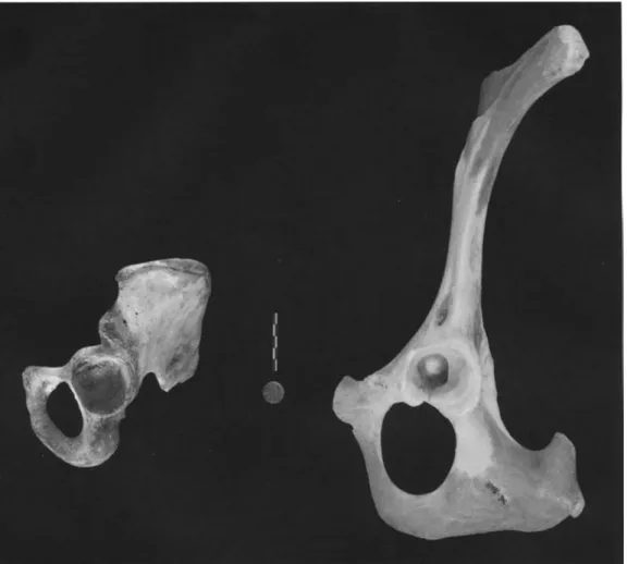
Fig. 3-12.A human left innominate (lateral view) is compared to cow's left innominate (lateral view).
42 Adams and Crabtree
Fig. 3-13. A human left pelvis (medial view) is compared to a cow's left pelvis (medial view).
~ ~ Fig.3-14.Humanlefttalusandcalcaneus(superiorviews)comparedtocow'sleftastragalus(plantarview)andcalcaneus(dorsalview).The humantalushasadistinctivehead,whilethecow'sastragalushasthe"doublepulley"formthatistypicalofallartiodactyls(even-toedungulates). Thecow'scalcaneusiselongated,andthedorsalsurfaceincludesanarticularfacetforthemalleolus,asmalltarsalbonethatistheevolutionary remnantofthedistalfibula.Thecowdoesnothaveaseparatefibula.
t
Fig.3-15.Thecow'srightmetacarpusandmetatarsus(dorsalviews)areshownontheleft.Thepalmar(volar)viewoftherightmetacarpusandthe plantarviewoftherightmetatarsusareshownontheright.Thesebonesaretheformedthroughthefusionofthethirdandfourthmetacarpalsand metatarsals.Eachofthetwodistalcondylesarticulateswithafirstphalanx.4 Human vs Bear
Fig. 4-00. A bear 's cranium and mandible (cranial view). The bear's upper dentition includes three incisors, dne canine, between two and four premolars, and two molars. The mandibular dentition includes three incisors, one canine, two to four premolars, and three molars.
45
46 Adams and Crabtree
Fig. 4-01. A human right humerus (anterior view) is compared to a bear's right humerus (cranial view). The lateral epicondylar crest (proximal to the lateral epicondyle) is well developed in the bear, as is the deltoid tuberosity.
Human vs Bear 47
Fig. 4-02.A humanright humerus (posterior view) is compared to a bear's right humerus (caudal view).
48 Adams and Crabtree
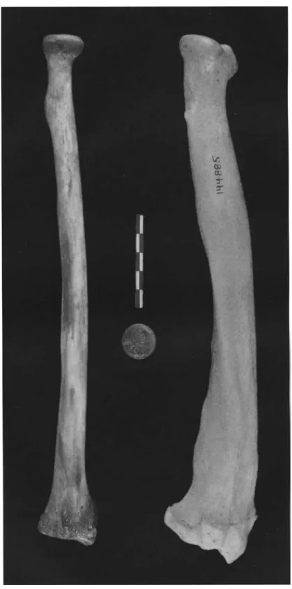
Fig. 4-03.A human right radius (anterior view) is compared to a bear 's right radius (caudal view).
Human skeletons are oriented with the palms forward, so that the radius and ulna are not crossed.
Quadrupedal animals are oriented with their paws facing the ground. This means that the proximal ulna is medial to the radius, while the distal ulna is on the lateral side.
Human vs Bear 49
Fig. 4-04.A human right radius (posterior view) is compared to a bear's right radius (cranial view).
50 Adams and Crabtree
Fig. 4-05. A human right radius (medial view) is compared to a bear's right radius (medial view).
Human vs Bear 51
Fig. 4-06. A human right ulna (anterior view) is compared to a bear's right ulna (cranial view). Note that the bear's olecranon process is larger and more well-developed .
52 Adams andCrabtree
Fig. 4-07. A human right ulna (posterior view) is compared to a bear's right ulna (caudal view).
Human vs Bear
Fig. 4-08.A human right ulna (lateral view) is compared to a bear's right ulna (lateral view).
53
54 Adams and Crabtree
Fig. 4-09. A human right ulna (medial view) is compared to a bear's right ulna (medial view).
Human vs Bear 55
Fig. 4-10.A human right femur (anterior view) is compared to a bear' s right femur (cranial view).
The distal condyles of the human femur show a distinctive asymmetry that is a result of "kneeing in"
or bringing the knees under the body. This is also referred to as the valgus knee.
56 Adams andCrabtree
Fig. 4-11. A human right femur (posterior view) is compared to a bear's right femur (caudal view).
The human femur shows the distinctive linea aspera which is not seen in quadrupeds.
Human vs Bear 57
Fig. 4-12.A human right tibia (anterior view) is compared to a bear' s right tibia (cranial view).
58 Adams and Crabtree
Fig. 4-13. A human right tibia (posterior view) is compared to a bear's right tibia (caudal view).
Human vs Bear
Fig. 4-14.A human right tibia (lateral view) is compared to a bear's right tibia (lateral view).
59
60 Adams and Crabtree
Fig. 4-15.A human right tibia (medial view) is compared to a bear's right tibia (medial view).
Human vs Bear 61
Fig. 4-16.A human right fibula (medial view) is compared to a bear 's right fibula (medial view).
62 Adams andCrabtree
Fig. 4-17. A human right fibula (lateral view) is compared to a bear's right fibula (lateral view).
0\ ~ Fig.4-18.Ahumanscapula(anteriorview)iscomparedtoabear'sscapula(medialview).Bothscapulaeareorientedastheywouldbeinahuman skeleton.
~ Fig.4-19.Ahumanscapula(posteriorview)iscomparedtoabear'sscapula(lateralview).Thespineofthebear'sscapuladividesthescapulainto roughlyequalhalves.
0'\ VI Fig.4-20.Ahumanrightinnominate(lateralview)iscomparedtoabear'spelvis(ventralview).Inthebear,thetwoinnomateshavefusedalong thepubicsymphysis.Thebroadiliumistypicalofhumanpelvesandisrelatedtobipedalism.Quadrupedshavelonger,narrowerilia.
0\ 0\
Fig.4-21.Ahumanleftinnominate(medialview)iscomparedtoabear'spelvis(dorsalview).Thetwobearinnominatesarefusedalongthepubic symphysis.
0\ '-l Fig.4-22.Ahumansacrum(anteriorview)iscomparedtoabear'ssacrum(ventralview).
0\ 00
Fig.4-23.Ahumansacrum(posteriorview)iscomparedtoabear'ssacrum(dorsalview).
Human vs Bear 69
Fig.4-24.A humansternum is compared to a complete bear's sternum plus the associated cartilaginous ribs. Note the differences in the shape of the human and the bear's manubrium.
70 Adams and Crabtree
Fig. 4-25. Human atlas (Cl , superiorview) is compared to a bear atlas (Cl, cranial view). Note that large wings on the bear atlas; these are typical of carnivores.
Fig. 4-26. Human axis (C2, lateralview) is compared to a bear atlas (C2, lateral view). The bear atlas is much longer and includes a large spinous process.
Human vs Bear 71
Fig. 4-27. Human left metacarpals 1-5 (anterior view) are compared to a bear's left metacarpals (volar view). The similarity in size and shape between human and bear metacarpals isfr~uentlynoted.
72 Adams andCrabtree
Fig. 4-28.Human left metatarsals 1-5 (superior view) are compared to a bear 's left metatarsals (dorsal view).
Human vs Bear 73
Fig. 4-29. A human left talus (superior view) is compared to a bear's left astragalus (plantar view).
Fig. 4·30. A human right calcaneus (superior view) is compared to a bear's right calcaneus (dorsal view). While these two bones are generally quite similar in form, note the differences in the shape of the sustentaculum tali.
5 Human vs Deer
Fig.5-00a.A cranium of a female white-tailed deer.Inmost deer species, the males have antlers while the females do not. The one exception is the reindeer ; both male and female reindeer have antlers .
75
76 Adams and Crabtree
Fig.S-OOb.A cranium of a male white-tailed deer. The dental formula for the upper dentition includes no incisors and canines, three premolars, and three molars. On the mandible, the dental formula is 3.1.3.3.
Human vs Deer 77
Fig. 5-01. A human right humerus (anterior view) is compared to a deer's right humerus (cranial view). Note the height of the greater tubercle on the deer's proximal humerus.
78 Adams and Crabtree
Fig. 5-02. A human right humerus (posterior view) is compared to a deer's right humerus (caudal view). Note the deeper olecranon fossa on the deer's distal humerus.
Human vs Deer 79
Fig. 5-03. A human right radius (anterior view) is compared to a deer 's right radius (caudal view).
The articulation with the ulna is visible on the lateral part of the caudal surface of the deer's radius.
80 Adams andCrabtree
Fig. 5-04. A human right radius (posterior view) IS compared to a deer's right radius (cranial view).
The human proximal radius has a distinct head and neck, while the deer radius has a slightly concave articular surface.
Human vs Deer 81
Fig. 5-05. A human right ulna (anterior view) is compared to a white-tailed deer 's right ulna (cranial view). Note the slender shaft of the deer's ulna.
82 Adams and Crabtree
Fig. 5-06. A human right ulna (posterior view) is compared to a white-tailed deer's right ulna (caudal view).
Human vs Deer 83
Fig. 5-07. A human right ulna is compared to a white-tailed deer 's right ulna (both lateral views).
Note the well-developed olecranon process on the deer' s ulna.
84 Adams andCrabtree
Fig. 5-08.A human left femur (anterior view) is compared to a deer 's left femur (cranial view). Note the well-developed greater trochanter in the deer 's femur.
Human vs Deer 85
Fig. 5-09. A human left femur (posterior view) is compared to a deer's left femur (caudal view). The linea aspera is visible on the human left femur. This is a distinctly human feature that is associated with the muscles used in bipedalism .
86 Adams andCrabtree
Fig. 5·10. A human left tibia (anterior view) is compared to a deer's left tibia (cranial view). The deer's distal tibia includes two parallel articular surfaces for articulation with the astragalus.
Human vs Deer
Fig. S-l1.A human left tibia (posterior view) is compared to a deer's left tibia (caudal view).
87
88 Adams andCrabtree
Fig. 5-12.A human left tibia (lateral view) is compared to a deer's left tibia (lateral view). Note the well-developed tibial tuberosity on the deer's tibia.
00 \D Fig.5-13.Ahumanrightscapula(anteriorview)iscomparedtoadeer'srightscapula(medialview).Bothbonesarepositionedastheywouldbeinahuman.
\Cl o Fig.5-14.Ahumanrightscapula(posteriorview)iscomparedtoadeer'srightscapula(lateralview).Notethelargeacromionprocessonthehuman scapula.Theglenoidcavityonthedeer'sscapulaisveryroundinshape(notpictured).
~
-
Fig.5-15.Ahumanrightinnominate(lateralview)iscomparedtoadeerrightinnominate(lateralview).\0 N Fig.5-16.Ahumansacrum(anteriorview)iscomparedtoadeer'ssacrum(ventralview).Thewingsofthedeersacrumarewideinrelationtothebody.
93
94 Adams and Crabtree
Fig. 5-18. Superior views of typical human cervical (top left), thoracic (middle left) , and lumbar (bottom left) vertebrae. Cranial views of typical white-tail deer cervical (top right), thoracic (middle right), and lumbar (bottom right) vertebrae.
\0 \Jl Fig.5-19.Humanlefttalusandcalcaneus(superiorviews)arecomparedtoawhite-taileddeer'sleftcalcaneus(dorsalview)and astragalus(plantarview).Thedeer'sastragalushasthetypical"double-pulley"formthatischaracteristicoftheartiodactyls.Thedorsal calcaneusincludesanarticulationforthemalleolus,asmalltarsalthatistheevolutionaryremnantofthedistalfibula.
96 Adams and Crabtree
Fig. 5-20.A white-tailed deer's metacarpus (volarview) and metatarsus (plantar view) are shown on the left. The dorsal viewsof the sameelements are shown on the right. These metapodia are composed of the fused third and fourth metacarpals and metatarsals. The deer metapodia have distinctive concave grooves along the plantar/volar surfaces that distinguish them from the metapodia of the bovids.
6 Human vs Pig
Fig. 6-00.A lateral view of an adult pig cranium . The pig dental formula is 3/3.1/1.4/4 .3/3.
97
98 Adams andCrabtree
Fig. 6-01. A human right humerus (anterior view) is compared to adult and juvenile pig right humeri (cranial views) . Note the large greater tubercle in the adult pig proximal humerus . The unfused epiphyses of the juvenile pig are also shown.
Human vs Pig 99
Fig.6-02.A human right humerus (posterior view) is compared to adult and juvenile pig right humeri (caudal views). The unfused epiphyses of the juvenile pig are also shown.
100 Adams and Crabtree
Fig. 6-03. A right human radius and ulna (anterior views) is compared to adult and juvenile right pig radii and ulnae (cranial views). In the adult pig, the radius is attached to the ulna. The unfused epi- physes of the juvenile pig radius are also shown.
Human vs Pig 101
Fig. 6-04.A right human radius and ulna (posterior views) are compared to adult and juvenile right pig radii and ulnae (caudal views). The unfused epiphyse s of the juvenile pig radius are also shown.
102 Adams and Crabtree
Fig. 6·05.A right human radius and ulna (lateral views) are compared to an adult right pig radius and ulna (lateral view). Note the large size of the pig's olecranon process .
Human vs Pig 103
Fig. 6-06.A human left femur (anterior view) is compared to adult and juvenile pig left femora (cra- nial views). Note the larger and more developed greater trochanter on the adult pig's femur. The unfused epiphyses of the juvenile pig are also shown.
104 Adams and Crabtree
Fig. 6-07.A human left femur (posterior view) is compared to adult and juvenile pig left femora (cau- dal views). Note the presence of the linea aspera on the human femur. The unfused epiphyses of the juvenile pig are also shown.
Human vs Pig 105
Fig. 6-08.A human left tibia (anterior view) is compared to adult and ju venile pig left tibiae (cranial views). The distal pig tibia has two parallel concave articul ar facets for articulation with the astra- galus . Note that the proximal epiphysis of the large pig is unfused. The unfused epiphy ses of the ju venile pig are also shown.
106 Adams and Crabtree
Fig.6-09.A human left tibia (posterior view) is compared to adult and juvenile pig left tibiae (caudal views). Note that the proximal epiphysis of the large pig is unfused. The unfused epiphyses of the juvenile pig are also shown .
Human vs Pig 107
Fig. 6-10. A human left tibia (lateral view) is compared to an adult pig left tibia (lateral view). Note that the proximal epiphysis of the large pig is unfused.
108 Adams andCrabtree
Fig. 6-11.A human right fibula(medial view) is comparedto a juvenile pig right fibula(medial view).
....
o \0 Fig.6-12.Ahumanrightscapula(anteriorview)iscomparedtoadultandjuvenilepigrightscapulae(medialviews).Notethatthelargehuman acromionprocessisnotentirelyblockedbythebodyofthescapulaandisvisibleintheanteriorview.- -
oFig.6-13.Ahumanrightscapula(posteriorview)iscomparedtoadultandjuvenilepigrightscapulae(lateralviews).Thepigscapulahasalarge tuberosityofthespinebutonlyarudimentaryacromionprocess.
.... ....
NFig.6-15.Ahumanleftinnominate(medialview)iscomparedtoadultandjuvenilepigleftinnominates(medialviews).Gapsin fusionofthepelvicbonesarevisibleonthejuvenilepig.
Human vs Pig 113
Fig. 6-16.Human axis (C2) is compared with an adult pig axis (C2). Both views are lateral.
Fig. 6-17.A human sternum (anterior view) is compared to two pig sternebrae .