저작자표시-비영리-동일조건변경허락 2.0 대한민국 이용자는 아래의 조건을 따르는 경우에 한하여 자유롭게
l 이 저작물을 복제, 배포, 전송, 전시, 공연 및 방송할 수 있습니다. l 이차적 저작물을 작성할 수 있습니다.
다음과 같은 조건을 따라야 합니다:
l 귀하는, 이 저작물의 재이용이나 배포의 경우, 이 저작물에 적용된 이용허락조건 을 명확하게 나타내어야 합니다.
l 저작권자로부터 별도의 허가를 받으면 이러한 조건들은 적용되지 않습니다.
저작권법에 따른 이용자의 권리는 위의 내용에 의하여 영향을 받지 않습니다. 이것은 이용허락규약(Legal Code)을 이해하기 쉽게 요약한 것입니다.
Disclaimer
저작자표시. 귀하는 원저작자를 표시하여야 합니다.
비영리. 귀하는 이 저작물을 영리 목적으로 이용할 수 없습니다.
동일조건변경허락. 귀하가 이 저작물을 개작, 변형 또는 가공했을 경우 에는, 이 저작물과 동일한 이용허락조건하에서만 배포할 수 있습니다.
의학박사 학위논문
양성 췌장 종양 환자에서의
내시경 초음파 유도하 고주파 치료의 유효성과 안전성 평가
Endoscopic ultrasound-guided
radiofrequency ablation for management of benign pancreatic tumors
울 산 대 학 교 대 학 원 의 학 과
최 준 호
[UCI]I804:48009-200000005258 [UCI]I804:48009-200000005258
Endoscopic ultrasound-guided
radiofrequency ablation for management of benign pancreatic tumors
지 도 교 수 서 동 완
이 논문을 의학박사 학위 논문으로 제출함
2017 년 12 월
울 산 대 학 교 대 학 원 의 학 과
최 준 호
최준호의 의학박사학위 논문을 인준함
심사위원 이 성 구 인
심사위원 서 동 완 인
심사위원 황 진 혁 인
심사위원 김 태 현 인
심사위원 박 도 현 인
울 산 대 학 교 대 학 원
2017 년 12 월
감사의 글
아직 부족한 것이 너무 많기 때문인지 언제나처럼 지난 시간들이 너무나도 아 쉽게 느껴집니다. 늘 처음 시작하는 마음가짐으로 저 자신과 지도해주신 교수님 께 부끄럽지 않은 사람이 되도록 최선을 다하도록 다짐합니다.
학위 과정 동안 저에게 관심과 도움을 주신 많은 분들께 짧은 글로 감사의 마 음을 전하는 것에 대해 죄송하게 생각합니다.
우선 부족한 저를 지도해 주시고, 학문의 길과 방향에 대해 항상 아낌없는 깨 우침을 주신 서동완 지도교수님께 진심으로 머리 숙여 감사드립니다. 교수님께서 해주셨던 말씀 한 마디, 한 마디가 학문적인 면뿐만 아니라, 생활 면에서도 저에 게는 큰 가르침이었습니다. 언제나 인자하신 미소로 많은 도움과 격려를 주시고 심사위원장을 흔쾌히 맡아주신 이성구 교수님께 감사드립니다. 그리고, 바쁘신 중에도 학위 논문 심사를 맡아주시고, 소중한 조언을 해주신 김태현 교수님, 황 진혁 교수님, 박도현 교수님께도 깊이 감사드립니다.
언제나 큰 사랑을 주시고 묵묵히 응원해주시는 사랑하는 부모님께 감사의 마 음을 전합니다.
박사학위를 받은 지금이 새로운 시작이라고 생각합니다. 더욱 연구에 정진하여 기대에 어긋나지 않은 학자가 되도록 노력하겠습니다. 감사합니다.
2017년 12월 최준호 올림
i Abstract
Background and Aims: Radiofrequency ablation (RFA) has been increasingly employed in
both experimental and clinical settings for the management of pancreatic lesions. This study aimed to evaluate the feasibility, safety, and efficacy of endoscopic ultrasound (EUS)-guided RFA in the management of benign solid pancreatic tumors.
Methods: In a single-center, prospective study, 10 patients with benign solid pancreatic tumors
underwent EUS-RFA using an 18-gauge or 19-gauge endoscopic RFA electrode. After inserting the RFA electrode into the mass, the radiofrequency generator was activated under real-time visualization to deliver 50 W of ablation power for 10 seconds. Depending on the tumor size, the procedure was repeated to adequately ablatethe tumor. Complete ablation was defined by the disappearance of enhancing tissue at the tumor site on the contrast-enhanced imaging studies.
Results: In 10 patients, 16 sessions of EUS-RFA were successfully performed. The patients
underwent a median of 1 (range, 1–3) RFA sessions. There were 7 cases of nonfunctioning neuroendocrine tumors, 1 case of an insulinoma, and 2 cases of solid pseudopapillary tumors;
the median largest diameter of the tumors was 20 mm (range, 8–28 mm). The anatomical locations of the pancreatic tumors were as follows: head (n=4), body (n=5), and tail (n=1).
During the clinical and imaging follow-ups (median 15 months, range 10–32 months), the postprocedure imaging showed complete ablation in 7 patients. The median EUS diameter of the tumors changed from 20 mm (IQR 15–24 mm) at the baseline to 6.5 mm (IQR 3.7–11.3) at the end of the follow-up. The patient with a symptomatic pancreatic insulinoma had symptom relief with biochemical improvement after treatment. In the 16 total ablation procedures, the procedure-related adverse events included one patient with abdominal pain (6.2%) and one with pancreatitis (6.2%).
ii
Conclusions: EUS-RFA may be a safe and potentially effective treatment option in selected
patients with benign solid pancreatic tumors. Multiple sessions may be required if there is a remnant or recurrent mass, and procedure-related adverse events must be cautiously monitored.
Key word:Endoscopic ultrasound, Pancreatic tumor, Ablation
iii Contents
Abstract··· i
List of Tables ··· iv
List of Figures ··· v
Introduction ··· 1
Methods ··· 2
Results ··· 6
Discussion ··· 14
References ··· 18
국문요약 ··· 21
iv List of Tables
Table 1. Baseline characteristics and outcomes (n=10) ··· 9
v List of Figures
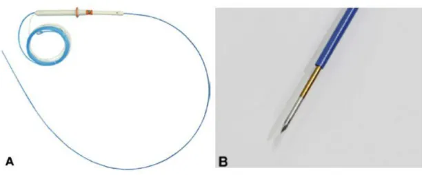
Figure 1. A, A radiofrequency ablation (RFA) electrode. An 19-gauge RFA electrode is composed of an electrode covered with protective tubing, a handle, and catheters for the cooling system. B, The exposed distal end of the electrode was needle-shaped and echogenic, and the length of the exposed tip of the electrode was 10 mm.··· 3
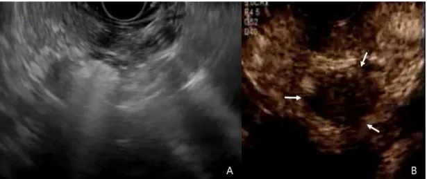
Figure 2. Imaging of EUS-guided radiofrequency ablation and follow-up contrast-enhanced EUS. A, EUS-guided radiofrequency ablation was performed from the duodenal bulb with 50 W of ablation power for 10 seconds and was repeated 4 times. B, After RFA, contrast-enhanced EUS showed a nonenhanced central necrotic portion (white arrow). ··· 5
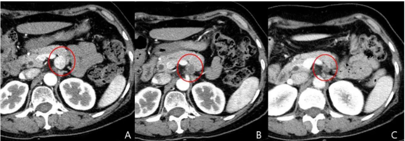
Figure 3. A, The abdominal CT showing a 19-mm hyperenhancing neuroendocrine tumor in the body of the pancreas (white arrow). B, The axial CT image from the 3-month follow-up revealing a nonenhancing hypodense lesion. C, The CT image from the 6-month follow-up depicting a decrease in the size of the ablated lesion. ··· 12
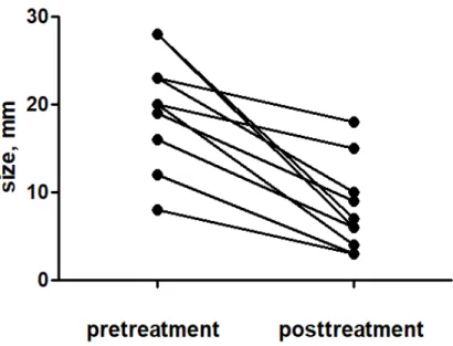
Figure 4. The changes in the EUS diameter of the pancreatic tumors after treatment. ··· 13
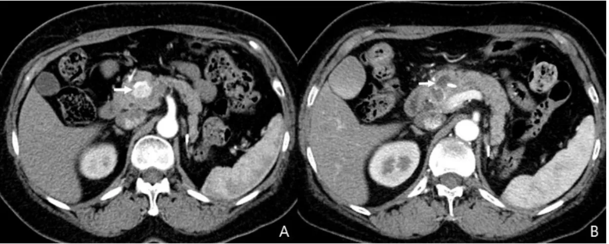
Figure 5. The abdominal CT image obtained before ablation shows a 2.3-cm neuroendocrine tumor in the head of pancreas (white arrow). B, The axial arterial phase CT image obtained 3 days after the second session of ablation showing a nonenhancing area of low attenuation with surrounding ring-like enhancement. ··· 1
1 INTRODUCTION
The widespread use of cross-sectional imaging has appreciably increased the number of incidental pancreatic tumors. In patients with sporadic benign solid pancreatic tumor, there are quite different views regarding the appropriate management strategy to adopt.1) Making a decision about whether to resect and ‘wait and watch‘ in the case of an incidentally detected pancreatic tumor is often challenging.2)Surgical resection is probably the best treatment for a pancreatic tumor, but surgery is not always successful in treating pancreatic tumors due to various factors, including the small size, adhesions caused by previous abdominal surgery, and the location of the tumor in a high-risk patients. Pancreatic surgery is associated with a substantial morbidity rate of 20% to 40% and a mortality rate of 2%.3,4) Therefore, a personalized management strategy is openly deployed, applying a risk-benefit analysis to each patient. In the light of the rising incidence of benign solid pancreatic tumors and the substantial proportion of patients in whom surgery is not feasible, new and effective forms of minimally invasive treatment are needed.3,5-9)
Radiofrequency ablation (RFA) is a safe and recognized method that has been used in recent years for the treatment of selected solid tumors of the liver, pancreas, adrenal gland, kidney, and lung and it has become a standard therapy for hepatocellular carcinoma.7)RFA can be applied percutaneously, intraoperatively, or recently endoscopically.10)An endoscopic RFA of the pancreas is performed by using an endoscopic ultrasound (EUS)-guided RFA probe which offers the combination of real-time visualization and precise localization with minimal invasiveness for the selective ablation of the targeted lesion. Experimental animal studies have reported on the safety and feasibility of EUS-RFA when applied to the pancreas.11,12)However, the translation of animal experiments into clinical human use has been limited. Song and
2
colleagues reported the technical feasibility and safety of EUS-RFA with an 18-gauge RFA electrode in six patients with unresectable pancreatic cancer.13)Thus, the logic of applying this ablative technology to the vexing problem of slow-growing benign solid tumors in the pancreas seems appealing.
Based on the preliminary experience,11,13)we assumed that EUS-RFA would be feasible and safe in patients with solid pancreatic tumors. Here, we report the outcomes of patients with benign solid pancreatic tumor treated with EUS-RFA.
PATIENTS AND METHODS
This was a single-center prospective pilot study conducted between October 2014 and September 2016. The study was approved by the Institutional Review Board at Asan Medical Center, and all patients signed a written informed consent forms prior to enrollment. This study was registered with the Clinical Research Information Service (CRIS) at the Korea National Institute of Health (NIH), which is a registry in the World Health Organization Registry Network (KCT0002467).
The inclusion criteria were (1) biopsy proven, benign solid pancreatic tumors smaller than 3 cm in patients who refused surgery or who had a high surgical risk (American Society of Anesthesiologists [ASA] physical status classification III or IV). The exclusion criteria were (1) advanced heart or lung disease precluding adequate sedation, (2) uncorrected coagulopathy, (3) a cardiac pacemaker, and (4) an inability to give informed consent.
All patients were discussed at a multidisciplinary tumor board prior to enrollment.
Equipments
The EUS-RFA system (STARmed Co., Seoul, Korea) consisted of an 18-gauge or 19-gauge prototype needle electrode comprised of a 7-Fr (18-gauge) or 5-Fr wire (19-gauge) and a working length of 140 cm (Fig. 1). In the early study period, a 18-gauge needle was used. In
3
the later study period, a newly-developed 19-gauge RFA needle (STARmed, Seoul, South Korea) was used for the procedure. The 19-gauge RFA needle is designed to offer more flexibility and passability in the pancreatic head and uncinate process. The inner metal part is insulated over its entire length except for the terminal 10 mm, which had a sharp conical tipe for energy delivery.
A radiofrequency generator (VIVA RF generator; STARmed Co.) with variable wattage settings was connected to the handle of the needle electrode. The temperature of the electrode was fixed at 90°C and modulated by an automatic power control unit. During ablation, the RF electrode was cooled and perfused internally with circulating chilled saline solution delivered via a pump.
Fig. 1. Modified 19-gauge monopolar radiofrequency electrode (STARmed Co., Koyang, Korea)
4
Endoscopic ultrasound-guided radiofrequency ablation
All patients were diagnosed using EUS-guided fine needle biopsy (FNB), with the aspirated materials examined by smear cytology and immunohistochemistry; after confirming the diagnosis, they underwent EUS-RFA.
The subjects were treated with EUS-RFA by an experienced endosonographer (D.W.S.), with the patient under conscious sedation using meperidine and midazolam and/or propofol. Broad spectrum prophylactic antibiotics were administered intravenously before each procedure.The needle electrode was passed under EUS guidance into the target lesion crossing a minimum of normal pancreatic parenchyma and avoiding the major vessels, or the pancreatic duct. The echogenic needle tip was positioned at the far end inside the target lesion. The energy was delivered after confirming the location of the tip of the needle electrode within the margin of the target lesion under EUS. Then, the RF generator was activated to deliver 50 W of ablation power for 10 seconds. Upon activation, after a lag period, echogenic bubbles gradually started appearing around the needle, indicating effective ablation at the site (Fig. 2).
The generator was then switched off. In larger lesions, the electrode may be repositioned under EUS visualization to ablate another zone along the same trajectory closer to the echoendoscope, while staying away from the gut wall. By using a fanning technique, additional needle passes can be made to further ablate different areas within the same lesion. Effort was made to first ablate the most technically demanding area, since visual artifacts may occur after applying RFA.
The next day, a simple abdominal radiograph and blood tests, including a complete blood count, liver function test, and serum amylase and lipase, were evaluated for adverse events. The adverse events were classified according to the published criteria.14
5
Fig. 2. Imaging of EUS-guided radiofrequency ablation and follow-up contrast-enhanced EUS.
A, EUS-guided radiofrequency ablation was performed from the duodenal bulb with 50 W of ablation power for 10 seconds and was repeated 4 times. B, After RFA, contrast-enhanced EUS showed a nonenhanced central necrotic portion (white arrow).
6 Main outcome measurement and study definitions
Initial treatment results were assessed within 1 week after RFA by contrast-enhanced EUS (CE-EUS) and/or contrast-enhanced computed tomography (CECT). A response to RFA was classified as radiologic complete response (CR, indicated by the absence of enhancing tissue at the tumor site) or incomplete response (when enhancing tissue was still observed at the tumor site). In order to achieve CR, the treatment course could include up to three sessions of RFA. The time frame between sessions was about 1 week, depending on the patient’s status. Radiologic CR achieved after a maximum of three iterative procedures spaced at 1 week were regarded as treatment success. These patients’ follow-up included CECT and CE-EUS every 3 to 6 months in the first year and then yearly thereafter.
The adverse events were classified according to the published criteria [14].RESULTS
The baseline characteristics are shown in Table 1. The anatomical locations of the pancreatic tumors were as follows: head (n=4), body (n=5), and tail (n=1). Two patients were ASA class III, and the others refused surgery. There were 7 cases of nonfunctioning neuroendocrine tumors (NETs), 1 case of an insulinoma, and 2 cases of solid pseudopapillary neoplasms (SPNs); the median largest diameter of the tumors was 20 mm (range, 8–28 mm).
The median
time between fine needle biopsy (FNB) diagnosis and the first ablation session was 40
days (interquartile range [IQR] 11–75 days). Tumor grading using a Ki-67 index could
be performed on 2 of the 10 tumors with adequate FNB specimens; one was G1 NET,
and the other was G2 NET.
One patient had symptomatic hypoglycemia, with the insulinoma confirmed by biochemical analysis.7 Table 1 Baseline characteristics and outcomes (n=10)
No. Age Se
x
Clinical presentation
Diagnosis Location Size of tumor, mm
Post- ablation size, mm
Electrode caliber, G
Response Sessions Treatment applications per session
Adverse event
1 34 M Hypoglycemia Insulinoma Head 12 3 19 Complete 1 3 -
2 21 F Incidental SPN Head 23 18 19 Incomplete 1 4 -
3 53 F Incidental SPN Tail 20 4 19 Complete 1 4 -
4 53 F Incidental NET Body 8 3 18 Complete 1 3 -
5 43 M Incidental NET Body 28 7 18 Incomplete 2 11 -
6 69 F Incidental NET Body 19 9 18 Complete 2 4 (1st), 7
(2nd)
-
7 70 M Incidental NET Body 20 15 18 Incomplete 1 4 -
8
8 40 M Incidental NET Body 16 6 19 Complete 1 4 -
9 69 F Incidental NET Head 28 6 18 Complete 3 6 (1st), 6
(2nd), 6 (3rd)
Abdominal pain
10 62 F Incidental NET Head 23 10 18 Complete 3 4 (1st), 3
(2nd), 4 (3rd)
Pancreatitis
SPN, solid pseudopapillary tumor; NET, neuroendocrine tumor
9
EUS-RFA was performed successfully in all 10 of the patients; 6 underwent one session of RFA, 2 underwent two sessions, and 2 patients were treated with three sessions. The median procedure times was 13 min (IQR 10-15 min). The needle and the RFA catheter exposed outside of the needle within the tumors were well visualized on real-time EUS imaging as hyperechoic reflexes of different diameter sizes in all of the procedures. During the RFA, a bright hyperechoic focus appeared around the needle electrode.
During follow-up (median 13 months, range 8–30 months), radiologic CR was achieved
in 70% of the patients (Fig.3). The median EUS diameter of the tumors changed from 20 mm (IQR 15–24 mm ) at the baseline to 6.5 mm (IQR 3.7–11.3) at the end of the follow-up, regardless of whether CR was achieved (Fig. 4). In addition, a change in the vascularity and central necrosis after EUS-RFA was demonstrated (Fig. 5). The one patient with an insulinoma showed complete relief from the hypoglycemia-related symptoms. That patient demonstrated early homogeneous enhancement of the tumor on CE-EUS before the EUS-RFA. After treatment, no enhancement was observed inside the tumor on the CH-EUS. Among the 7 patients with nonfunctioning NETs, CR was observed in 5 patients, whereas 2 of the patients had persistent NETs. In the two patients with SPNs, the tumor was almost completely ablated in 1 patient (no. 3 in Table 1), but the other one demonstrated persistent SPN (no. 2 in Table 1). This patient refused surgery.
10
Fig. 3. A, The abdominal CT showing a 19-mm hyperenhancing neuroendocrine tumor in the body of the pancreas (red circle). B, The axial CT image from the 3-month follow-up revealing a nonenhancing hypodense lesion. C, The CT image from the 6-month follow-up depicting a decrease in the size of the ablated lesion.
11
Fig. 4. The changes in the EUS diameter of the pancreatic tumors after treatment.
12
Fig. 5. A, The abdominal CT image obtained before ablation shows a 2.3-cm neuroendocrine tumor in the head of pancreas (white arrow). B, The axial arterial phase CT image obtained 3 days after the second session of ablation showing a nonenhancing area of low attenuation with surrounding ring-like enhancement.
13
Of the 16 ablation procedures, the procedure-related adverse events included 1 case of abdominal pain (6.2%) and 1 of pancreatitis (6.2%). Acute pancreatitis developed in 1 patient (no. 10 in Table 1) after the first session of RFA. The serum amylase and lipase levels were elevated to 850 and 2740 U/L, respectively, 1 day after the procedure.
The patient with
pancreatitis responded well to medical treatment, requiring intravenous fluid
resuscitation. Seven days after initial RFA, this patient underwent prophylactic
pancreatic duct stenting prior to the second session of RFA to prevent post-procedure
pancreatitis.
14 DISCUSSION
This pilot study demonstrated that EUS-RFA is a relatively safe and potentially effective treatment modality in selected cases of benign solid pancreatic tumors.
In this series, EUS- RFA was successfully completed in 100% of the cases, with a radiologic CR in 7 of the 10 patients (70%).
After the procedures, the ablated lesions displayed a change in vascularity, with central necrosis.In previous animal experiments, the size of the ablation area was 2.5 cm when the RFA was delivered with 50 W of ablation power for a duration of 5 minutes.11) Recently, we reported the use of EUS-RFA in 6 patients with unresectable pancreatic cancer.13)In that study, an 18- gauge RFA electrode was inserted into the pancreatic lesion to deliver 20–50 W of ablation power for 10 seconds.13)Those patients experienced mild abdominal pain without any major adverse events.13)Based on those results, we determined the duration and ablation power for the ablation of pancreatic tumors. In this series, EUS-RFA was successfully completed in 100%
of the cases, with a complete ablation in 7 of the 10 patients (70%).
In the present study, the patients responded gradually to the treatment, which was consistent with the progressive shrinkage of the tumor volume that was induced by the RFA over time.
There was a 58.9% reduction in the area [median pre-RFA diameter of 20 mm (IQR 15–24 mm) vs. median post-RFA diameter of 6.5 mm (IQR 3.7–11.3)]. The different percentages of cytoreduction can be partly attributed to the number of sessions performed. EUS-RFA can be added to the treatment to reduce the tumor burden (equivalent to surgical debulking), often with a palliative intent.
Complete tumor necrosis is the goal of any treatment modality, including emerging therapies such as ethanol ablation and RFA. Regardless of achieving complete ablation, the cytoreduction after treatment suggest that this treatment was effective in controlling the tumor
15
burden. In order to achieve complete necrosis, the treatment paradigm for RFA in pancreatic tumors requires a sufficient ablation margin beyond the visible tumor margin to ensure ablation of the infiltrative and/or microscopic disease that may not be visible by current imaging methods. However, this principle must be balanced against the need to preserve functional pancreas and to prevent pancreatitis.
In a recent study of 139 patients who underwent surgical resection of incidental pancreatic NETs, the complete surgical resection rate was 87%; however, 44% of those patients experienced postoperative adverse events.6,15) Despite advances in surgery, the perioperative morbidity of a pancreatic resection is still substantial, even in large volume centers. The success of RFA in hepatic tumors along with the need for non-surgical alternatives for pancreatic tumors prompted the investigators to attempt RFA in the pancreas.7In this series, procedure-related adverse events were observed in 12.4% of the patients, including only one case of acute pancreatitis. No major complications resulted from the multiple sessions of EUS- RFA performed in our patients, and the treatment was generally well tolerated. EUS provides the real-time visualization of the application and treatment, thus offering a reassuring safety net for the examiner as well as immediate observation of the results achieved.7) The preliminary experiences of RFA suggest that the iatrogenic injuries might be limited by technical precautions, such as a reduction of the ablation temperature (< 90℃), the maintenance of a safety margin from the major vessels or from the duodenum, and the use of a step-up approach in cases of large-sized lesions.16In addition, an inner cooling system helps to prevent the charring of the electrode surface enabling the efficient transmission of heat.7,16)
Thermal injuries to the upstream main pancreatic duct during RFA may be associated with severe pancreatitis.17)In this series, there was one case of pancreatitis after the first session of RFA, thus requiring endoscopic pancreatic stenting. The tumor was persistent in this patient and required a repeat EUS-RFA. Before the second session of EUS-RFA, we performed
16
prophylactic pancreatic stenting to prevent pancreatitis. A physician may be more prone to place a prophylactic pancreatic stent, particularly if the patient has any prior history of postprocedure pancreatitis or the tumor was located close to main pancreatic duct. Whether prophylactic pancreatic stenting reduce the risk of post-RFA pancreatitis in tumors close to the main pancreatic duct needs to be further evaluated.
Though incidentally discovered, non-functioning PNETs <2 cm may be monitored by using scheduled radiologic examinations (watch-and-wait policy), a small tumor size alone does not guarantee that a lesion is benign because malignant potential is not negligible. The relentless pursuit of an early diagnosis and treatment with less invasive modality may result in many paradigm shifts in gastroenterology field after a long period of controversy. For example, endoscopic ablation of Barrett’s esophagus and colon polypectomy are representative innovations that were ultimately accepted as standard of care. EUS-guided ablation may have the potential to become one of the standard therapy for some pancreatic tumors, but needs to be refined.
Despite the growing interests in EUS-RFA, there is still a lack of standardization and dose- effect dosimetry studies to evaluate the relationship between the energy and duration of application and the zone of ablation produced to safely and effectively ablated the targeted tumor.7,10,13,18) Before the utilization of EUS-RFA, there must be an appropriate indication, discussion of the risks and benefits with the patients, and careful endoscopic planning.
This study had several limitations. First, the sample size was small, with only 10 patients having follow-up imaging. In addition, this was a single arm, non-comparative study, with no comparison to a “watch and wait” approach conducted. A third limitation was that no surgical or histological samples of the treated tumor were obtained. The treatment response was assessed mainly by using contrast-enhanced computed tomography CT and CE-EUS, and the
17
precision of CT in the measurement of small tumors may be questioned. Given that benign solid pancreatic tumors are slow to grow, and to attain malignant transformation, a longer follow-up period of at least 5―10 years is needed to evaluate the final outcomes of RFA, including potential inhibition of cancer development. In addition, the investigators who assessed the treatment response were not be blinded to the treatment; therefore, bias from that source cannot be excluded.
In selected cases, EUS-RFA offers effective local control of benign solid pancreatic tumors combined with a relatively low risk of procedure-related complication and no mortality. The main eligibility criteria may be related to the size, location, and tumor stage. It could be attempted for the treatment of benign solid pancreatic tumors in patients who are at a high surgical risk. Refinements in the RFA electrode and generator are needed to deliver a well- defined localized area of tissue ablation, without causing pancreatitis or ductal injuries.
18 REFERENCES
1. Falconi M, Eriksson B, Kaltsas G, Bartsch DK, Capdevila J, Caplin M, et al.ENETS Consensus Guidelines Update for the Management of Patients with Functional Pancreatic Neuroendocrine Tumors and Non-Functional Pancreatic Neuroendocrine Tumors. Neuroendocrinology2016;103:153-71.
2. Gaujoux S, Partelli S, Maire F, D'Onofrio M, Larroque B, Tamburrino D, et al.
Observational study of natural history of small sporadic nonfunctioning pancreatic neuroendocrine tumors. J Clin Endocrinol Metab2013;98:4784-9.
3. Choi JH, Seo DW, Song TJ, Park DH, Lee SS, Lee SK, et al. Long-term outcomes after endoscopic ultrasound-guided ablation of pancreatic cysts. Endoscopy2017.
4. Allen PJ, D'Angelica M, Gonen M, Jaques DP, Coit DG, Jarnagin WR, et al. A selective approach to the resection of cystic lesions of the pancreas: results from 539 consecutive patients. Ann Surg2006;244:572-82.
5. Park DH, Choi JH, Oh D, Lee SS, Seo DW, Lee SK, et al. Endoscopic ultrasonography-guided ethanol ablation for small pancreatic neuroendocrine tumors:
results of a pilot study. Clin Endosc2015;48:158-64.
6. Paik WH, Seo DW, Dhir V, Wang HP. Safety and Efficacy of EUS-Guided Ethanol Ablation for Treating Small Solid Pancreatic Neoplasm. Medicine (Baltimore) 2016;95:e2538.
7. Lakhtakia S, Seo DW. Endoscopic ultrasonography-guided tumor ablation. Dig Endosc2017;29:486-94.
8. Choi JH, Oh D, Lee JH, Park JH, Kim KP, Lee SS, et al.Initial human experience of endoscopic ultrasound-guided photodynamic therapy with a novel photosensitizer and a flexible laser-light catheter. Endoscopy2015;47:1035-8.
9. Oh HC, Seo DW, Song TJ, Moon SH, Park DH, Soo Lee S, et al. Endoscopic
19
ultrasonography-guided ethanol lavage with paclitaxel injection treats patients with pancreatic cysts. Gastroenterology2011;140:172-9.
10. Rossi S, Viera FT, Ghittoni G, Cobianchi L, Rosa LL, Siciliani L, et al.
Radiofrequency ablation of pancreatic neuroendocrine tumors: a pilot study of feasibility, efficacy, and safety. Pancreas2014;43:938-45.
11. Kim HJ, Seo DW, Hassanuddin A, Kim SH, Chae HJ, Jang JW, et al. EUS-guided radiofrequency ablation of the porcine pancreas. Gastrointest Endosc2012;76:1039- 43.
12. Goldberg SN, Mallery S, Gazelle GS, Brugge WR. EUS-guided radiofrequency ablation in the pancreas: results in a porcine model. Gastrointest Endosc1999;50:392- 401.
13. Song TJ, Seo DW, Lakhtakia S, Reddy N, Oh DW, Park DH, et al.Initial experience of EUS-guided radiofrequency ablation of unresectable pancreatic cancer.
Gastrointest Endosc2016;83:440-3.
14. Cotton PB, Eisen GM, Aabakken L, Baron TH, Hutter MM, Jacobson BC, et al.A lexicon for endoscopic adverse events: report of an ASGE workshop. Gastrointest Endosc2010;71:446-54.
15. Haynes AB, Deshpande V, Ingkakul T, Vagefi PA, Szymonifka J, Thayer SP, et al.
Implications of incidentally discovered, nonfunctioning pancreatic endocrine tumors:
short-term and long-term patient outcomes. Arch Surg2011;146:534-8.
16. Keane MG, Bramis K, Pereira SP, Fusai GK. Systematic review of novel ablative methods in locally advanced pancreatic cancer. World J Gastroenterol2014;20:2267- 78.
17. Elias D, Baton O, Sideris L, Lasser P, Pocard M. Necrotizing pancreatitis after radiofrequency destruction of pancreatic tumours. Eur J Surg Oncol2004;30:85-7.
20
18. Lakhtakia S, Ramchandani M, Galasso D, Gupta R, Venugopal S, Kalpala R, et al.
EUS-guided radiofrequency ablation for management of pancreatic insulinoma by using a novel needle electrode (with videos). Gastrointest Endosc2016;83:234-9.
21 국문요약
배경: 고주파 치료는 초음파를 보면서 전극이 부착된 바늘을 종양 내에 삽입하여, 고주파를 통하여 순간적으로 고열이 발생시켜 종양을 괴사시키는 방법으로 동물 실험 및 소규모 임상 시험이 시행된 바 있다. 본 연구에서는 양성 고형성 췌장 종양 치료에 대한 내시경초음파 유도하 고주파 치료법의 안정성 및 유효성을 평 가하고자 한다.
방법: 양성 고형성 췌장 종양이 확인되어 내시경초음파 유도하 고주파 치료법을 시행받기로 한 10 명의 환자들이 연구에 참여하였다. 내시경초음파 유도 아래고 주파를 발생시키는18게이지 또는 19 게이지 전기침을 종양부위에 삽입해 발전기 에 연결된 전극으로부터 50 W고부하의 교류를 10초간 흐르게 하여 전기침 주변 의 종양조직을 괴사시킨다. 종양 크기에 따라 충분한 괴사가 이루어질 수 있도록 고주파 열 치료 횟수를 반복한다. 종양 완전 괴사의 판정은 조영증강 전산화 단 층 촬영 및 조영증강 내시경초음파에서 종양 내에 조영 증강되는 부위가 없는 경우로 정의하였다.
결과: 10 명의 참여자에게 총 16 회의 내시경초음파 유도하 고주파 치료를 성공 적으로 시행하였다. 참여대상 중 비기능성 췌장 내분비종양이 7 례, 인슐린종이 1 례, 고형 가유두상 종양이 2 례이었다. 종양 크기의 중앙값은 20 mm (범위, 8–
28 mm) 이다. 종양 위치는 췌장 두부가 4 례, 체부가 5 례, 미부가 1 례이었다.추 적관찰 도중 (중앙값 15 개월, 범위 10-30 개월), 시술 후 완전 반응은 7 례에서 확인되었다. 내시경 초음파에서 관찰된 종양 크기의 중앙값은 20 mm (IQR 15-24 mm)에서 6.5 mm (IQR 3.7-11.3 mm)로 유의하게 감소하였다. 인슐린종으로 확인된 1 명은 시술 후 저혈당 증상의 개선 및 생화학 검사상에서도 호전이 확인되었다.
총 16 회의 시술과정에서, 시술과 연관된 합병증은 2 례에서 확인되었고, 1 례는 중등도의 췌장염, 1 례는 복통이었다.
22
결론: 선택적인 양성 췌장 종양 환자군에서 내시경 초음파 유도하 고주파 치료는 기술적으로 타당하고, 비교적 안전하게 실행할 수 있었다. 그러나 불완전 제거가 된 환자에서는 반복적인 시술이 필요할 수 있고, 시술 후 췌장염 등의 합병증 발 생에 대해 주의가 필요하다.
중심 단어: 내시경초음파, 췌장 종양, 소작