저작자표시-비영리-변경금지 2.0 대한민국 이용자는 아래의 조건을 따르는 경우에 한하여 자유롭게
l 이 저작물을 복제, 배포, 전송, 전시, 공연 및 방송할 수 있습니다. 다음과 같은 조건을 따라야 합니다:
l 귀하는, 이 저작물의 재이용이나 배포의 경우, 이 저작물에 적용된 이용허락조건 을 명확하게 나타내어야 합니다.
l 저작권자로부터 별도의 허가를 받으면 이러한 조건들은 적용되지 않습니다.
저작권법에 따른 이용자의 권리는 위의 내용에 의하여 영향을 받지 않습니다. 이것은 이용허락규약(Legal Code)을 이해하기 쉽게 요약한 것입니다.
Disclaimer
저작자표시. 귀하는 원저작자를 표시하여야 합니다.
비영리. 귀하는 이 저작물을 영리 목적으로 이용할 수 없습니다.
변경금지. 귀하는 이 저작물을 개작, 변형 또는 가공할 수 없습니다.
Master’s Thesis of Science in Agriculture
CRISPR/Cas9 System for Efficient Genome Editing in
Filamentous Fungi Monascus ruber Based on PacBio SMRT Sequencing
PacBio SMRT 염기서열분석 기술 기반 Monascus ruber대상
CRISPR/Cas9 시스템 개발
August 2021
Hye Ree Yoon
Department of International Agricultural Technology Graduate School of International Agricultural Technology
Seoul National University
i
Abstract
The genus Monascus has been used in the production of food components, natural pigments, and food supplements with positive effects on human health.
As a result of beneficial effects, secondary metabolites produced by Monascus spp. have received worldwide attention and used industrially in recent years. It has been proved that Monascus spp. can synthesize various secondary metabolites: Monascus pigments, monacolin K, and citrinin. Monascus pigments have been used as natural food colorants and possess a wide range of biological functions. In addition, monacolin K lowers cholesterol by inhibiting HMG-CoA reductase. Even though Monascus spp. produce beneficial secondary metabolites, some strains can secrete citrinin, which has been found to be nephrotoxic, hepatoxic, and carcinogenic. Thus, the Monascus fermented food products have been concerned and controversial.
With the advance of fungal metabolic engineering, the formation of secondary metabolites in filamentous fungi could be regulated by genetic engineering. The aim of this study was to establish CRISPR/Cas9 system in Monascus spp. based on PacBio SMRT sequencing. In Chapter 2, the whole genome sequence of Monascus ruber was generated. The total length of 25.9 Mb was obtained using PacBio RSII sequencer with de novo assembly. As a result of genome assemblies with long reads from PacBio SMRT sequencing, the whole genome sequence of M. ruber consisted of 13 contigs with 9,639 predicted genes. Furthermore, citrinin biosynthetic gene clusters were mostly lost, while beneficial secondary metabolites, monacolin K and Monascus pigments, biosynthetic gene clusters were present in M. ruber, indicating this strain serves as a promising industrial strain without citrinin production. To validate M. ruber could be characterized as a citrinin-free strain, HPLC analysis was performed and citrinin was not detected
ii
in M. ruber. With the genetic analysis of the function of biosynthetic related gene clusters, comprehensive insight into secondary metabolites of Monascus spp. was discussed in Chapter 2.
In Chapter 3, CRISPR/Cas9 system was established in M. ruber to precisely engineer MpigI and MpigI’, putative negative transcriptional regulators. In vitro transcribed sgRNAs were adopted for transformation in the Cas9 expressed transfomrants to target MpigI and MpigI’. Based on Sanger sequencing results, six putative mutants were obtained. The mutants generated from the Cas9- mediated cleavage with dual sgRNAs were able to produce increased Monascus pigment production compared to the wild-type strain since induced- downregulation of MpigI and MpigI’ leads the increase in Monascus pigment production. Further analysis of mutants validated that CRISPR/Cas9 system was successfully established in M. ruber. This study was the first report of CRISPR/Cas9 system in M. ruber.
Keywords: Monascus ruber; Monascus pigments; Fungal metabolic engineering; PacBio SMRT sequencing; CRISPR/Cas9 system
Student number: 2019-27916
iii
Contents
Abstract ··· i
Contents ··· iii
List of Tables ··· vii
List of Figures ··· viii
Chapter 1 Research background ··· 1
1. Fungi ··· 1
1.1. Filamentous fungi ··· 1
1.2. Monascus spp. ··· 2
1.3. Red yeast rice ··· 2
2. Secondary metabolites ··· 3
2.1. Secondary metabolites in Monascus spp. ··· 3
2.1.1. Monascus pigments ··· 5
2.1.2. Monacolin K ··· 7
2.1.3. Citrinin ··· 9
3. Genome editing ··· 9
3.1. Definition and DNA repair pathway ··· 9
3.1.1. Non-homologous end joining (NHEJ) pathway ··· 10
3.1.2. Homology directed repair (HDR) pathway ··· 10
iv
3.2. CRISPR/Cas9 system ··· 10
3.3. CRISPR/Cas9 system in different species of filamentous fungi ··· 13
4. Overall objectives ··· 15
Chapter 2 Whole genome sequence of Monascus ruber isolated from Korean traditional fermented food ··· 16
1. Introduction ··· 16
2. Materials and methods ··· 18
2.1. Strain and culture conditions ··· 18
2.2. DNA extraction ··· 18
2.3. Genomic sequencing and assembly ··· 19
2.4. Citrinin analysis ··· 19
3. Results and discussion ··· 21
3.1. Genome sequence and assembly ··· 21
3.2. Comparison with other publicly available Monascus genomes ··· 24
3.3. Identification of secondary metabolite gene clusters ··· 26
3.3.1. Monascus pigments biosynthesis ··· 28
3.3.2. Monacolin K biosynthesis ··· 31
3.3.3. Citrinin biosynthesis ··· 33
4. Conclusions ··· 35
v Chapter 3
CRISPR/Cas9 system in filamentous fungi Monascus ruber ··· 36
1. Introduction ··· 36
2. Materials and methods ··· 39
2.1. Strains, plasmid, primers, and culture conditions ··· 39
2.2. Preparation of in vitro transcriptional sgRNA ··· 41
2.3. Protoplast preparation and transformation ··· 41
2.4. DNA extraction and PCR analysis of putative M. ruber transformants ··· 42
2.5. Analysis of secondary metabolites ··· 44
2.5.1. Monascus pigment analysis ··· 44
2.5.2. Extraction and analysis of monacolin K ··· 44
2.5.3. Citrinin analysis ··· 45
2.6. RNA extraction and RT-PCR analysis ··· 45
3. Results and discussion ··· 46
3.1. Establishment of Cas9 expressed transformants ··· 46
3.2. Designing dual sgRNA for disrupting MpigI and MpigI’ ··· 49
3.3. PCR screening of M. ruber ∆MpigI and ∆MpigI’ mutants ··· 49
3.4. Sequencing analysis of ∆MpigI and ∆MpigI’ mutants ··· 53
3.5. Comparison of the wild-type M. ruber strain and the mutants of ∆MpigI and ∆MpigI’ ··· 61
3.5.1. Fungal growth ··· 61
3.5.2. Colony morphology ··· 61
vi
3.5.3. Monascus pigment production ··· 65
3.5.4. Monacolin K analysis ··· 68
3.5.5. Citrinin analysis with M. purpureus BCRC 31541 ··· 68
3.6. RT-PCR analysis of ∆MpigI and ∆MpigI’ mutants ··· 71
4. Conclusions ··· 73
References ··· 74
Abstract in Korean ··· 87
Acknowledgements ··· 89
vii
List of Tables Chapter 1
Table 1.1. Currently studied filamentous fungi with CRISPR/Cas9 system ··· 14
Chapter 2
Table 2.1. General features of M. ruber genome ··· 22 Table 2.2. Results of assembly ··· 23 Table 2.3. Gene annotation information ··· 27
Chapter 3
Table 3.1. Primers used in this study ··· 40
viii
List of Figures Chapter 1
Fig. 1.1. Secondary metabolites of Monascus spp. ··· 4
Fig. 1.2. Main pigments produced by Monascus spp. ··· 6
Fig. 1.3. HMG-CoA reductase inhibitor pathway ··· 8
Fig. 1.4. CRISPR/Cas9 system with two different DSBs repair pathways ··· 12
Chapter 2
Fig. 2.1. Comparison of M. ruber and M. purpureus YY-1 genomic locations ··· 25Fig. 2.2. Biosynthetic pathway of Monascus pigments ··· 30
Fig. 2.3. Biosynthetic pathway of Monacolin K ··· 32
Fig. 2.4. Schematic representation of (a) gene clusters of citrinin in Monascus spp. (b) gene clusters of citrinin in M. ruber (c) HPLC results of M. ruber and M. purpureus BCRC 31541 ··· 34
Chapter 3
Fig. 3.1. Strategies for the establishment of CRISPR/Cas9 system in M. ruber ··· 43Fig. 3.2. PCR analysis of cas9 gene integration into the genome ··· 48
Fig. 3.3. Schematic representation of (a) the gene clusters of Monascus pigments and dual target sites in (b) MpigI and (c) MpigI’ loci ··· 51
ix
Fig. 3.4. PCR amplification of the target regions in (a) MpigI and (b) MpigI’ locus
using primers flanking the cleavage sites ··· 52
Fig. 3.5. Sequence analysis of the mutant ∆MpigI16-7 ··· 55
Fig. 3.6. Sequence analysis of the mutant ∆MpigI16-15 ··· 56
Fig. 3.7. Sequence analysis of the mutant ∆MpigI16-17 ··· 57
Fig. 3.8. Sequence analysis of the mutant ∆MpigI16-22 ··· 58
Fig. 3.9. Sequence analysis of the mutant ∆MpigI’14-5 ··· 59
Fig. 3.10. Sequence analysis of the mutant ∆MpigI’14-7 ··· 60
Fig. 3.11. Colony diameter of the wild-type M. ruber (green line), ∆MpigI16-7 (yellow line), ∆MpigI16-15 (blue line), ∆MpigI16-17 (orange line), ∆MpigI16-22 (dark gray line), ∆MpigI’14-5 (khaki line), and ∆MpigI’14- 7 (light gray line) ··· 63
Fig. 3.12. Colony morphology of the wild-type, ∆MpigI16-7, ∆MpigI16-15, ∆MpigI16-17, ∆MpigI16-22, ∆MpigI’14-5, and ∆MpigI’14-7 ··· 64
Fig. 3.13. Pigment analysis of (a) yellow (b) orange, and (c) red pigment of wild-type M. ruber (green line), ∆MpigI16-7 (yellow line), ∆MpigI16-15 (blue line), ∆MpigI16-17 (orange line), ∆MpigI16-22 (dark gray line), ∆MpigI’14-5 (khaki line), and ∆MpigI’14-7 (light gray line) ··· 66
Fig. 3.14. Pigment of wild-type, ∆ MpigI16-7, ∆MpigI16-15, ∆MpigI16-17, ∆MpigI16-22, ∆MpigI’14-5, and ∆MpigI’14-7 ··· 67
Fig. 3.15. Monacolin K analysis of the wild-type and mutants ··· 69
Fig. 3.16. HPLC-FLD chromatograms of wild-type M. ruber strain (brown line) and M. purpureus BCRC 31541 strain (blue line) compared to standard citrinin 50 µg/mL (pink line) ··· 70
x
Fig. 3.17. RT-PCR analysis of expression of genes associated with Monascus pigments ··· 72
1
Chapter 1.
Research background
1. Fungi
Fungi are a large and diverse group of eukaryotes (Wistreich, 2007). Since many species of fungi can tolerate the wide range of pH and temperature, fungi can live in various environments, such as water, plants, and foods (Frac et al., 2017; Li et al., 2017). Remarkably, fungi play a substantial role as a source of natural product bioactive compound (Alberti et al., 2017). Since the discovery of penicillin in the 1920s, fungal bioactive compounds ranging from antibiotics to statins have been prescribed to save the lives of people (Meyer et al., 2016). These fungal bioactive compounds, termed secondary metabolites, are predominantly produced by filamentous fungi (Keller, 2019).
1.1. Filamentous fungi
Filamentous fungi play vital roles in different industries: pharmaceutical, agriculture, and food (Shi et al., 2017). Because of their considerable economic value of many of their metabolites, filamentous fungi have been widely applied in the production of natural pigments, antibiotics, and cholesterol medications (Dufosse et al., 2014; Nielsen et al., 2017; Shi et al., 2017). Although filamentous fungi produce beneficial secondary metabolites, they can produce other metabolites such as mycotoxins, which can cause harmful or toxic effects on livestock and humans (Salazar-Cerezo et al., 2020).
2
1.2. Monascus spp.
Van Tieghem (1884) firstly screened Monascus spp. in red yeast rice and characterized them. The filamentous fungi Monascus spp. have been routinely used in the production of fermented foods in eastern Asia (Chen et al., 2015).
Their most famous fermented food product is known as red yeast rice, and it has been used as a food supplement to enhance the color and delicacy of meat, fish, and soybean products as part of the Chinese cuisine (Ma et al., 2000). Also, it is recognized as a folk medicine for the rejuvenation of the body and the improvement of blood flow (Zhu et al., 2019). As a result of beneficial effects on human health, the secondary metabolites produced by Monascus spp. have received worldwide attention and been used industrially in recent years (Chen et al., 2015). The most common industrial strains are known as M. ruber, M.
purpureus, and M. pilosus (Cheng et al., 2013; Hsu et al., 2011; Lian et al., 2015)
1.3. Red yeast rice
Red yeast rice, termed as Hongguk (in Korean) or Hongqu (in Chinese), is produced by fermenting the steamed rice with Monascus strains (Chen and Hu, 2005). First of all, cooked nonglutinous rice is steamed to a state of semi- gelatinization (Ma et al., 2000). After inoculation of Monascus into the rice for fermentation, the rice grains are incubated at 25-28 ℃ and regularly flipped until pigment becomes deep red (Lin et al., 2008). The red yeast rice has been characterized as a folk medicine for improving food digestion and blood circulation (Ma et al., 2000). Previous studies have validated that red yeast rice could lower cholesterol level in blood due to the presence of cholesterol synthase inhibiter, which is known as HMG-CoA reductase (Ma et al., 2000). With the
3
rising prevalence of hyperlipidemia becomes a worldwide public health concern, Monascus fermented products have received more attention than before.
2. Secondary metabolites
2.1. Secondary metabolites in Monascus Spp.
It has been proved that Monascus spp. can synthesize various secondary metabolites: Monascus pigments, monacolin K and citrinin (Chen et al., 2015;
Endo 1979; Feng et al., 2012).
4
Fig. 1.1. Secondary metabolites ofMonascus spp.
5
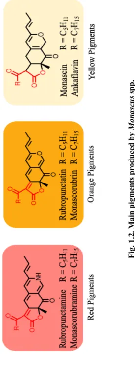
2.1.1. Monascus pigments
Monascus pigments are polyketide components, also called azaphilones, which are compounds with an oxygenated bicyclic nucleus and a quaternary center (Chen et al., 2015; Kim and Ku, 2018; Patacova, 2013). The main pigments produced by Monascus spp. are six compounds: rubropunctamine and monascorubramine (red pigments), rubropunctatin and monascorubrin (orange pigments) and monascin and ankaflavin (yellow pigments) (Kim and Ku, 2018).
The orange pigments, rubropunctatin and monascorubrin, are formed by the esterification of polyketide chromophore with beta-ketoacid (from the fatty acid synthase pathway) (Chen et al., 2017; Liu et al., 2018). Then, the red pigments are generated when the orange pigments react with amino group (NH3) (Chen et al., 2017). In contrast, the yellow pigments are formed by reduction of the orange pigments (Chen et al., 2017). Not only Monascus pigments have been used as natural food colorants, but also have several biological functions, leading to antioxidant, antitumor, antimicrobial, antimutagenic properties, and potential antiobesity activities (Chen et al., 2015; Feng et al., 2012). Since the major Monascus pigments structurally belong to azaphilones that inhibit other enzyme activities, several beneficial biological functions are available (Patakova, 2013).
6
Fig. 1.2. Main pigments produced by Monascus spp.
7
2.1.2. Monacolin K
Endo (1979) firstly isolated monacolin K from the culture of M. ruber.
Monacolin K (lovastatin) lowers cholesterol by inhibiting HMG-CoA (5- hydroxy-3-methylglutaryl-coenzyme A) reductase, which is known as key and rate liming enzyme for cholesterol synthesis (Klimek et al., 2009). The mechanism of cholesterol synthesis is described in Fig. 1.3.
Cardiovascular diseases, particularly coronary artery disease, are one of the global leading causes of mortality (Liao, 2002). The strong independent predictors of coronary artery disease are increase in total cholesterol, LDL- cholesterol and triglycerides concentrations, as well as decrease in HDL- cholesterol (Lin et al., 2015). Statins, which are widely prescribed class of drugs to lower cholesterol, work by competitively inhibiting the rate limiting enzyme, HMG-CoA reductase, to treat hyperlipidemia (Ward et al., 2019). With the prevalence of hyperlipidemia, Monascus fermented food products have been regarded as the hypolipidemic function food, which is having a cholesterol- lowering effect (Lee and Pan, 2012). Lin et al. (2015) reported that M. purpureus Went rice significantly reduced LDL-cholesterol, total cholesterol, triglycerides, and apolipoprotein B levels, and well tolerated in patients with hyperlipidemia.
Therefore, Monascus fermented food product has been validated to lower cholesterol as a functional food.
8
Fig. 1.3. HMG-CoA reductase inhibitor pathway (Slater and Macdonald, 1988).
9
2.1.3. Citrinin
Even though Monascus spp. produce beneficial metabolites and have been used widely in various industries, some strains can secrete mycotoxin citrinin, which was found to be nephrotoxic, hepatoxic, and carcinogenic (Chen et al., 2015; Li et al., 2013). Due to safety concern and controversy over citrinin production in Monascus spp., some countries reinforced the restriction on citrinin in Monascus fermented products. The European Union countries (2019, November 7) reported that the maximum level of citrinin in food supplements was lowered from 2000 𝜇g/kg to 100 𝜇g/kg based on rice fermented with Monascus spp. In eastern Asia, the limit varies in different countries: 50 𝜇g/kg in Korea and 200 𝜇g/kg in Japan (Fu et al., 2007; Kim et al., 2007; Patacova, 2013). Thus, it is important to identify non- citrinin producing strain to be used as promising industrial strain.
3. Genome editing
3.1. Definition and DNA repair pathway
Genome editing enables to manipulate the gene of interest in which DNA is deleted, inserted or replaced via sequence specific manner (Liu et al., 2015;
Zheng et al., 2017). The first approach in genome editing begins with introducing double-strand breaks (DSBs) at the target site of specific gene. Since DSBs lead the frequency of homologous recombination, DSBs have been introduced to edit genome in fungal molecular biology artificially and precisely (Krappmann, 2007).
When DSBs at the target site of gene take place, damaged DNA is repaired by either non-homologous end joining (NHEJ) pathway or homologous direct recombination (HDR) pathway (Schuster and Kahmann, 2019). The significant difference between two repair pathways is that NEHJ joins DNA without
10
homology while HDR requires stretches of homologous or homologous sequence (Krappmann, 2007).
3.1.1. Non-homologous end joining (NHEJ) pathway
During NHEJ pathway, nucleotide insertions and deletions (indels) or frameshifts are introduced during the process of repair (Sansbury et al., 2019). In most filamentous fungi, NHEJ is predominant DNA repair mechanism (Schuster and Kahmann, 2019).
3.1.2. Homologous direct recombination (HDR) pathway
During HDR pathway, homologous DNA fragment (donor DNA) to the target sequence is required (Schuster and Kahmann, 2019). Thus, this repair pathway is accomplished by combining Cas9 endonuclease and sgRNA with the transformation of donor DNA (Schuster and Kahmann, 2019).
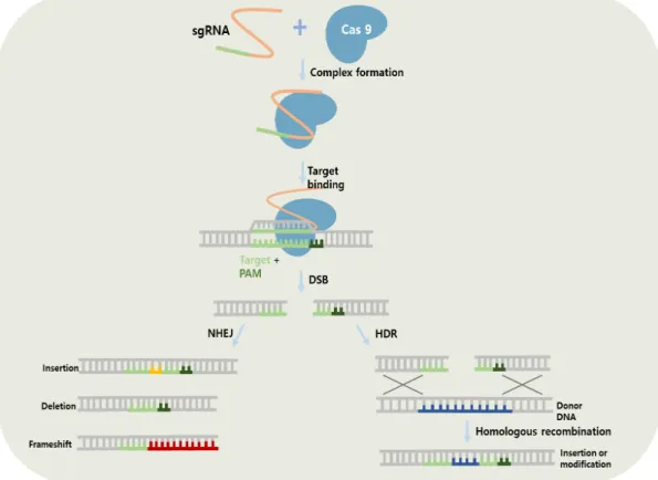
3.2. CRISPR/Cas9 system
The bacterial immune mechanism CRISPR (Clustered regulatory interspaced short palindromic repeats)/Cas9 system has become a powerful genome editing technology due to its high efficiency with convenience (Nodvig et al., 2015; Pohl et al., 2016; Wang et al., 2018). The two important components are involved in CRISPR/Cas9 system: Cas9 endonuclease and single-guide RNA (sgRNA) (Pohl et al., 2016). The Cas9 is from the bacterium Streptococcus pyogenes, and the sgRNA contains a 20-nucleotide sequence (Schuster and Kahmann, 2019). To successfully establish CRISPR/Cas9 system in the target site of genome, both Cas9 endonuclease and the sgRNA should be present in the nuclease (Schuster and Kahmann, 2019). Thus, Cas9 recognizes a protospacer adjacent motif (PAM) sequence and then catalyzes DSBs in the target site (Liu et al., 2015; Shi et al.,
11
2017). Fig. 1.4 describes the mechanism of CRISPR/Cas9 system with two different DSBs repair pathways
Genetic transformation of fungi meets with many difficulties. Due to complex of cell wall structures, different transformation strategies are required for different fungal species (Li et al., 2017). Several transformation strategies have been employed in filamentous fungi for applying CRISPR/Cas9 system: Poly- ethylene glycol (PEG) mediated transformation, agrobacterium-mediated transformation, and electroporation (Schuster and Kahmann, 2019).
12
Fig. 1.4. CRISPR/Cas9 system with two different DSBs repair pathways.
13
3.3. CRISPR/Cas9 system in different species of filamentous fungi
CRISPR/Cas9 system has been established in several filamentous fungi due to their beneficial effects of secondary metabolites on human health, including Aspergillus spp., Fusarium oxysporum, Neurospora crassa, Penicillium chrysogenum, Trichoderma reesei (Pohl et al., 2016; Schuster and Kahmann, 2019; Wang et al., 2018). Table 1.1. represents currently studied filamentous fungi with CRISPR/Cas9 genome editing system.
14
Species Repair pathway Application References
Alternaria alternata NHEJ single gene disruption Wenderoth et al., 2017; Igbalajobi et al., 2019
Aspergillus aculeatus NHEJ single gene disruption Nodvig et al., 2015 Aspergillus brasiliensis NHEJ single gene disruption Nodvig et al., 2015
Aspergillus carbonarius NHEJ/HDR single gene disruption Nodvig et al., 2015; Weyda et al., 2017
Aspergillus fumigatus NHEJ/HDR single gene disruption/
multiple gene disruption
Fuller et al., 2015; Weber et al., 2017;
Zhang et al., 2016
Aspergillus lucheunsis NHEJ/HDR single gene disruption Nodvig et al., 2015; Nodvig et al., 2018
Aspergillus nidulans NHEJ/HDR single gene disruption/
multiple gene disruption
Nodvig et al., 2015; Nodvig et al., 2018
Aspergillus niger NHEJ/HDR single gene disruption/
multiple gene disruption
Nodvig et al., 2015; Nodvig et al., 2018
Aspergillus oryzae NHEJ/HDR single gene disruption/
multiple gene disruption
Katayama et al., 2015; Nodvig et al., 2018
Beauveria bassiana NHEJ/HDR single gene disruption/
multiple gene disruption
Chen et al.,2017 Fusarium fujikuroi NHEJ/HDR multiple gene disruption Shi et al., 2019 Fusarium oxysporum NHEJ/HDR single gene disruption Wang et al., 2018
Mucor circinelloides NHEJ/HDR single gene disruption/
multiple gene disruption
Nagy et al., 2017
Myceliophthora thermophila NHEJ/HDR single gene disruption/
multiple gene disruption
Liu et al., 2017 Neurospora crassa NHEJ/HDR single gene disruption Matsu-Ura et al., 2015 Penicillium chrysogenum NHEJ/HDR single gene disruption Pohl et al., 2016 Sclerotinia sclerotiorum NHEJ/HDR single gene disruption Li et al., 2018 Talaromyces atroroseus NHEJ single gene disruption Nielson et al., 2017 Trichoderma reesei NHEJ/HDR single gene disruption Liu et al., 2015
Ustilago maydis NHEJ single gene disruption/
multiple gene disruption
Schuster et al., 2018
Table 1.1. Currently studied filamentous fungi with CRISPR/Cas9 system (Schuster and Kahmann, 2019; Song et al., 2019).
15
4. Overall objectives
In recent years, perspective on foods has been thoroughly changed. Beyond their nutritional values, foods should provide additional health benefits.
Functional foods can promote development and protect against chronic disease risk (Hasler, 2002). Thus, global functional food markets are expected to increase because unhealthy and western food consumption give rise to demand for functional foods (Hasler, 2002).
Monascus fermented food products, especially red yeast rice, caught more attention than ever on account of its validated efficacies to lower cholesterol level and other beneficial effects on human health. However, safety over Monascus fermented food product is still concerning and controversial due to citrinin, which limits Monascus fermented food products to be consumed. In consequence, it is important to investigate citrinin-free strain to be used as industrial strain.
In Chapter 2, the whole genome sequence of M. ruber was obtained using PacBio SMRT sequencing with de novo assembly. Based on genome assembly and annotation, the gene clusters of secondary metabolites and biosynthetic pathways were further discussed.
In Chapter3, CRISPR/Cas9 genome editing system was established in M. ruber.
Interestingly, M. ruber possessed two different MpigI loci, characterized as putative negative regulators of the biosynthesis of Monascus pigments. With this precise genome editing, the target sites of two different MpigI (MpigI and MpigI’) loci were successfully engineered by the Cas9-mediated cleavage with dual sgRNAs and then induced by NHEJ repair pathway.
16
Chapter 2.
Whole genome sequence of Monascus ruber isolated from Korean traditional fermented food
1. Introduction
The history of DNA sequencing began with Fredrick Sanger. Sanger et al.
(1977) invented the first generation of DNA sequencing, termed Sanger sequencing, with chain-terminating dideoxynucleotides (ddNTPs). From 1977, Sanger sequencing led to many genetic discoveries and dominated for three decades until emerging Next-generation sequencing (NGS) (Dunn et al., 2018).
Compared to Sanger sequencing, NGS was able to sequence large number of genes and entire genome at once (Dunn et al., 2018). Both Sanger sequencing and NGS have been utilizing lately, but high-throughput sequencing has been challenging. Relatively short read length of NGS generated sequences often has resulted assemblies with a large number of contigs (Brede et al., 2020; Smits 2019).
As an alternative, Single Molecule Real Time (SMRT) DNA sequencing, developed by Pacific Biosciences (PacBio), has become the first commercially available for reads that are much longer than NGS or even Sanger sequencing (Chin et al., 2013). Thus, PacBio SMRT sequencing results reduced error rates with long read sequencing since this sequencing utilizes circular consensus sequencing where DNA polymerase repeatedly replicates hairpin-ligated
17
amplicons, facilitating de novo assembly and genome finishing (Whon et al., 2018).
In recent years, there has been substantial attempt to the annotation and sequencing of filamentous fungal genomes (Jones, 2007). Several studies have been reported the whole genome sequence of Monascus spp. Liang et al. (2018) reported the complete genome sequence of M. purpureus YY-1 and provided the first comprehensive prediction of the biosynthetic pathway of Monascus pigments. This study was the first publicly available genome sequence of Monascus species and the whole genome sequence of M. purpureus YY-1 was sequenced using NGS. Total length of 24.1 Mb was obtained with 7,491 genes in M. purpureus YY-1, which was known as one of the most widely used industrial stains for food colorant production in China.
The whole genome of M. ruber using PacBio RSII sequencer with high quality de novo assembly was generated to obtain trustworthy assembly and annotation in Chapter 2. As a result of genome assemblies with long reads from PacBio SMRT sequencing, the whole genome sequence of M. ruber was obtained and annotated. Then, the function of genes involved in secondary metabolites was further analyzed.
18
2. Materials and methods
2.1. Strain and culture conditions
The strain M. ruber was isolated from Korean traditional fermented food, and the strain M. purpureus BCRC 31541 was purchased from Bioresources Collection and Research Center (Taipei, Taiwan). Both strains were cultured on potato dextrose agar (PDA) medium at 30 ℃ for hyphae collection. For liquid culture, potato dextrose broth (PDB) medium was used.
2.2. DNA extraction
Genomic DNA of M. ruber was extracted through the glass beads method as previously described with slight modifications (Aamir, 2015). The fungal mass obtained from PDA was placed into 2 mL tube containing sterile glass beads and lysis buffer (100 mM Tris HCl pH 8.0, 50 mM EDTA, 3% (w/v) SDS). Tubes containing fungal mass were vortexed vigorously for 20 min. After homogenization process, RNase A was added and incubated at 37 ℃ for 15 min.
Then, equal volume of phenol: chloroform: isoamyl alcohol (25:24:1) was added to the supernatant and centrifuged 12,000 rpm for 15 min. The upper aqueous layer was taken, and then equal volume of 100% ethanol was added. The mixture was kept at -20 ℃ for 30 min and centrifuged 12,000 rpm for 10 min. The precipitated DNA pellets were washed with 70% ethanol and dissolved in TE buffer. The quantity and purity of DNA were determined using the NanoVue Plus Spectrophotometer (GE Health Care Co., Nordrhein-Westfalen, Germany) and sent Macrogen (Seoul, Republic of Korea) for sequencing.
19
2.3. Genomic sequencing and assembly
The sample was prepared according to a protocol for sequencing on the PacBio Sequel system. The DNA templates were sequenced using PacBio RS II sequencer, and de novo assembly was performed using CANU (v1.7) software. It was accomplished by mapping single pass reads to seed reads, which represented the longest portion of the read length distribution. Subsequently, a consensus sequence of the mapped read was generated, resulting in long and highly accurate fragments of the target genome. Then, reads were corrected and filtered since some reads did not provide extra information for constructing the genome. In addition, the reads that had too high or too low overlaps were filtered. With the overlapping data, they contained information of each contig, so contigs were constructed with higher quality through the self-mapping step. After complete genome was assembled, the locations of genes were identified. Then, their functions were annotated. Maker (v.2.31.8) was used to predict the location while Protein BLAST+ was performed with UniProt Swiss-Prot (201806).
2.4. Citrinin analysis
Citrinin was analyzed though HPLC as Feng et al. (2014) described. The supernatant was centrifuged for 10 min at 12,000 rpm and then filtered through 0.22 𝜇m membrane filter. HPLC was performed on UltiMate 3000 BioRS system (Thermo Fisher Scientific, Walthan, MA, USA). For stationary phase, the column of inertsil ODS-3 (4.6 mm × 250 mm, i.d., 5 𝜇𝑚) was used. For mobile phase, a mixture of ACN, water and 0.5% ortho-phosphoric acid in the ratio of 70:25.5:4.5 (v/v/v) was carried out at a flow rate of 0.8 mL/min. The injection volume was 10 𝜇L and the column temperature was set at 30 ℃. The citrinin
20
contents were monitored by a fluorescence detector at 331 nm excitation wavelength and at 500 nm emission wavelength.
21
3. Results and discussion
3.1. Genome sequence and assembly
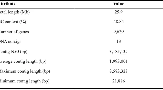
The whole genome sequence of M. ruber was generated using the PacBio RSII sequencer and de novo genome assembly was conducted with CANU (v1.7) software. Table 2.1. represents the general features of whole genome sequence of M. ruber. After filtering process, PacBio SMRT long-read sequencing yielded 737,395 reads with an N50 value of 14.9 kb. Overlapping reads that originated from the same region of the genome could be joined together to form contigs.
With CANU bioinformatics software, the overall genome assembly was improved while simultaneously reduced runtime (Korean et al., 2015). In consequence, the total length of 25.9 Mb in 13 contigs was generated with GC content of 48.84 %. The N50 value was increased from 14.9. kb to 3.1 Mb, so CANU performed high quality de novo assembly using PacBio library preparation. The length of maximum contig was approximately 3.6 Mb and average length of contigs was about 2.0 Mb. As a result, the consensus sequence with higher quality was generated. The assembly results were summarized in table 2.2.
After genome was analyzed, the locations of protein genes were predicated, and function were annotated. Maker (v2.31.8) was performed to predict the location while protein BLAST+ (v2.6.0) was performed with UniProt Swiss-Prot (201806). The total number of 9,639 genes were predicted and annotated. For RNA, 154 tRNAs, and 41 rRNAs were predicted.
22
Table 2.1. General features of M. ruber genome.
Attribute Value
Total length (Mb) 25.9
GC content (%) 48.84
Number of genes 9,639
DNA contigs 13
Contig N50 (bp) 3,185,132
Average contig length (bp) 1,993,001
Maximum contig length (bp) 3,583,328
Minimum contig length (bp) 21,886
23
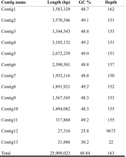
Table 2.2. Results of assembly.
Contig name Length (bp) GC % Depth
Contig1 3,583,328 48.7 162
Contig2 3,570,546 49.1 151
Contig3 3,344,543 48.8 153
Contig4 3,185,132 49.2 151
Contig5 2,672,239 49.0 151
Contig6 2,300,501 48.8 157
Contig7 1,932,116 48.8 150
Contig8 1,891,921 49.2 152
Contig9 1,567,545 48.3 153
Contig10 1,494,082 48.3 155
Contig11 317,868 49.2 155
Contig12 27,316 25.8 9673
Contig13 21,886 30.2 22
Total 25,909,023 48.84 163
24
3.2. Comparison with other publicly available Monascus genomes
A few publicly available whole genome sequences of Monascus spp. were obtained from NGS in previous studies. According to Liang et al. (2018), M.
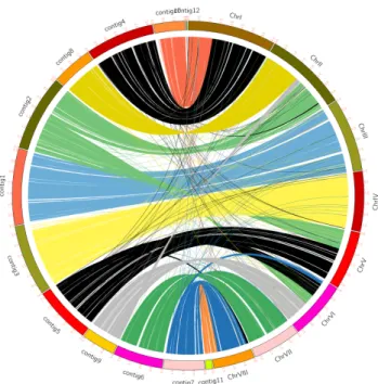
purpureus YY-1 has been widely used in food colorant in China, and genome size of this strain was reported 24.1 Mb with 7,491 genes. Fig. 2.1 represents comparison of M. ruber and M. purpureus YY-1 genomic locations.
In addition, Higa et al. (2020) reported that genome sizes of M. pilosus NBRC4520, M. purpureus NBRC4478, and M. ruber NCBR4485 were around 24 Mb from Illumina Miseq: 8,647 genes M. pilosus NBRC4520, 8,891 genes in M. purpureus NBRC4478, and 8,645 genes in M. ruber NCBR4485
Among publicly available whole genome sequences of Monascus spp., M.
ruber isolated from Korean fermented traditional food possessed additional number of genes and increase in genome size.
25
Fig. 2.1. Comparison of M. ruber and M. purpureus YY-1 genomic locations.
The left side of the outer ring depicts the number of contigs in M. ruber and the right side of the outer ring depicts the number of chromosomes in M. purpureus YY-1. The internal ribbons in different colors connect regions of homologous sequence shared between the two strains.
26
3.3. Identification of secondary metabolite gene clusters
Table 2.3. represents the gene and function of each gene that are involved in secondary metabolite biosynthesis. With the advantage of PacBio SMRT sequencing, only 13 contigs were assembled in M. ruber. In addition, all genes related in secondary metabolite biosynthesis were in contig 2. Two different MpigI loci were found from the results of gene annotations, demonstrating two different MpigI were present in M. ruber. While gene clusters of Monascus pigments and monacolin k were present, gene clusters of citrinin were almost lost, suggesting M. ruber could be a promising industrial strain without citrinin production.
27
contig Gene ID Start End Strand Gene Product Function
contig 2 LOCUS_001523-RA 490661 496669 + MpigJ Fatty acid synthase subunit alpha
Pigments biosynthesis contig 2 LOCUS_001524-RA 496680 498135 - MpigI Negative regulatory factor Pigments
biosynthesis contig 2 LOCUS_001525-RA 498265 499088 - MpigI Negative regulatory factor Pigments
biosynthesis contig 2 LOCUS_001526-RA 499551 500660 + MpigH Dehydrogenase Pigments
biosynthesis contig 2 LOCUS_001527-RA 500771 501592 - MpigG Oxidoreductase Pigments
biosynthesis contig 2 LOCUS_001528-RA 501913 503307 - MpigF Amine oxidase Pigments
biosynthesis contig 2 LOCUS_001529-RA 503789 504817 + MpigE Aryl-alcohol
dehydrogenase Pigments biosynthesis contig 2 LOCUS_001530-RA 504957 506324 - MpigD 3-O-acetyltransferase Pigments
biosynthesis contig 2 LOCUS_001531-RA 506932 507991 + MpigC Dehydrogenase Pigments
biosynthesis contig 2 LOCUS_001532-RA 50814 510123 - MpigB Transcriptional activator Pigments
biosynthesis contig 2 LOCUS_001533-RA 512402 520544 + MpigA Polyketide synthase Pigments
biosynthesis contig 2 LOCUS_001576-RA 645879 646832 - mokC_0 Dihydromonacolin L
monooxygenase mokC
Monacolin K biosynthesis contig 2 LOCUS_001577-RA 646862 647595 - mokC_1 Dihydromonacolin L
monooxygenase mokC Monacolin K biosynthesis contig 2 LOCUS_001578-RA 647921 657623 + mokA Lovastatin nonaketide
synthase
Monacolin K biosynthesis contig 2 LOCUS_001579-RA 658028 658354 - - hypothetical protein
contig 2 LOCUS_001580-RA 661952 662158 + - hypothetical protein
contig 2 LOCUS_001581-RA 662581 663372 - mokD Esterase mokD Monacolin K biosynthesis contig 2 LOCUS_001582-RA 663824 665050 + mokE Deshydrogenase mokE Monacolin K biosynthesis contig 2 LOCUS_001583-RA 665143 666347 - lovD_1 Acyltransferase LovD Monacolin K biosynthesis contig 2 LOCUS_001584-RA 667488 670846 - mokG 3-hydroxy-3 methylglutaryl
coenzymeA reductase mokG
Monacolin K biosynthesis contig 2 LOCUS_001585-RA 671858 673272 + mokH Transcription factor
mokH Monacolin K
biosynthesis contig 2 LOCUS_001586-RA 675002 677151 + mokI Efflux pump mokI Monacolin K biosynthesis contig 2 LOCUS_001587-RA 679695 687775 + mokB Lovastatin diketide synthase Monacolin K biosynthesis
Table 2.3. Gene annotation information.
28
3.3.1. Monascus pigments biosynthesis
Fig. 2.2 describes a schematic pathway of Monascus pigments biosynthesis.
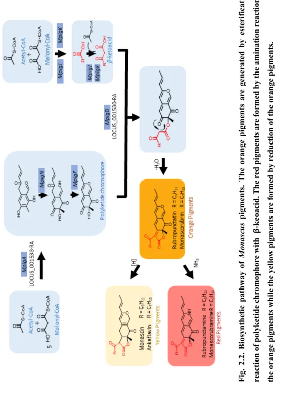
The biosynthesis of Monascus pigments begin with the condensation of Acetyl CoA and Malonyl-CoA that led the formation of hexaketide chromophore by polyketide synthase (Hajjaj et al., 2000; Chen et al., 2017). Monascus pigments are chemical reactions by the esterification of polyketide chromophore with β- ketoacid (Liu et al, 2018). MpigD, which encodes 3-O-aetyltransferase, leads the formation of the orange pigments by the esterification (Hajjaj et al., 2000; Chen et al., 2015). It is generally accepted that the reduction reaction of the orange pigments forms the yellow pigments, while the amination reaction of the orange pigments forms red pigments (Chen et al., 2017).
Multiple studies have been conducted the gene clusters of Monascus pigments biosynthesis. Liu et al. (2018) found that MpigA encoded the polyketide synthase and catalyzed the biosynthesis of key aromatic ring intermediate. Thus, inactivation of MpigA (the homolog of MpKS5) led an abolishment of pigment and displayed albino phenotype in M. purpureus mutant W 13, validating MpigA was engaged in pigment production (Balakrishnan et al., 2013). Xie et al. (2013) reported that deletion of pigR (MpigB) led to the loss of pigment production in M.
ruber M7, while its overexpression activated a 12-fold higher yield of pigments, suggesting pigR was a positive regulator. Liu et al. (2013) found that disruption, complementation, and overexpression of MpigE in M. ruber M7 had very little effects, and deletion of MpigE yielded four kinds of yellow Monascus pigments and very little red pigments, suggesting the MpigE could be related in the conversion of different pigment compositions.
29
Biosynthesis of Monascus pigments is very complex. Even though several genes related to Monascus pigment biosynthesis were manipulated through either disruption or overexpression of the target gene, MpigI, a putative negative regulatory factor, was not fully understood while MpigB, a positive regulatory factor, was reported in M. ruber M7. Therefore, further studies are needed to investigate the function of MpigI.
30
Fig. 2.2. Biosynthetic pathway ofMonascus pigments. The orange pigments are generated by esterification reaction of polyketide chromophore with𝛃-keoacid. The red pigments are formed by the amination reaction of the orange pigments while the yellow pigments are formed by reduction of the orange pigments.
31
3.3.2. Monacolin K biosynthesis
Monacolin K, also known as lovastatin, lowers the cholesterol level by inhibiting the HMG-CoA reductase which is the rate limiting step in cholesterol synthesis (Klimek et al., 2009).
Chen et al. (2008) found that disruption in mokA led the complete loss of monacolin K production in M. pilosus BCRC38072, suggesting that mokA was responsible for monacolin k nonaketide synthase. In addition, Sakai et al. (2009) reported that disruption in mokB did not produce monacolin K although monacolin J was accumulated in M. pilosus NCRC4480, indicating that mokB was responsible for biosynthesis of side-chain diketide moiety. Chen et al. (2010) confirmed that transformants showed a 1.7-fold higher yield of monacolin K than the wild-type M. pilosus BCRC38072 strain. Monacolin K biosynthetic genes in the transformants were overexpressed than the wild-type through RT-PCR analysis, indicating the mokH upregulated the transcription and was responsible for transcription factor.
32
Fig. 2.3. Biosynthetic pathway of monacolin K. Once monacolin J is synthesized, the binding of 2-methylbutanoic acid to monacolin J leads the formation of monacolin K.
33
3.3.3. Citrinin biosynthesis
According to previous studies, citrinin production was controlled by either optimization of culture condition or application of genetic engineering (Liang et al., 2018). The optimization of culture condition was traditional strategy to control citrinin level, and it was hard to block citrinin production completely (Liang et al., 2018). Thus, manipulation of citrinin biosynthetic related genes could be more practical way to eliminate citrinin concentration by applying genetic engineering. Fig. 2.4 (a) describes the gene clusters of citrinin in Monascus spp. Knockout of pksCT gene was reported to abolish citrinin biosynthesis in M. ruber and M. purpureus (Shimizu et al., 2005; Jia et al., 2010;
He and Cox 2016).
As shown in Fig. 2.4 (b), citrinin gene clusters of M. ruber were mostly lost, and this strongly suggests that M. ruber could not be able to produce citrinin during liquid-state fermentation. To validate this, HPLC analysis was performed to detect citrinin and M. purpureus strain was used as a control strain. Fig. 2.4 (c) represents the HPLC analysis of M. ruber and M. purpureus BCRC 31541. While M. purpureus BCRC 31541 produced approximately 238.26 ± 15 µg/mL of citrinin on day 18 of cultivation of citrinin, M. ruber was not able to produce citrinin. The growth between strain M. ruber and M. purpureus BCRC 31541 did not have any significant differences. Thus, both results from genome sequence and HPLC analysis demonstrated that M. ruber could be served as a promising industrial strain.
34
Fig. 2.4. Schematic representation of (a) gene clusters of citrinin in Monascus spp.
(b) gene clusters of citrinin in M. ruber (c) HPLC results of M. ruber and M.
purpureus BCRC 31541. Each plot represents the average of triplicates and error bar depicts the standard deviation.
ays.
35
4. Conclusions
In Chapter 2, the whole genome sequence of M. ruber was obtained. Citrinin biosynthetic gene clusters were mostly lost, while beneficial biosynthetic gene clusters of secondary metabolites, monacolin K and Monascus pigments, were present in M. ruber. To confirm gene clusters of citrinin were mostly lost in M.
ruber, HPLC analysis was performed and then citrinin was not detected at all.
The results indicated that M. ruber could be served as a promising industrial strain without citrinin production.
The total length of 25.9 Mb was generated using PacBio RSII sequencer and de novo assembly. This genome consists of 13 contigs with 9,639 predicted genes.
With the genetic analysis of gene clusters based on whole genome sequence, secondary metabolites of M. ruber was discussed in Chapter 2.
36
Chapter 3.
CRISPR/Cas9 system in filamentous fungi Monascus ruber
1. Introduction
In Chapter 2, gene clusters involved in secondary metabolites of M. ruber were studied and discussed. Chapter 2 concluded that citrinin related gene clusters were mostly lost in M. ruber, suggesting M. ruber could be a citrinin-free strain.
According to the results from genome sequence, two different MpigI loci were observed in M. ruber. In Chapter 3, CRISPR/Cas9 system was employed to precisely manipulate two different MpigI, termed MpigI and MpigI’
Several studies have been conducted to understand the pigment biosynthetic genes in Monascus spp. Balakrishnan et al. (2012) reported that mppR1 (the homologous of MpigB in M. ruber) and mppR2 (the homologous of MpigI in M.
ruber) genes in M. purpureus were regulatory genes encoded transcription factors for pigment biosynthesis. In addition, Huang et al. (2017) found that H2O2
stimulated the pigment production via up-regulation of transcript levels, but mppR2 was significantly down-regulated and negatively correlated to pigment production. Chen et al. (2015) also reported that the function of MpigI was negative regulatory factor. Thus, several studies have been demonstrated the function of MpigI could be putative negative regulator, which suppresses the production of pigment, so further studies were necessary to confirm whether MpigI suppresses the pigment production or not.
37
Since filamentous fungi have been proved to produce valuable natural products, CRISPR/Cas9 system has been established in several filamentous fungi, including Aspergillus spp., Fusarium oxysporum, Neurospora crassa, Penicillium chrysogenum, Trichoderma reesei (Pohl et al., 2016; Schuster and Kahmann, 2019; Wang et al., 2018). Furthermore, CRISPR/Cas9 system has been employed to engineer the pigment production of bikaverin, fungal red pigment with antimicrobial and antitumor activities, in Fusarium oxysporum. Wang et al.
(2018) reported that Bik 1 was mutated by using CRISPR/Cas9 system, and introduced mutants were unable to produce the red pigment. This study confirmed that Bik 1 was responsible for the synthesis of red pigment. Thus, pigment production of secondary metabolite gene cluster in filamentous fungi could be induced by CRISPR/Cas9 system.
In Chapter3, CRISPR/Cas9 system, precise genome editing, was applied to genetically engineer the specific target sites in M. ruber. The genes involved in regulation of Monascus pigments were manipulated. Monascus pigments were widely used as natural food colorants in various industries and possessed several biological functions, leading to antioxidant, antitumor, antimicrobial, antimutagenic properties and potential antiobesity activities. The mutants with increase in Monascus pigments could be beneficial to wide range of industries.
To increase the Monascus pigment production in M. ruber, CRIPSR/Cas9 system was employed to inactivate MpigI with dual sgRNAs. According to previous studies, gene expression of MpigI was downregulated rather than upregulated under cultivation conditions for high pigment production. Thus, CRISPR/Cas9- induced mutations in MpigI were expected to upregulate the transcription of pigment biosynthesis. CRISPR/Cas9 system was employed to overproduce Monascus pigments by inactivating MpigI. Dual sgRNAs were manufactured to
38
inactivate MpigI. After PEG-mediated transformation, randomly selected mutants, and wild-type M. ruber strain were compared to validate the increase in pigment production though analysis of secondary metabolites, sequencing, and gene expression.
39
2. Materials and methods
2.1. Strains, plasmid, primers, and culture conditions
The Escherichia coli TOP10 strain (Invitrogen Co, Carlsbad, CA, USA) was served as the general cloning host. The propagation of plasmid was grown in Luria-Bertani (LB) medium at 37 ℃ with 50 𝜇g/ml ampicillin. The strain M.
ruber was isolated from Korean traditional fermented food, and the strain M.
purpureus BCRC 31541 was purchased from Bioresources Collection and Research Center (BCRC) in Taiwan. Both strains were cultured on PDA medium at 30 ℃ for hyphae collection. For protoplast regeneration and transformation resistance, molten regeneration medium (0.1% (w/v) yeast extract, 0.1% (w/v) casein enzymatic hydrolysate, 1 M sucrose, 1.6% (w/v) Agar) was prepared for the selection with 200 𝜇g/mL hygromycin B. The plasmid pFC 332 (#87845) containing the Cas9 expressing cassette was purchased from Addgene Inc (Watertown, MA, USA). Primer synthesis were performed by Bioneer Corporation (Daejeon, Republic of Korea), and primers used in this study are listed in table 3.1.
40
Primers Sequence (5’ to 3’) Description
F_ M. ruber CTCAATGCTTGGTCGTCTCG random PCR of
M. ruber R_M. ruber TGACTACCTCTCTCCGGGAA
F_cas9 pFC332 GAAGTATAGCATCGGGCTGG flanking regions of
cas9 R_cas9 pFC332 TGACTAAGGTCGATACGGGT
F_MpigI TCAGCCGTAGCCCAGAGGTG amplification of MpigI
R_MpigI ATGTGGATTGGCCTGCTCTTCG
F_MpigI’ GCAGAAACTGCCTCACCAGG amplification of
MpigI’
R_MpigI’ ATCTCACTGACCTGCCAGGC
F_sgRNA 1 TAATACGACTCACTATAGGACTCGACTGCTCCCTCCACAGTTTTAGAGCTAGAA in vitro transcription of
sgRNA F_sgRNA 2 TAATACGACTCACTATAGGGTAAGCGCACCTCTGGGCTAGTTTTAGAGCTAGAA
F_sgRNA 3 TAATACGACTCACTATAGGTCTCCTCCCTGCTCACACAAGTTTTAGAGCTAGAA F_sgRNA 4 TAATACGACTCACTATAGGAGCTCCTTCAAAGCCAGCTGGTTTTAGAGCTAGAA
R_sgRNA AGCACCGACTCGGTGCCACTTTTTCAAGTTGATAACGGACTAGCCTTATTTTAACTTGCTATTTCTAGCTCTAAAAC
F_MpigI_del TTCAGGTAGAGCAGCAGCGT flanking regions of
MpigI R_MpigI_del GGGCAGATTGATCGTCCGAT
F_MpigI’_del TGGCCGCTGCGAAGGGAATA flanking regions of
MpigI’
R_MpigI’_del ATCGTGCACAGCGTGGCATTC
Oligo dt 18 TTTTTTTTTTTTTTTTTT cDNA synthesis
GAPDH_F GAGATCAAGCAGGCCATCAAG RT-PCR analysis of
GAPDH GAPDH_R GTAACCCCACTCGTTGTCGT
F_MpigI_RT AAACACATTCAGCCGTAGCC RT-PCR analysis of
MpigI R_MpigI_RT ATAGGCATGTCGTCGGAAAC
F_MpigI’_RT GGCGGACATAGGTGCACG RT-PCR analysis of
MpigI’
R_MpigI’_RT CAGCAGCAGCTCCTTCAAAG
F_MpigA_RT CCTGAATGGGTGCAACGAGTAC RT-PCR analysis of
MpigA R_MpigA_RT TATGTACCGCCTCTGCACTG
Table 3.1. Primers used in this study.
41
2.2. Preparation of in vitro transcriptional sgRNA
To efficiently inactivate the target genes, dual sgRNAs were designed to guide Cas9 to simultaneously cleave two sites, spaced 438 bp apart in MpigI and 362 bp apart in MpigI’. PAM sequence should be present in the 3’end of the target DNA sequence. The 20 nucleotides upstream of PAM sequence (5’-20NGG-3’) in MpigI and MpigI’ were designed with the SnapGene Tool. The sgRNA DNA templates were assembled and transcribed by using the MEGAscript T7 kit (Thermo Fisher Scientific, Walthan, MA, USA) according to the manufacture’s protocol. Following in vitro transcription, enzymatic reactions were purified with MEGAclaer kit (Thermo Fisher Scientific, Walthan, MA, USA). The quantity and purity of sgRNA was measured using the NanoVue Plus Spectrophotometer (GE Health Care Co., Nordrhein-Westfalen, Germany). The purified sgRNAs were stored at -80 ℃, and 100 𝜇g of synthesized sgRNA fragments were transformed into the Cas9 expressed transformants.
2.3. Protoplast preparation and transformation
The PEG-meditated transformation method was followed with slight modifications (Turgeon et al., 2010). The wild-type of M. ruber strain was inoculated on PDA and incubated at 30 ℃ for 7 days. Asexual spores (conidia) of the plates were harvested by gently scraping the agar with sterile distilled water.
The spores were filtered through 40 𝜇m nylon filter and then filtered spore suspension was inoculated in 100 mL of PDB for 18 h at 30 ℃. The collected mycelia were washed with sterile 0.7 M NaCl. Subsequently, the mycelia were suspended in enzyme solution (0.7 M NaCl, 2% (w/v) Cellulase, 1% (w/v) lysing enzyme and 1% (w/v) Driselase) and then incubated at for 2 h at 30 ℃ with shaking of 80 rpm. Protoplast release was checked every 30 min by microscopic
42
observation. Released protoplasts were carefully filtered two times, first using sterile Miracloth and then 40 𝜇m nylon filter. Then protoplasts were washed three times with STC buffer (1 M sucrose, 50 mM Tris-HCl and 50 mM CaCl2
pH 8.0). Finally, the protoplasts were resuspended in 500 𝜇L STC buffer and adjusted their density to approximately 107-108. About 10 𝜇g of linearized plasmid DNA and 100 𝜇g of sgRNAs were added into 100 𝜇L of protoplast suspension and incubated on ice for 20 min. Then, freshly prepared PEG buffer (60% (w/v) PEG 4000, 50 mM Tris-HCl and 50 mM CaCl2 pH 8.0) was added.
After transformation, protoplasts were incubated on molten regeneration medium at 30 ℃ for 3-5 days. Finally, transformants were randomly selected and cultured on PDA for further analysis.
2.4. DNA extraction and PCR analysis of putative M. ruber transformants
The genomic DNA was extracted same as described in Section 2.2. in Chapter 2. To confirm integration of Cas9, PCR amplification was performed with AccuPower Taq PCR premix (Bioneer Co., Daejeon, Republic of Korea) and evaluated though gel electrophoresis. Gene disruption in individual transforments were confirmed by Sanger sequencing.
43
Fig. 3.1. Strategies for the establishment of CRISPR/Cas9 system in M. ruber. Cas9 is delivered as plasmid while sgRNA is synthesized following in vitro transcription. PEG-mediated transformation method is applied.
44
2.5. Analysis of secondary metabolites 2.5.1. Monascus pigment analysis
The supernatant was removed via centrifugation, and the fermentation samples were taken every 2 days. After filtration, the absorbance of three Monascus pigments (yellow, orange, and red) was determined by using UV-1800 spectrophotometer (Shimadzu Co., Kyoto, Japan). Optical density (OD) values at 410, 465, and 505 nm were measured to determine the yields of pigments.
2.5.2. Extraction and analysis of monacolin K
Prior to monacolin K analysis, monacolin K was extracted with minor modifications, according to the previously reported methods (Lin et al., 2018).
The wild-type and mutants were inoculated on PDB for 14 days. The mycelium was dried by oven heating at 60 ℃ and finely grounded into powder.
Approximately 0.1 g of preparations were extracted with 4 mL of 75% ethanol for 10 min on an ultrasonic bath and subsequently centrifuged at 4200 rpm for 10 min. The supernatant was filtered through 0.22 𝜇m membrane filter. Monacolin K was analyzed though HPLC as previously described (Feng et al., 2014). HPLC was performed on UltiMate 3000 BioRS system (Thermo Fisher Scientific, Walthan, MA, USA). For stationary phase, the column of inertsil ODS-3 (4.6 mm
× 250 mm, i.d., 5 𝜇𝑚) was used. For mobile phase, a mixture of ACN, water and 0.5% ortho-phosphoric acid in the ratio of 60:37:3 (v/v/v) was carried out with the flow rate of 1.0 mL/min. The injection volume was 20 𝜇L and the column temperature was set at 25 ℃. The monacolin K contents were monitored by a DAD detector at 237 nm.
45
2.5.3. Citrinin analysis
Citrinin was analyzed same as described in section 2.4. in Chapter 2.
2.6. RNA extraction and RT-PCR analysis
Mycelia of the wild-type M. ruber strain and mutants from 7-day-old cultures were homogenized in liquid nitrogen and grounded into a powder in a pre-chilled mortar and pestle. Then, processed with Universal RNA extraction kit (Bioneer Co., Daejeon, Republic of Korea) following the manufacture’s protocol. The quantity and purity of RNA samples were measured using the NanoVue Plus Spectrophotometer (GE Health Care Co., Nordrhein-Westfalen, Germany), and cDNA was synthesized with AccuPower RT premix kit (Bioneer Co., Daejeon, Republic of Korea). For cDNA synthesis, 1 µg of RNA was reverse-transcribed in a 20 µL