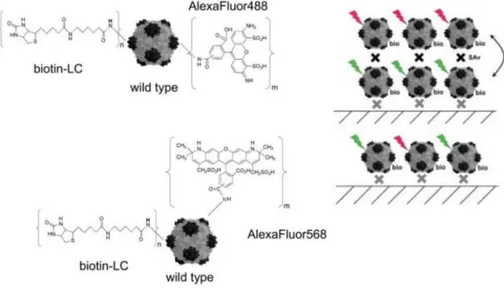
These conjugations of aphibodies and fluorescent protein are formed post-translationally, allowing simple and rapid binding between aphibodies and fluorescent proteins. ESI-TOF MS : Electrospray Ionization - Time of Flight Mass Spectrometry eYFP : Enhanced Yellow Fluorescent Protein.
Introduction
Targeted Cell Imaging and Drug delivery
Folate acid-conjugated magnetite nanoparticles target BT-20 breast cancer cells in vitro studies.21 The hormone-releasing hormone (LHRH) receptor is another target that has been extensively investigated for the targeted delivery of imaging agents and the treatment of localized breast cancer. Iron oxide nanoparticles conjugated with LHRH and interacted with MDA-MB-435S.luc breast cancer cells and demonstrated the accumulation of targeted nanoparticles in tumor tissues.22.
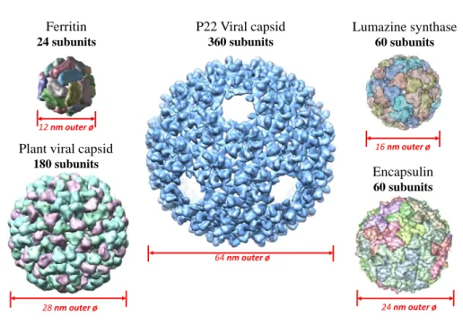
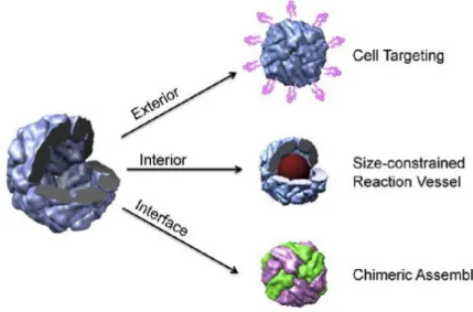
High-ordered Structure
Therefore, protein-based nanoparticles such as protein cages can be used to make a uniform high-order structure while maintaining the characteristics of a multilayer arrangement, or a complex assembly can be used by adding different functions to each protein cage. . in many fields. It demonstrated that CCMV can be used as a multifunctional nanoplatform to improve the signal in the imaging system.38.
Protein-based Nanoplatforms
- Protein Cage Architectures
- Fusion Protein using Monomeric Proteins
Live fluorescence cell images of SK-BR-3 cells showed the positions of the free AlDox (red), eYFP-SC (yellow), and AlDox-loaded GST-HER2Afb:eYFP (AlDox-GST-HER2Afb:eYFP), respectively ( Figure 3.10). The cytotoxicity of AlDox-GST-HER2Afb:eYFP was investigated using 3-(4,5-dimethylthiazol-2-yl)-2,5-diphenylatetrazolium bromide (MTT) cell viability assay and the results are given in Figure 3.16.
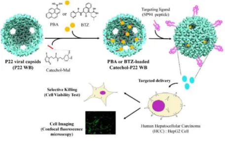
Encapsulin Protein Cage Nanoparticles as a Targeted Delivery Nanoplatform
Materials and Methods
Reactions were dialyzed against phosphate buffer (50 mM sodium phosphate, 100 mM sodium chloride, pH 7.5) overnight to remove unreacted F5M. Subsequently, 10 mol equivalent of SP94 peptide was incubated with E_LH42C123-SMCC with vigorous shaking overnight and dialyzed against the same buffer (50 mM sodium phosphate, 100 mM sodium chloride, pH 6.5) for another night.
Results and Discussion
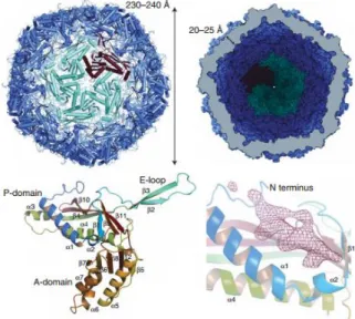
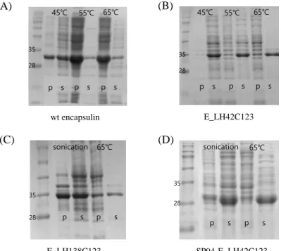
As alternative candidates, loop 42 (E_LH42) or 138 (E_LH138) in encapsulin, six consecutive linker histidines were introduced into wt encapsulin, resulting in unusual thermal stability (Figures 2.2 B and C). This result indicated that the loop regions of residues 42 and 138 were exposed to the outer or inner surface (Figures 2.3 A and B). The molecular weight of the dissociated subunit was measured to confirm the amino acid substitution (Figure 2.5 A, middle).
Although no size change such as dissociation or aggregation was observed under the TEM (Figure 2.5 B, middle), purified E_LH42C123 eluted much later than wt encapsulinine in SEC (Figure 2.5 C, middle and bottom), indicating that the hydrodynamic diameter becomes smaller then wt. encapsulin. The increase in the ratio at 280/260 nm was observed, indicating that there was no obvious nucleic acid association with cage (Figure 2.5 C, center). Their intactness with a uniform size of approximately 23 nm in diameter was confirmed using TEM (Figure 2.5 B, top).
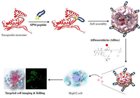
While SP94-fE_LH42C123 bound well to HepG2 cells, fE_LH42C123 without SP94-peptide bound little (Figure 2.7) indicating that the genetically introduced SP94 peptide was successfully presented on the surface of encapsulin and allowed them to bind specifically to the cells.

Conclusions
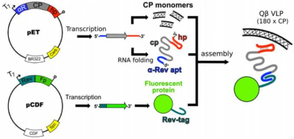
Cell Imaging Modular Toolkits using SpyTag/SpyCatcher
Materials and Methods
Cell growth continued at 30ºC (except ST-HER2Afb/ST-EGFRAfb, 37ºC) for 12-15 h after IPTG induction and cells were harvested by centrifugation at 5000 rpm for 10min at 4ºC. The pelleted cells were resuspended in 35 ml of pH 6.5 phosphate buffer (50 mM sodium phosphate and 100 mM sodium chloride) and sonicated for 10 min in 30 s intervals after adding lysozyme to the resuspended solution. All types of proteins were then centrifuged at 12000 g for 1 hour at 4ºC and purified by IMAC (1 mL HisTrap FF column / GE HealthCare).
MDA-MB-468 cells were grown in Leibovitz's L-15 medium with 10% FBS, 1% antibiotic-antimycotic, 25 mM HEPES, and sodium bicarbonate. AFPC (affibody:fluorescent protein conjugates) samples were processed for 1 hour at 37ºC and cells were fixed with 4% paraformaldehyde in PBS in case of live cell imaging. Before sealing, cells were washed twice with PBS and nuclei were stained with DAPI.
Cells were treated with 200 µL of medium containing 0.5 mg/mL MTT for 4 h, and then, the medium was removed and resuspended with 200 µL of dimethyl sulfoxide (DMSO) to dissolve formazan crystals formed by viable cells.
Results and Discussion
ST-GST-HER2Afb was purified using the same methods as those for ST-HER2Afb. To follow the post-target binding event, we treated SK-BR-3 cells with GST-HER2Afb:eYFP and visualized their positions according to incubation times (Figure 3.11). ST-GST-HER2Afb only without AlDox showed no red fluorescence signal, whereas AlDox-GST-HER2Afb:eYFP showed strong red fluorescence signals (Dox) throughout all time courses (Figure 3.14).
Red fluorescence signals could be observed not only in the nucleus but also in the cytosol, and the morphology of SK-BR-3 cells treated with AlDox-GST-HER2Afb:eYFP was significantly changed already at an early time (30 min) (Figure 3.14 ). While the viability of SK-BR-3 cells treated with AlDox-GST-HER2Afb:eYFP or free AlDox significantly decreased as AlDox concentrations increased, GST-HERAfb:eYFP cells were unaffected. While free Dox effectively killed MCF10A cells at nearly the same levels as SK-BR-3 cells, only about 20% of MCF10A cells were killed after treatment with AlDox-GST-HER2Afb:eYFP (Figure 3.15).
Dose-dependent cytotoxicity profiles of AlDox-GST-HER2Afb:eYFP, GST-HER2Afb:eYFP and free Dox towards SK-BR-3 (A) and MCF10A (B).

Conclusions
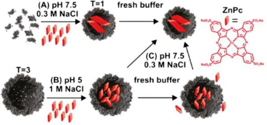
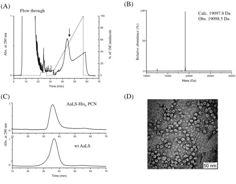
After sample injection, the column was equilibrated with binding buffer A and AaLS-His6 PCN was eluted with a linear gradient of 5-100% elution buffer B at a flow rate of 1 mL/min. A His6 affinity tag was genetically introduced to the C-terminus of PCN wt AaLS. No obvious dissociation of AaLS-His6 PCN from immobilized Ni was observed even after buffer washing, suggesting that multiple His-tags on the surface of AaLS-His6 PCN interacted with chelated Ni ions.
Since the surface of the QCM sensor chip itself is quite rough at the atomic level (Figure 4.7), but has the same properties as the ACM gold sensor chip, the LbL assemblies of AaLS PCN derivatives were measured through AFM. These studies clearly suggest that engineered encapsulin can be one of the specific targeted therapeutic delivery platforms. E.; Douglas, T., Virus-like particle nanoreactors: programmed encapsulation of the thermostable CelB glycosidase inside the P22 capsid.
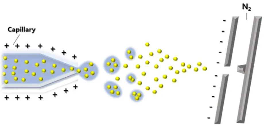
The speed of the ion depends on the mass-to-charge ratio (heavier ions with the same charge reach slower speeds, although ions with a higher charge will also increase in speed).
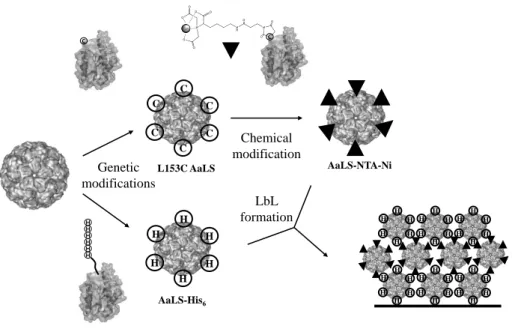
Layer-by-layer Assemblies with Protein Cage via Simple His-tag/metal Recognition
Materials and Methods
The resulting AaLS-His6 PCNs and L153C AaLS PCNs were purified by immobilized ion metal affinity chromatography (IMAC) and heat precipitation followed by size exclusion chromatography as previously described.34. Complexes of Ni ion and maleimido-NTA were formed by mixing NiSO4 and maleimide-NTA with 1:1 ratio at room temperature for an hour. Then, L153C AaLS PCNs were incubated with 10 mol equivalents of maleimido-NTA-Ni ion complexes at room temperature with vigorous shaking overnight.
The subunit masses of genetically and chemically modified AaLS PCNs were analyzed using an ESI-TOF mass spectrometer (Xevo G2 TOF, Waters) coupled to a Waters UPLC and an autosampler. AaLS-His6 and AaLS-NTA-Ni PCNs were introduced at concentrations of approximately 100 μg/ml in HEPES buffer (10 mM HEPES, 100 mM NaCl, pH 7.4), respectively. In this process, Au/Si substrates were immersed in AaLS. -His6 PCN solution for 25 min, slowly taken out of solution in vertical direction, blow-dried with nitrogen gas and then rinsed with DI water, resulting in the monolayer coating of AaLS-His6 PCN on Au/Si substrates.
By repeating these procedures alternately with AaLS-NTA-Ni PCN and AaLS-His6 PCN solution, bilayer and trilayer coated samples were obtained.
Results and Discussion
To investigate the cage integrity of AaLS-His6 PCNs, size exclusion chromatography (SEC) and transmission electron microscopy (TEM) were used. AaLS-His6 PCNs eluted at the same position as AaLS PCNs on the SEC (Figure 4.2 C). The binding affinity between AaLS-His6 and AaLS-NTA-Ni was investigated using quartz crystal microbalance (QCM) measurements.
While the AaLS-His6 PCN frequency of the monolayer QCM sensor dramatically decreased after the introduction of AaLS-NTA-Ni PCN (dashed line), the wt AaLS PCN frequency of the monolayer QCM sensor remained unchanged (solid line). These data indicated that the Ni ions on the surface of AaLS-NTA-Ni and the His-tags on the surface of AaLS-His6 PCN directly interact and enable the formation of regular LbL assemblies and stabilize the multilayer nanostructure. The root mean square (RMS) roughness (Rq) value showed 1.45 nm, indicating that the deposition of AaLS-His6 PCN resulted in a uniform monolayer (Figure 4.8 A) without significant solidification.
Sequential layer formations were achieved by alternating deposition of AaLS-NTA-Ni PCN and AaLS-His6.
Conclusions
Concluding Remarks
E.; Douglas, T., Use of the internal cavity of the P22 capsid for site-specific initiation of atom transfer radical polymerization. In particular, the choice of solvent used for electrospraying the polymer depends mainly on two factors. This time will depend on the velocity of the ion, and is therefore a measure of its mass-to-charge ratio.
Since the high selectivity of the antibody is used, only the substance to be measured can be accurately measured, and the interference caused by the interfering substance can be minimized. Since the wave is on the boundary of the conductor and the external medium (air, water or vacuum for example), these oscillations are very sensitive to any change of this boundary, such as the adsorption of molecules to the conducting surface. 12. Functional groups on the ligand that can be used for coupling include NH2, SH, CHO and COOH.13 CM5 is regenerated by selective dissociation of the analyte from the covalently immobilized ligand.
Conditions should be chosen to achieve complete dissociation of the analyte without affecting the binding characteristics of the ligand. We have also shown that molecules with different functions can be ligated using the spittag outer surface of the cages. Based on the results so far, it is possible to test the activity of the enzyme to see if it can be used in the assay, and the number of enzymes can be checked to see how the enzyme activity changes.
Encapslin Protein Cage as a Targeted Delivery Nanoplatform