저작자표시-비영리-변경금지 2.0 대한민국 이용자는 아래의 조건을 따르는 경우에 한하여 자유롭게
l 이 저작물을 복제, 배포, 전송, 전시, 공연 및 방송할 수 있습니다. 다음과 같은 조건을 따라야 합니다:
l 귀하는, 이 저작물의 재이용이나 배포의 경우, 이 저작물에 적용된 이용허락조건 을 명확하게 나타내어야 합니다.
l 저작권자로부터 별도의 허가를 받으면 이러한 조건들은 적용되지 않습니다.
저작권법에 따른 이용자의 권리는 위의 내용에 의하여 영향을 받지 않습니다. 이것은 이용허락규약(Legal Code)을 이해하기 쉽게 요약한 것입니다.
Disclaimer
저작자표시. 귀하는 원저작자를 표시하여야 합니다.
비영리. 귀하는 이 저작물을 영리 목적으로 이용할 수 없습니다.
변경금지. 귀하는 이 저작물을 개작, 변형 또는 가공할 수 없습니다.
의학박사 학위논문
L-arginine처리를 통한 암세포의 시스플라틴 반응성 개선 및 Argininosuccinate synthetase
발현정도와 연관된 단독작용 효과로서의 세포사멸유도
Exogenous L-arginine ameliorates cisplatin-sensitivity in various cancer cells and alone induces apoptosis
depending on the degree of endogenous Argininosuccinate synthetase expression
울산대학교 대학원 의 학 과
김 하 일
[UCI]I804:48009-200000286271 [UCI]I804:48009-200000286271
Exogenous L-arginine ameliorates cisplatin- sensitivity in various cancer cells and alone induces apoptosis depending on the degree of endogenous
Argininosuccinate synthetase expression
지도교수 심 주 현
이 논문을 의학박사 학위논문으로 제출함 2020년 02월
울산대학교 대학원 의 학 과
김 하 일
김하일의 의학박사학위 논문을 인준함
심사위원장 이한주 (인) 심사위원 김강모 (인) 심사위원 류민희 (인) 심사위원 유수종 (인) 심사위원 심주현 (인)
울산대학교 대학원
2020년 02월
i Abstract
The association is well known between cisplatin-resistance and the loss of endogenous argininosuccinate synthetase (ASS1) expression in various cancer cells. Except for the on- target effect of cisplatin resistance related with epigenetic silencing of ASS1, other resistance mechanisms in cancer cells is currently unclear. ASS1 is a key enzyme and rate-limiting step in arginine synthesis, there is a possibility of the signaling alteration triggered by different ASS1 expression level or by cellular arginine availability. In other words, there is potential to ameliorate the cisplatin sensitivity via alteration of intracellular signaling or metabolic change driven by ASS1 itself or arginine. Given this circumstance, we aim to investigate how the ASS1 expression level and arginine supply effect on cisplatin-resistance and survival in cancer cells in terms of cellular signaling. We found not only exogenous arginine supply restores cisplatin- sensitivity in cancer cells but also arginine supply alone could negatively associate with cancer cell survival by alteration of certain signaling molecules according to ASS1 expression level.
Keywords
Cisplatin-resistance, Argininosuccinate synthetase 1, L-Arginine, Apoptosis, Signaling change
ii Contents
English abstract ··· ⅰ
List of Figures and Tables ··· ⅲ
Introduction ··· 1
Methods ··· 3
Results ··· 6
Discussion ··· 24
References ··· 31
Korean abstract ··· 35
iii
List of Figures and Tables
Figure 1. The restoration of endogenous ASS1 ameliorate cisplatin-sensitivity in various cancer cells···7
Figure 2. MTS analysis for various cancer cells treated with 5 and 10 mM L-arginine with or without 5 and 10 μM cisplatin···11
Figure 3. Proliferative assay related with ASS1 expression levels. ···15
Figure 4. Nitric oxide formation analysis according to ASS1 expression level ···19
Figure 5. Dose-dependent effect of arginine supply for glycolysis pathway enzymes according to ASS1 expression level···21
Figure 6. Western blot analysis for cancer cell lines in terms of ASS1 expression···23
Table 1. Summary of pro-apoptotic signaling molecule change in cancer cells···24
1 Introduction
Arginine is a multi-functional amino acid required for protein synthesis and modification, involved in the synthesis of many metabolites that intertwined with intracellular signaling even associated with cell survival (1, 2). argininosuccinate synthetase (ASS1) is a key enzyme and rate-limiting step in arginine synthesis, the downregulation of ASS1 results a dependence on extracellular arginine, is known as arginine auxotrophy (3-5).
There has been reported the association with chemoresistance and poor clinical outcomes in the arginine auxotrophic cancer cells such as hepatocellular carcinoma (HCC), melanoma, mesothelioma, pancreatic cancer, prostate cancer, renal cell carcinoma, sarcoma, and small cell lung cancer (5, 6).
Previous studies showed that the loss of ASS1 expression via epigenetic silencing by changing the methylation status of the promotor region has been linked to cisplatin resistance and clinical outcome in ovarian cancer, and HCC (7, 8). Unfortunately, the mechanism of cisplatin resistance was composed not only on-target resistance (epigenetic silencing) but also the other resistance mechanisms in terms of cellular signaling and metabolism (9). Nevertheless, the actual effect for ASS1 loss in cancer is currently unclear, the loss of ASS1 expression with cisplatin resistance were reported various cancers including malignant pleural mesothelioma, glioblastoma, bladder cancer, and in some lymphoid malignancies (5, 10). Although, there is potential to ameliorate the cisplatin sensitivity via alteration of the level of ASS1 expression or supply of arginine, the changing effect of the resistance and cell survival were not yet explored (11, 12).
This study aims to investigate how the ASS1 expression level and arginine supply
2
effect on cisplatin resistance and survival in cancer cells in terms of cellular signaling.
3 Methods
Cancer cell lines and lentiviral transfection
The human HCC cells SNU 398, Huh7, SNU 475, SUN 449, Hep3B, PLC/PRF/5 and human pancreatic cancer cell lines, AsPC1, MiaPaca2 were obtained from the Korean Cell Line Bank. Cell lines were cultured in Dulbecco’s Modified Eagle Medium (DMEM;
Welgene, South Korea). Culture media was supplemented with 10% fetal bovine serum (FBS; GIBCO, Gaithersburg, MD, USA) and cells were at 37 °C in humidified 5% CO2. The recombinant lentivirus was produced in ASS1-low cells (defined by both cellular mRNA and protein expression level) using Lipofectamine 2000 according to the manufacturer’s instructions.
Cell viability assay
To examine the sensitivity of HCC cell lines to cisplatin and Arginine, cytotoxicity was measured using the MTS assay (Promega, Fitchburg, WI, USA). Cells were plated in 96-well plates at 2 × 10³/well for each cell line. Cells were exposed to different concentrations of cisplatin (5μM, 10μM) and arginine (5 mM, 10 mM) for 72 hours.
Twenty microliters of MTS solution were added to each well containing 100μL of culture medium and the cells were then incubated for 2 h at 37 °C. Absorbance at 490 nm was measured using a Sunrise™ microplate reader with Magellan™ software (Tecan, Seestrasse 103, Mannedorf, Switzerland). Viability was expressed as a percentage of viability in untreated cells. The concentration of cisplatin and arginine resulting in 50% growth inhibition (IC50) was calculated using GraphPad Prism 7.0 software.
4
mRNA extraction and RT-PCR, protein expression of ASS1
Total cellular RNA was extracted using TRIzol (Thermo Fisher Scientific, Waltham, MA, USA) according to the manufacturer’s instructions. The isolated RNA was resuspended in RNAse-free water and RNA concentration was measured at 260 nm using a NanoDrop 2000 UV–VIS Spectrophotometer (Thermo Scientific, USA).
Reverse transcription (RT) was carried out in a 20 μL reaction mixture using a first strand cDNA synthesis kit (Invitrogen, Waltham, MA, USA) according to the manufacturer’s instructions. RT-PCR was performed using AccuPower PCR premix (Bioneer, South Korea) with addition of first-strand cDNA via thermocycling in a Perkin-Elmer9600 thermal cycler (Perkin-Elmer). The probed according to standard methods with 32P-labeled cDNA against GFP, actin and human p53. For protein analysis, total protein was extracted with 50mMTris 150 mM NaCl 0.1% Triton X-100 0.1 mM DTT plus proteinase inhibitors. Protein (10–50 Xg) was separated by 10%
SDS PAGE and immunoblotted according to standard methods with rabbit polyclonal antibodies against GFP (Abcam, Cambridge, U.K.) or X-actin (Sigma).
Nitric oxide measurement
Measurement of nitric oxide (NO) generation by Griess Reagent system (Promega Nitrite assay system). Nitrite measurement by using a multichannel pipettor, dispense 50ul of the sulfanilamide solution to all experimental samples and wells containing the dilution series for the Nitrite standard reference curve. Incubate 5-10 minutes at room temperature, protected from light. Incubate at room temperature for 5-10minutes, measure absorbance within 30 minutes in a plate reader absorbance at 540nm
5 Western blot analysis
Cellular expression level of ASS1 with or without cisplatin and arginine mediated alteration of signaling pathways was examined by western blot analysis. Cells were lysed in RIPA buffer (InTron, South Korea) supplemented with protease inhibitor (Sigma-Aldrich, USA). The resultant lysate was centrifuged at 12,500 rpm for 20 min at 4 °C and supernatants were collected. The protein concentration was measured by BCA assay (Promega, USA). After SDS-PAGE and transfer, membranes were blocked with 5% non-fat milk for 1 h and incubated with primary antibodies overnight at 4 °C, and then incubation with secondary antibodies (Cell Signaling, USA) for 1 h at room temperature. Target protein bands were detected using ECL reagents (GE, Fairfield, CT, USA).
Statistical analysis
GraphPad Prism version 7.0 was used to test results for statistical significance.
Differences in ASS1 levels between groups were analyzed using an unpaired two- tailed t-test. The F test was used to determine whether variances were equal or unequal. A one-way ANOVA was used for multiple group comparisons. A p value <
0.05 was set as a level of statistical significance. In determining statistical significance for ASS1 protein levels after cisplatin treatments or for arginine and cisplatin combination analysis, each drug concentration was compared to the untreated, or zero drug, sample to attain a p value for that particular drug concentration.
6 Result
The restoration of ASS1 gene expression in HCC cell lines via lentiviral vector lead to overcome cisplatin resistance
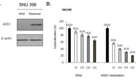
Previous studies demonstrated ASS1-low expressed cancer cell lines had cisplatin resistance (9). We first examined the changing effect of cisplatin-sensitivity by ASS1 restoration in ASS1-low cells. For verifying the endogenous expression level of ASS1, mRNA and protein level were examined in various cell lines. We selected SNU 398 for investigating ASS1 restored effect (Figure 1A).
Next step, we designed ASS1 delivery system via lentiviral vector (Figure 1B).
Lentiviral vectors were transfected to selected ASS1-low cells, then we confirmed the ASS1 expression level by Western blot (Figure 1C). To examine the change of cisplatin sensitivity, ASS1 restored cells compared with control cells by MTS assay (Figure 1D,All Ps<0.001 versus control). We conducted the examination by dose- dependent manner of cisplatin, the ASS1 restored cells showed increasing sensitivity for cisplatin in terms of cell survivals, overcoming the resistance. To these result, ASS1 restoration in ASS1-low cells recovered cisplatin sensitivity, in HCC cell lines.
7
8
Figure 1. The restoration of endogenous ASS1 ameliorate cisplatin-sensitivity in HCC cell line. (A) Baseline endogenous ASS1 expression level in cancer cell lines. (B) ASS1 gene delivery via lentivirus vector, GFP-tagged ASS1 proteins confirmed in visual, and (C) Western blot analysis after transfection showed the restoration of ASS1 gene in ASS1-low cancer cell line. (D) After delivery ASS1 gene, ASS1 restored cancer cells that overcame those cisplatin-resistance traits in MTS (All Ps<0.001 versus control).
9
L-arginine with cisplatin treatment in various cancer cells show that L-arginine not only ameliorate cisplatin-sensitivity but also alone could negatively effect on tumor cells survival depending on the degree of endogenous ASS1 expression
We confirmed that restoration of cellular expression of ASS1 in cancer cells could overcome the cisplatin resistance. The cellular arginine level controlled by ASS1 expression level, then we hypothesized that additional supply of arginine contributes to ameliorate the cisplatin sensitivity (4, 9).
The cancer cells were treated with 5 and 10mM L-arginine with or without 5 and 10μM cisplatin, followed analysis by MTS assay. As expected, the ASS1-expressed cell lines, SNU 449 and PLC/PRF/5 are sensitive to cisplatin than ASS1-low cells, as dose dependent manner. Interestingly, the additional supply of arginine accelerates anti- proliferative effect on these cell lines. Somewhat differently, ASS1-low cells showed that the exogenous arginine under the cisplatin treatment act like dose-dependent manner, not arginine alone(Figure 2A).
From the MTS assay results, we hypothesized that L-arginine mono-supplement could negatively effect on tumor cells survival depending on endogenous ASS1 expression level. Firstly, as expected, ASS1 low-cancer cells showed a slightly rise of proliferation without cisplatin treatment. However, in some ASS1-low expression cells, SNU 398 and Huh 7, showed sharp decreased in their proliferation after additional treatment of cisplatin, the anti-proliferative effect beat the proliferative effect as dose- dependent manner of cisplatin. Secondly, ASS1 expressed-cancer cells showed the anti-proliferative effect as a dose-dependent manner of arginine supply, more prominent in co-treatment of cisplatin as a dose dependent manner (Figure 2A).
10
To validate this arginine-dependent anti-proliferative effect on cancer cells, we screened serials of cancer cell lines by same treatment method as above. The trend of cancer cells proliferation by arginine dose, broadly classified by the degree of cisplatin-sensitivity (Figure 2B).
11
12
13
Figure 2. MTS analysis for cancer cell lines treated with 5 and 10mM L-arginine with or without 5 and 10μM cisplatin. (A) ASS1-expressed cancer cells are sensitive to cisplatin than ASS1-low cells, as dose dependent manner. Additional supply of arginine accelerates cytotoxic effect on these cell lines, even ASS1-low cells showed as a dose-dependent manner except for non-cisplatin treated cells. (B) Screened the serials of cancer cell lines by same treatment method. The trend of cancer cells proliferation by arginine dose, broadly classified by the degree of cisplatin-sensitivity. (P-value for MTS: (* <0.05), (** <0.001))
14
Not only the anti-proliferative effect but also the metastatic potential of cancer cells could be inhibited by arginine supply
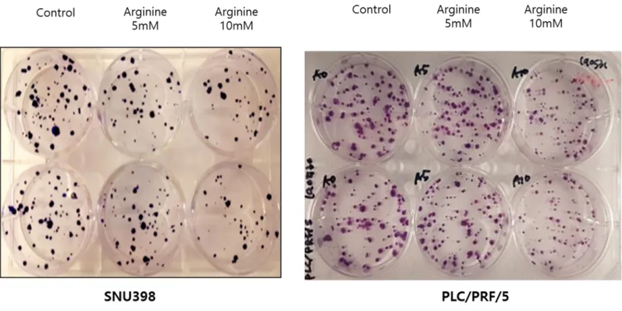
We conducted migratory and colony-forming assay for evaluating metastatic potential of HCC and other various cancer cell lines in each of different arginine supply and ASS1 expression level.
Figure 3Ashowed the migration assay of Huh 7 and SNU 449, represent ASS1 low- and expressed cells, respectively, with different dose of arginine supply. SNU 449 inhibited by arginine supply dose dependent manner, otherwise Huh 7 showed no effect versus control group. Colony-forming assay for ASS1 low- (SNU 398) and expressed (PLC/PRF/5) cancer cells were evaluated, ASS1 expressed cells showed negative effect by arginine supplement than ASS1 low-expressed cells (Figure 3B).
15
16
Figure 3. Proliferative assay related with ASS1 expression levels (A) The migration assay to Huh7 and SNU 449, represent ASS1 low
17
and normally expressed cancer cells, respectively. SNU 449 inhibited by arginine supply dose dependent manner, otherwise Huh 7 showed grossly no effect versus control group. It suggests that SNU 449 inhibited by those metastatic potential by arginine supply. (B) Colony-forming assay to SNU398 and PLC/PRF/5, represent ASS1 low and normally expressed cancer cells, respectively. Both cells showed that anti-proliferative effect by arginine supply as dose dependent manner.
18
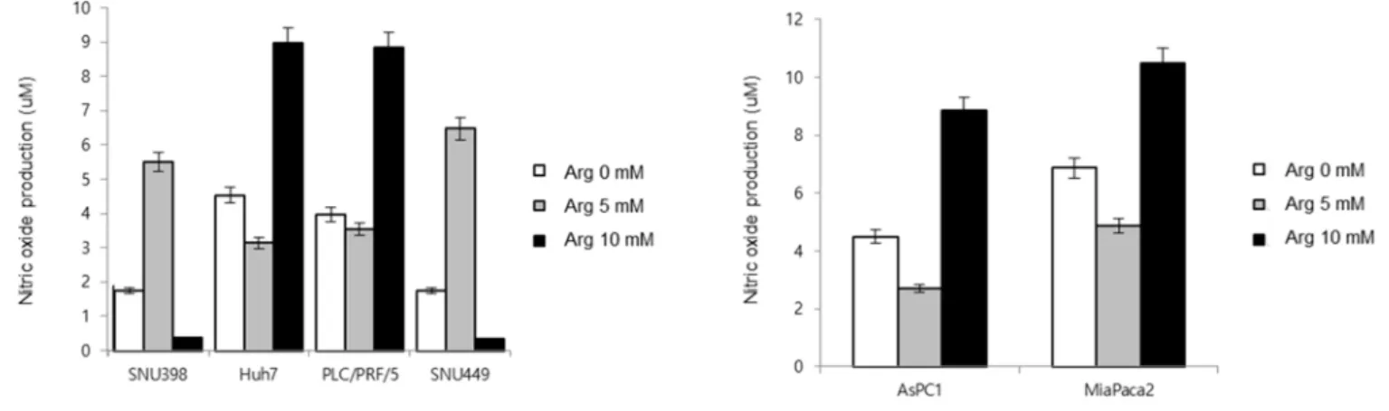
Exogenous L-arginine supply showed no consistent effect on Nitric oxide formation by the degree of ASS1 expression level in cancer cells
For evaluating the cause of cell death in terms of cellular signaling pathway or metabolic alteration, we measured cellular Nitric oxide (NO) by arginine supplement at the different ASS1 expression levels. Whether ASS1 level effect on the ability of NO formation in cancer cells, ASS1 low- and expressed- cells were converted into opposite traits in terms of ASS1 level, respectively. Huh 7(ASS1-low) restored the ASS1 expression by lentiviral vector, on the other hand, Hep 3B (ASS1-expressed) inhibit their ASS1 expression by knock-out (Figure 4A). The result suggested that the NO formation heavily influenced by cellular expression of ASS1 in cancer cells.
In general, early phase (within 60 min) NO changes reflect the effect of arginine supply by i-NOS, we evaluated early phase NO level in ASS1 expressed- and low- cancer cell lines. Disappointingly, dose-up treatment of arginine showed that there was no consistent trend of NO formation in terms of ASS1 expression level (Figure 4B). Amon ASS1-low HCC cell lines, SNU 398 showed NO formation in low-dose arginine (5mM), not high-dose (10mM) arginine supply. However, other ASS1-low cells, Huh 7 showed that the response of NO level by high-dose arginine. Unexpectedly, SNU 449, as a high ASS1 cell, showed NO formation in low-dose arginine. Two pancreatic tumor cell lines also examined, AsPC1 and MiPanc2, response high dose of arginine only in early phase. There could exist cell-specific trait, ASS1 level was not simply affecting on NO formation by arginine supply level.
19
20
Figure 4. Nitric oxide (NO) formation analysis according to ASS1 expression level (A) Huh 7(ASS1 low expressed cell) restored the ASS1 expression by lentiviral vector, Hep 3B (ASS1 expressed cell) inhibit their ASS1 expression by knock-out. The result suggested that the NO formation heavily influenced by cellular expression of ASS1 in cancer cells. (B) Dose-up treatment of arginine showed that there was no consistent trend of NO formation in terms of ASS1 expression level in early phase (within 60 min)
21
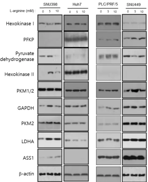
The relevance of the glycolysis pathway and ASS1 expression level showed no consistent changing effect in terms of an alteration of ASS1 expression level
Figure 5 showed that arginine-derived changing effect of glycolytic enzyme expression level. The results did not show consistent effect on glycolysis pathway enzymes on western blot analysis.
Figure 5. Dose-dependent effect of arginine supply for glycolysis pathway enzymes according to ASS1 expression level
22
The cancer cells survival was associated with arginine supply and cellular signaling, could be classified by endogenous ASS1 expression level
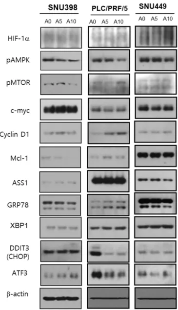
To examine which signaling molecules are altered their expression under arginine supply condition, cancer cells were evaluated in terms of ASS1 expression level. Each of apoptosis-related molecular level were examined by western blot (Figure 6). ASS1- low cancer cells (SNU 398) showed the decrease in expression level of pmTOR and Mcl-1. On the other hand, ASS1-expressed cells (PLC/PRF/5, SNU 449) showed the decrease of pAMPK expression level, not pmTOR and Mcl-1.
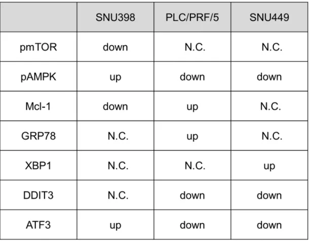
The expression level of ER stress related proteins was also evaluated. The ASS1-low cancer cells showed up-regulation of ATF3, on the other hand, ASS1-expressed cancer cells showed downregulation of ATF3 and DDIT3. There was no change on GRP78 and XBP1 protein in both cell lines (Table 1). These results suggested that the relationship of apoptosis with arginine supply is quite different from ASS1 low and ASS1 expressed cancer cells.
23
Figure 6. Western blot analysis for various cancer cells in terms of ASS1 expression.
Each of apoptosis-related molecular level were screened. ASS1- low cancer cell (SNU 398) showed the decrease in expression level of pmTOR and Mcl-1. On the other hand, ASS1 expressed cells (PLC/PRF/5 and SNU 449) showed the decrease of pAMPK expression level, not pmTOR and Mcl-1.
24
Table 1. Summary of pro-apoptotic signaling molecule change in cancer cells
2 4 Discussion
There has been reported that various cancer showed the ASS1 null-expression, related with aberrant metabolism, chemo-therapeutic resistance, and patients survival (5). Although, molecular mechanism of cisplatin resistance is related with an intertwined signaling pathway, the low-expressed level of ASS1 confers to cisplatin- resistance ovarian cancer and HCC in previous report (7, 8, 11). There are no studies for directly evaluating “off-target” effect (i.e. molecular circuitries that deliver compensatory survival signals even though they are not directly activated by cisplatin) on cisplatin resistance with or without arginine supply (9). We demonstrated that restoration of ASS1 expression by delivery vector (not epigenetic restoration) ameliorates the cisplatin sensitivity in various cancer cells. It suggested that a considerable proportion of cisplatin resistance was engaged the off-target mechanism.
In other words, ASS1-related cellular signaling or metabolic changes in cytoplasm could be affected on cisplatin-resistance, not just the nuclear module (9, 13).
The restoration of endogenous ASS1 leads to increase the cellular arginine levels.
We hypothesized that arginine related signaling pathway could be involved cisplatin- resistance by changes in the cytoplasmic module, the “off-target” of cisplatin resistance, because arginine is not just metabolic substrate or precursors of biosynthesis, it involves cellular signaling by itself or its metabolites (4, 6, 14, 15). We examined the dose-dependent effect of arginine supply in ASS1 low- and constitutional-expressed cell lines concurrently treated with cisplatin.
In our study, there were two interesting findings: 1) combination treatment cisplatin with arginine in cancers cells showed that accelerating effect those of anti-proliferative effect than cisplatin only treatment cells, even arginine supply alone showed
26
significant anti-proliferative effect. 2) In ASS1-low cells, although arginine mono- supply showed increased proliferation as expected, on the other hand, the anti- proliferative effect beat the proliferative effect by combination treatment of cisplatin and arginine as a dose-dependent manner. These results consist with in some part of MTS assay, arginine supply to cancer cells could inhibit their proliferation according to ASS1 expression level. Overall, the arginine supply could act as a cisplatin sensitizer, and even induced signaling change in cancer cells. Most of previous studies conducted by therapeutic approach that inhibition of arginine influx by enzymatic molecule, ADI-PEG20 and rhArg-PEG, targeted ASS1 low expression cancers (6, 15). These studies based on expectations of potential anti-cancer effect by arginine deprivation reached to suboptimal results, because of drug resistance and unrevealed mechanism (3, 15). There are few studies for the effect of additional supply of arginine for cancer cells, reported signal modulating effects with promote cell proliferation in breast and endometrial tumor cells, on the other hands, anti- proliferative effect on gastric cancer (16-18). So far there has been no report that exogenous arginine supply could act as a cisplatin sensitizer, even negative effects on cancer cell survival by the degree of ASS1 expression level.
To explore if these results were a general phenomenon in cancer cell lines, we examined MTS analysis with various tumor cell lines (Figure 2B). The results were varied, however, there was a consistent trend about dose-dependent of arginine supply. Our result showed when ASS1 expressed in cancer cells, arginine ameliorates cisplatin-sensitivity. In some aspect, ASS1 low-expressed cancer cells also promote their proliferation by arginine mono-supplementation as normal tissue. However, the effect of arginine in terms of cisplatin-sensitivity, concurrent treatment could effectively
27
suppress cancer cells even ASS1 low- cancer cells. These results suggest that combination treatment of arginine with cisplatin for cancer treatment simply enhancing their treatment effect, even cisplatin-resistance cancer cells. These findings indicated that arginine changes cellular signaling pathway as off-target manner.
Next, we examined whether cellular signaling changes or metabolic alterations were different between ASS1-low and ASS1-expressed cancer cells in the situation of arginine supply. There were some reports to proposed signal alteration mechanism by arginine supply via formation of NO by Akt/PI3k signaling, lysosomal sensing lead to mTOR signaling change, altered metabolism via changing glycolysis enzyme expression level (changing Warburg effect), and pre-apoptotic effect by mitochondrial membrane disturbances (19). However, these arginine-driven signaling changes were not correlated with off-target resistance mechanism of cisplatin known so far (9, 13).
To validate cellular metabolic and signaling changes, we examined following additional experiments: NO formation, alteration of glycolysis enzyme expression, cell survival apoptosis signaling molecules, and ER stress-related proteins.
Cellular NO formation is closely related to cancer cell survival by connected with various signaling molecules (20-23). There has been various studies that NO related with cell survival signaling via mTOR, down-stream of Akt/PI3k signaling, not as a direct toxic effect by ROS effect (19, 24). Firstly, we checked NO levels, the NO detected in early phase (after 30-60 min of arginine supply) could be the meaningful (protective or cytotoxic) signaling effect for arginine supply, changing arginine level effects by i-NOS. Exogenous arginine could induce i-NOS no matter how intracellular arginine level, explained the phenomenon as “Arginine paradox” (22). In our study did not demonstrate the certain effect on arginine in NO formation. Figure 4Ashowed
28
that restoration or knock-out of ASS1 expression directly effect on NO formation, these results suggested that to be formed to some amount of NO need endogenous ASS1 expression exclusively, not the changing of arginine. ASS1-low expression cancer cells do not mean the zero expression of ASS1. Our results not enough to explain the phenomenon of arginine dependence of cancer cell survival and ASS1 expression by NO analysis. Future study is needed to verify time-dependent effect of arginine supply and NO-related signaling alteration, and relationship of cancer cell survival.
Secondly, we checked the glycolysis pathway. Our results indicated that ASS1 expression level dose not specifically effect on glycolysis pathway enzymes, because there was no certain alteration of related enzymes. There were some hypothetical approaches evaluate the relationship between ASS1 and glycolysis pathway, called Warburg effect. The Akt/PI3k pathway via NO (the most representative signaling alteration) to accelerating glycolysis was experimented previous study (23), We hypothesized that, the changing ASS1 expression level accelerating NO signaling and Akt/PI3k activated, could lead to inhibitory effect on mTOR signaling as well as Warburg effect in certain cell lines. However, there was no alteration of glycolytic pathway enzymes by ASS1 expression level. In these results, we assumed that exogenous arginine supply was also related to some survival signaling, as off-target of the cisplatin, not just alteration of the energy metabolism in cancer cells.
In terms of arginine supply alone for cell survival signaling molecules, the results showed that there was some difference of the changes by ASS1 expression level.
There were some studies that report for “off-target” signaling molecules for cisplatin resistance, including relevance of autophagy or unfolded protein response (UPR) (25-
29
27). In our study, ASS1 low-expressed cells showed the decrease in expression level of pmTOR and Mcl-1 and increased in ATF3. ASS1 expressed cells showed the decrease of pAMPK expression level, not pmTOR and Mcl-1. AMPK and mTOR signaling pathway associated with cellular energy sensing and transcriptional regulation of autophagy (28-30). The downregulation of ATF3 related with ER stress and formation of UPR (26). Taken together, ameliorating the effect of cisplatin sensitivity, arginine effect on different way of “off-target” between ASS1 low-expressed and expressed tumor cells. Additionally, endogenous ASS1 expression level could be a predictive maker not only the trait of cisplatin-sensitivity but also the different off- target cisplatin resistance effect in the presence of arginine supply.
This study has several limitations. First, we focused on off-target effect of cisplatin resistance. For that reason, our study was not fully demonstrated the effect of exogenous arginine supply to cancer cells. However, we demonstrated that the alteration of cellular signaling could reflect off-target effectiveness of cisplatin resistance as well as anti-proliferative effect by arginine alone. Second, previous in vivo studies showed the various range of plasma arginine for evaluating anti-cancer treatment effect versus side effect (31, 32). We used certain dose of arginine (5mM, 10mM) and fixed cell harvest time (72 hours after treatment). Although we could not verify the dose-dependent anti-proliferative effect of arginine mono supply, we believe that our study use a reasonable dose for alteration for signaling, not direct cytotoxic effect by arginine, comparing previous studies (33, 34). Third, we could not verify all the metabolite and those pathways related to arginine. Theoretically, arginine and their altered signaling effect were numerous: Urea cycle, Pentose phosphate pathway, polyamine synthesis and regulation pathway, and arginine transporter with their
30
sensing mechanism (1, 2, 35-38). However, in terms of therapeutic approach for cancer treatment, we suggest one simple possibility for ameliorates cisplatin sensitivity via arginine supply that relatively harmless and easily applicable. Future study is needed for verifying the effectiveness in vivo.
In summary, we found not only exogenous arginine supply ameliorate cisplatin- sensitivity in cancer cells but also alone could negatively associate with cancer cell survival by alteration of certain signaling molecule according to the ASS1 expression level. Our finding suggest that exogenous arginine supply could be utilized for inducing “metabolic addiction” in the situation for cisplatin resistance cancer cells by the off-target effect.
31 Reference
1. Morris SM, Jr. Enzymes of Arginine Metabolism. J Nutr. 2004;134.
2. Morris SM, Jr. Arginine Metabolism Revisited. J Nutr. 2016;146(12):2579S- 86S.
3. Riess C, Shokraie F, Classen CF, Kreikemeyer B, Fiedler T, Junghanss C, et al. Arginine-Depleting Enzymes - An Increasingly Recognized Treatment Strategy for Therapy-Refractory Malignancies. Cell Physiol Biochem. 2018;51(2):854-70.
4. Jahani M, Noroznezhad F, Mansouri K. Arginine: Challenges and opportunities of this two-faced molecule in cancer therapy. Biomed Pharmacother.
2018;102:594-601.
5. Delage B, Fennell DA, Nicholson L, McNeish I, Lemoine NR, Crook T, et al.
Arginine deprivation and argininosuccinate synthetase expression in the treatment of cancer. Int J Cancer. 2010;126(12):2762-72.
6. Albaugh VL, Pinzon-Guzman C, Barbul A. Arginine-Dual roles as an onconutrient and immunonutrient. J Surg Oncol. 2017;115(3):273-80.
7. Helleman J, Jansen MP, Span PN, van Staveren IL, Massuger LF, Meijer-van Gelder ME, et al. Molecular profiling of platinum resistant ovarian cancer. Int J Cancer.
2006;118(8):1963-71.
8. McAlpine JA, Lu H-T, Wu KC, Knowles SK, Thomson JA. Down-regulation of argininosuccinate synthetase is associated with cisplatin resistance in hepatocellular carcinoma cell lines: implications for PEGylated arginine deiminase combination therapy. BMC Cancer. 2014;14.
9. Galluzzi L, Senovilla L, Vitale I, Michels J, Martins I, Kepp O, et al. Molecular mechanisms of cisplatin resistance. Oncogene. 2012;31(15):1869-83.
32
10. Long Y, Tsai WB, Wang D, Hawke DH, Savaraj N, Feun LG, et al.
Argininosuccinate synthetase 1 (ASS1) is a common metabolic marker of chemosensitivity for targeted arginine- and glutamine-starvation therapy. Cancer Lett.
2017;388:54-63.
11. Nicholson LJ, Smith PR, Hiller L, Szlosarek PW, Kimberley C, Sehouli J, et al. Epigenetic silencing of argininosuccinate synthetase confers resistance to platinum-induced cell death but collateral sensitivity to arginine auxotrophy in ovarian cancer. Int J Cancer. 2009;125(6):1454-63.
12. Scott L, Lamb J, Smith S, Wheatley DN. Single amino acid (arginine) deprivation: rapid and selective death of cultured transformed and malignant cells. Br J Cancer. 2000;83(6):800-10.
13. Galluzzi L, Vitale I, Michels J, Brenner C, Szabadkai G, Harel-Bellan A, et al.
Systems biology of cisplatin resistance: past, present and future. Cell Death Dis.
2014;5:e1257.
14. Patil MD, Bhaumik J, Babykutty S, Banerjee UC, Fukumura D. Arginine dependence of tumor cells: targeting a chink in cancer's armor. Oncogene.
2016;35(38):4957-72.
15. Qiu F, Huang J, Sui M. Targeting arginine metabolism pathway to treat arginine-dependent cancers. Cancer Lett. 2015;364(1):1-7.
16. Ma Q, Wang Y, Gao X, Ma Z, Song Z. L-arginine reduces cell proliferation and ornithine decarboxylase activity in patients with colorectal adenoma and adenocarcinoma. Clin Cancer Res. 2007;13(24):7407-12.
17. Nanthakumaran S, Brown I, Heys SD, Schofield AC. Inhibition of gastric cancer cell growth by arginine: molecular mechanisms of action. Clin Nutr.
2009;28(1):65-70.
33
18. Caso G, Mcnurlan MA, McMillan ND, Eremin O, Garlick PJ. Tumour cell growth in culture: dependence on arginine. Clinical Science. 2004;107.
19. Vannini F, Kashfi K, Nath N. The dual role of iNOS in cancer. Redox Biol.
2015;6:334-43.
20. Lind DS. Arginine and Cancer. 2004.
21. Stuehr DJ. Enzymes of the L-Arginine to Nitric Oxide Pathway. 2004.
22. Shin S, Mohan S, Fung HL. Intracellular L-arginine concentration does not determine NO production in endothelial cells: implications on the "L-arginine paradox".
Biochem Biophys Res Commun. 2011;414(4):660-3.
23. Caneba CA, Yang L, Baddour J, Curtis R, Win J, Hartig S, et al. Nitric oxide is a positive regulator of the Warburg effect in ovarian cancer cells. Cell Death Dis.
2014;5:e1302.
24. Tejedo JR, Cahuana GM, Ramirez R, Esbert M, Jimenez J, Sobrino F, et al.
nitric oxide triggers the phosphatidylinositol 3-kinase/Akt survival pathway in insulin- producing RINm5F cells by arousing Src to activate insulin receptor substrate-1.
Endocrinology. 2004;145(5):2319-27.
25. Ron D, Walter P. Signal integration in the endoplasmic reticulum unfolded protein response. Nat Rev Mol Cell Biol. 2007;8(7):519-29.
26. Sano R, Reed JC. ER stress-induced cell death mechanisms. Biochim Biophys Acta. 2013;1833(12):3460-70.
27. Yadav RK, Chae SW, Kim HR, Chae HJ. Endoplasmic reticulum stress and cancer. J Cancer Prev. 2014;19(2):75-88.
28. Dalle Pezze P, Ruf S, Sonntag AG, Langelaar-Makkinje M, Hall P, Heberle AM, et al. A systems study reveals concurrent activation of AMPK and mTOR by amino acids. Nat Commun. 2016;7:13254.
34
29. Shin HJ, Kim H, Oh S, Lee JG, Kee M, Ko HJ, et al. AMPK-SKP2-CARM1 signalling cascade in transcriptional regulation of autophagy. Nature.
2016;534(7608):553-7.
30. Garcia D, Shaw RJ. AMPK: Mechanisms of Cellular Energy Sensing and Restoration of Metabolic Balance. Mol Cell. 2017;66(6):789-800.
31. Tepaske R, te Velthuis H, Oudemans-van Straaten HM, Heisterkamp SH, van Deventer SJH, Ince C, et al. Effect of preoperative oral immune-enhancing nutritional supplement on patients at high risk of infection after cardiac surgery: a randomised placebo-controlled trial. The Lancet. 2001;358(9283):696-701.
32. Grimble GK. Adverse Gastrointestinal Effects of Arginine and Related Amino Acids. The Journal of Nutrition. 2006;137.
33. Boger RH. The Pharmacodynamics of L-Arginine. The Journal of Nutrition.
2007;137.
34. Suzuki T, Morita M, Hayashi T, Kamimura A. The effects on plasma L-arginine levels of combined oral L-citrulline and L-arginine supplementation in healthy males.
Biosci Biotechnol Biochem. 2017;81(2):372-5.
35. Szefel J, Danielak A, Kruszewski WJ. Metabolic pathways of L-arginine and therapeutic consequences in tumors. Adv Med Sci. 2019;64(1):104-10.
36. Haines RJ, Pendleton LC, Eichler DC. Argininosuccinate synthase: at the center of arginine metabolism. Int J Biochem Mol Biol. 2011;2.
37. Keshet R, Szlosarek P, Carracedo A, Erez A. Rewiring urea cycle metabolism in cancer to support anabolism. Nat Rev Cancer. 2018;18(10):634-45.
38. Chantranupong L, Scaria SM, Saxton RA, Gygi MP, Shen K, Wyant GA, et al.
The CASTOR Proteins Are Arginine Sensors for the mTORC1 Pathway. Cell.
2016;165(1):153-64.
35 국문 요약
제목: L-arginine처리를 통한 암세포의 시스플라틴-반응성 개선 및
Argininosuccinate synthetase 발현정도와 연관된 단독작용 효과로서의
세포사멸유도
연구 배경 및 목적: Argininosuccinate synthetase (ASS1) 은 세포내 arginine 생성의 key enzyme이며, 내인성 ASS1 발현이 소실된 암세포는 항암화학치료, 특히 시스플라틴 내성과 연관되어 있음이 잘 알려져 있다. ASS1 유전체의 후성 유전학적 차이 이외의 세포내 신호전달이나 대사와 연관된 내성기전은 명확히 규명된 바 없는 바, 본 연구에서는 ASS1 발현정도가 다양한 여러 종류의 암세포에 대해 시스플라틴 및 arginine처리를 통하여 시스플라틴-민감도 개선효과나 세포신호전달 변화, 그리고 세포 사멸과의 연관성을 밝히고자 하였다.
연구 방법: ASS1의 발현정도가 다른 암세포주를 이용하여, 세포내 ASS1 발현정도, 시스플라틴 처리 및 arginine 처리를 통하여 시스플라틴 민감도 변화정도와, 세포의 생존, 그리고 세포대사나 신호전달과 관련된 단백질 발현 변화를 확인한다.
36
결과: 다양한 암세포주에서 ASS1 발현이 정상에 가까울수록 arginine 공급을
통해 cisplatin에 대한 민감도를 증가시켜, 암세포 사멸이 증가함을 확인하였고,
ASS1 발현이 유지되는 암세포일수록 arginine 단독처리와 세포사멸의
연관관계가 높음을 확인하였다. 암세포는 내인성 ASS1발현정도에 따라 외부 arginine처리에 대해 서로 다른 세포신호전달체계와 연관되어 보인다.
고찰: ASS1 발현정도는 cisplatin 내성정도와 관련을 보이며, 이는 cisplatin의 후성 유전학적 target(on-target) 뿐만 아니라, 세포내 신호전달변화와 연관된 off- target의 효과가 상당부분 작용하는 것으로 보인다. ASS1 발현이 낮은 암세포에서는 Arginine 공급을 통해 cisplatin의 암세포 사멸효과를 개선시킬 수 있었고, ASS1 이 발현되는 암세포에서는 단독처리만으로도 세포 사멸 효과가 관찰되었다. 이는 ASS1 발현정도에 따라 서로 다른 세포내 신호전달변화와 연관된 것으로 보이며, 이에 대해 in vivo 실험을 통한 추가 증명이 필요하다.
중심단어: Cisplatin resistance, Argininosuccinate synthetase, L-Arginine, Apoptosis, Signaling change
37
L-arginine
처리 를통 한암 세포 의시 스플 라틴 반응 성개 선및
Argininosuccinate synthetase
발현 정도 와연 관된 단독 작용 효과 로서 의세 포사 멸유
도2 0 1 9
김 하 일