저작자표시-비영리-변경금지 2.0 대한민국 이용자는 아래의 조건을 따르는 경우에 한하여 자유롭게
l 이 저작물을 복제, 배포, 전송, 전시, 공연 및 방송할 수 있습니다. 다음과 같은 조건을 따라야 합니다:
l 귀하는, 이 저작물의 재이용이나 배포의 경우, 이 저작물에 적용된 이용허락조건 을 명확하게 나타내어야 합니다.
l 저작권자로부터 별도의 허가를 받으면 이러한 조건들은 적용되지 않습니다.
저작권법에 따른 이용자의 권리는 위의 내용에 의하여 영향을 받지 않습니다. 이것은 이용허락규약(Legal Code)을 이해하기 쉽게 요약한 것입니다.
Disclaimer
저작자표시. 귀하는 원저작자를 표시하여야 합니다.
비영리. 귀하는 이 저작물을 영리 목적으로 이용할 수 없습니다.
변경금지. 귀하는 이 저작물을 개작, 변형 또는 가공할 수 없습니다.
의학박사 학위논문
중증 경동맥 협착증 환자에서 ABCA1과 타겟 miRNA의 발현에 대한 분석 연구
Expression of miRNA targeting ABCA1 among patients with significant carotid artery stenosis
울산대학교대학원 의학과
정선정
[UCI]I804:48009-200000507231 [UCI]I804:48009-200000507231
Expression of miRNA targeting ABCA1 among patients with significant carotid artery stenosis
지도교수 조용필
이 논문을 의학박사 학위 논문으로 제출함
2021년 8월
울산대학교대학원 의학과
정선정
영문요약
Expression of miRNAs Targeting ABCA1 Among Patients with Significant Carotid Artery Stenosis
Seonjeong Jeong1, Ji Hye Jun2, Jae Yeon Kim2,3, Hee Jung Park2, Gi Jin Kim2,3, Yong-Pil Cho1 From the 1 Division of Vascular Surgery, Department of Surgery, University of Ulsan College of Medicine, 2 Department of Biomedical Science, CHA University, and 3Research Institute of Placental Science, CHA University
Background
Carotid artery stenosis is a dynamic process associated with an increased risk of cardiovascular events. However, knowledge of biomarkers useful for identifying and quantifying high-risk carotid plaques associated with the increased incidence of cerebrovascular events is insufficient. Therefore, the objectives of this study were to evaluate the expression of ABCA1 and validate its target miRNA candidates in human carotid stenosis arteries to identify its potential as a biomarker.
Methods
Between July 2019 and April 2020, 50 patients with carotid artery stenosis who underwent CEAs with tissue and blood sampling were consecutively enrolled in this prospective study.
In human carotid stenosis arterial tissues and plasma, the expression of ABCA1 and its target miRNAs (miRNA-33a-5p, 33b-5p, and 148a-3p) were evaluated by quantitative real time- polymerase chain reaction (qRT-PCR), immunohistochemistry, and enzyme-linked
immunosorbent assay (ELISA). To evaluate the specific serum biomarkers of the patients with significant carotid artery stenosis, we compared the serum biomarkers of patients with significant carotid artery stenosis (n = 50) with those of normal subjects without carotid artery stenosis or other atherosclerosis risk factors (n = 6).
Result
ABCA1 was highly expressed in the common carotid artery, whereas its expression was lower in the internal region due to injury by stenosis (p < 0.05). In arterial tissues, the expression of miR-33a-5p and 33b-5p was not different, whereas the level of miR-148a-3p was significantly increased (p < 0.05). The expression of ABCA1 was significantly decreased in the plasma of stenosis patients but its expression was not different in arterial tissues (p < 0.05). However, significantly more target miRNAs were secreted by stenosis patients than normal patients (p
< 0.05).
Conclusion
In carotid artery stenosis, histopathological transitions such as the accumulation of plaque and shrinkage of the arterial wall occurred as well as inflammatory reactions. ABCA1 was decreased by stenosis and the formation of plaques while its target miRNAs (miRNA-33a-5p, 33b-5p, and 148a-3p) were significantly increased in patient plasma but not in arterial tissues.
Therefore, miRNA-33a-5p, 33b-5p, and 148a-3p represent possible biomarkers of carotid artery stenosis by directly targeting ABCA1.
목차
영문요약... i
표, 그림 및 약어목록... v
서론...1
연구방법...2
1. 대상환자군 모집 ... 2
2. 시행된 수술의 과정과 조직 검체의 처리... 3
3. 임상결과의 정의 4. ABCA1 을 타켓하는 miRNAs 의 예측 5. ABCA1 효소결합면역흡착검사 6. 실시간 중합효소 연쇄반응의 정량적 분석...4
7. 면역조직화학검사 8. 클로닝...5
9. 통계분석법 결과...6
1. 환자군의 특성... 6
2. ABCA1 을 타켓하는 miRNAs 의 선택과 검증 ... 10
3. 경동맥병변 부위의 조직병리학적 분석 ... 11
4. 경동맥병변 부위의 염증 상태 ... 12
5. ABCA 1과 타겟 miRNA의 발현...13
고찰... 16
결론... 18
참고문헌... 20
국문요약... 26
표, 그림 목차 및 약어 목록 Table 1. Baseline demographic and clinical characteristics of the study population...8
Table 2. miRNAs targeting ABCA1 ... 13
Figure 1. Diagram of the study design ...6
Figure 2. Urine exosome characterization and size ... 10
Figure 3. Histopathological transition in the carotid stenosis artery... 12
Figure 4. Inflammation in carotid artery stenosis ... 13
Figure 5. Expression of ABCA1 and its target miRNAs in carotid artery stenosis ... 14
Figure 6. Expression of ABCA1 and its target miRNAs in the plasma of carotid artery stenosis patients ... 15
Figure 7. Correlation of ABCA1 and its target miRNAs in the plasma of carotid artery stenosis patients ... 16
CAS, carotid artery stenosis;
CEA, carotid endarterectomy;
CBC, complete blood count;
DUS, Doppler ultrasound;
MRA, magnetic resonance angiography;
MRI, magnetic resonance imaging;
CVD, cardiovascular disease;
CKD, chronic kidney disease;
ECA, external carotid artery;
HDL, high-density lipoprotein;
ICA, internal carotid artery;
LDL, low-density lipoprotein;
TIA, transient ischemic attack;
NC, negative control;
Col I, collagen I;
α-SMA, alpha-smooth muscle actin;
IL-6, interleukin-6;
IL-10, interleukin-10;
IFN-γ, interferon-gamma;
TNF-α, tumor necrosis factor-alpha.
Introduction
The higher degree of carotid artery stenosis is linked to a higher risk of ipsilateral neurologic events, and significant carotid artery stenosis (greater than 50% diameter reduction) accounts for about 30% of ischemic strokes with an annual risk of ipsilateral stroke of 1–3%.1-3Carotid artery stenosis—mainly due to carotid atherosclerosis is a dynamic process associated with an increased risk of cardiovascular events, which persists over time even in cases of asymptomatic mild carotid artery stenosis.4Currently, a widely accepted concept is that carotid plaques go through a remodeling process, and sometimes atherosclerosis with even low-grade carotid artery stenosis may result in cerebrovascular events.5,6Thus, plaque characteristics other than the degree of stenosis alone may be important for identifying subjects at high risk of stroke.7 However, there is little knowledge on biomarkers useful for identifying and quantifying high-risk carotid plaques associated with the increased incidence of cerebrovascular events.
MicroRNAs (miRNAs) are single-stranded small non-coding RNAs of approximately 19 to 28 nucleotides which participate in regulating expression of genes through binding to the 3’ untranslated region (3’-UTR) of target messenger RNAs (mRNAs).8miRNAs are important in a variety of biological mechanisms such as differentiation, development, and cell proliferation.9miRNA is transcribed by RNA polymerases II or III to primary miRNA with a cap and a poly-A tail. Then, it is processed into short 70-nt stem-loop structures known as pre-miRNAs by a protein complex. Pre-miRNAs are processed into miRNA duplexes by DICER. The miRNA duplex is accompanied by the protein argonaute (AGO) and forms an RNA-induced silencing complex (RISC). This complex targeted mRNA regulates gene expression by translational repression or mRNA degradation.10They are processed in the blood as subcellular foci, where some of the mRNAs are stored or de-capped and degraded.11 miRNAs are secreted from the cell by exosomes and microvesicles.12These are found in different types of body fluids such as saliva, serum/plasma, urine, cerebrospinal fluid, and synovial fluid.13 Therefore, the analysis of miRNA can be used to identify biomarkers for pathophysiological changes and diseases.14
Atherosclerosis is a chronic inflammatory process of the arterial wall associated with various atherosclerosis risk factors.15Higher low-density lipoprotein (LDL) and lower high-density lipoprotein (HDL) levels are considered important contributors to the development of atherosclerosis and cardiovascular disease.16The ATP-binding cassette transporter 1 (ABCA1) is an integral cell-membrane protein that regulates the high-density lipoprotein (HDL) biogenesis by the transport of excess cellular free cholesterol.17The interaction of apoA-I with ABCA1 stimulates signal transduction involved in both the anti-inflammatory and lipid efflux mechanisms mediated by ABCA1.18 Increased ABCA1 activity induces a significant decline in the size of atherosclerotic lesion, likely due to increased efflux and changes in level and composition of HDL.19However, limited studies to date have examined the role of miRNAs in the progression of carotid atherosclerosis and carotid stenosis.
Therefore, the objectives of this study were to evaluate the expression of ABCA1 and validate its target miRNA candidates in human carotid artery stenosis to identify possible biomarkers.
Methods and Materials
Study Design and Population
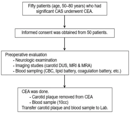
This study was performed prospectively with approval from the Institutional Review Board (2019-0873) of Asan Medical Center. All information pertaining to subjects and all human samples were used in compliance with Korean legislation, and all human participants provided written informed consent. This study employed human carotid plaques and blood samples from patients (age, 50–80 years) who had significant carotid bifurcation stenosis (i.e., ≥70% in asymptomatic patients and ≥50% in symptomatic patients) based on the North American Symptomatic Carotid Endarterectomy Trial criteria) on Doppler ultrasound (DUS) imaging and underwent carotid endarterectomy (CEA) (Figure 1 and Supplementary Figure 1).2 To evaluate the specific serum biomarkers of the patients with significant carotid artery stenosis, we compared the serum biomarkers of patients with significant carotid artery stenosis (n = 50)
with those of normal subjects without carotid artery stenosis or other atherosclerosis risk factors (n = 6). The normal subjects also provided written informed consent. The demographic characteristics, risk factors of interest, clinical characteristics, outcomes, DUS findings of carotid diameter reduction, plaque morphology, and plaque area were recorded for all consecutive patients in an Excel (Microsoft Corp., Redmond, WA, USA) database and analyzed.
Index Procedures and Tissue Sampling
The surgical procedures were performed as previously detailed.20A conventional endarterectomy was performed with patch angioplasty in the standard fashion under general anesthesia and routine carotid shunting. The carotid plaques removed during CEA and the blood samples taken before CEA in 50 patients were analyzed. The internal carotid region was harvested as a disease sample and the common carotid region was considered a normal sample for comparison with the internal region. Postoperatively, the patients were given dual antiplatelet therapy (low dose aspirin plus clopidogrel) with a statin in combination with stringent blood pressure control and close observation in an intensive care unit for at least 24 hours.21All patients were followed up both clinically and by magnetic resonance imaging with angiography or carotid DUS before discharge.
Clinical Outcomes
The clinical outcomes were defined as a fatal or nonfatal stroke or a transient ischemic attack ipsilateral to the CEA during the perioperative period (within 30 days after CEA). Neurologic events were defined as previously detailed.4,20,22
Prediction of miRNAs Targeting ABCA1
The miRNA targets were predicted using a 95% context percentile based on theTargetScan6.2 database (http://www.targetscan.org/vert_71/, accessed March 2018).23
ABCA1 ELISA Analysis
The levels of ABCA1 were analyzed by enzyme-linked immunosorbent assay (ELISA). The concentrations were quantified using the human ABCA1 (Abcam Inc., Cambridge, MA, USA) ELISA kit in strict accordance with the manufacturer’s instructions and detected using a microplate reader (BioTek, Winooski, VT, USA). All reactions were analyzed in triplicate.
Quantitative Real-time Chain Reaction (qRT-PCR)
Total RNA was isolated from the human carotid stenosis arteries using TRIzol (Invitrogen, Carlsbad, CA, USA). Also, miRNA from plasma was extracted using the miRNeasy Serum/Plasma Kit (Qiagen, Hilden, Germany). Reverse transcription was performed with 500 ng of total RNA and Superscript III reverse transcriptase (Invitrogen). Complementary DNA (cDNA) was amplified by PCR. In the case of cDNA synthesis for miRNAs, we used the miR-X miRNA First-Strand Synthesis kit (Takara Bio, Kusatsu, Shiga, Japan). Real-time PCR was performed using SYBR Master Mix (Roche, Basel, Switzerland) and a CFX Connect™ Real-Time System (Bio-Rad, Hercules, CA, USA). The gene expression was normalized to that of human glyceraldehyde 3-phosphatedehydrogenase (GAPDH) and U6 for miRNA expression. The sequences of the primers are shown in Supplementary Table 1. All reactions were performed at least in triplicate.
Immunohistochemistry
To analyze the expression of ABCA1, α-smooth muscle actin (SMA), and CD56 in carotid stenosis artery tissues, we used ABCA1 (Novus, Centennial, CO, USA), α-SMA (Dako, Santa Clara, CA, USA), and CD56 (Abcam, Cambridge, UK) antibodies, respectively. The arterial sections were incubated with 3% H2O2 in methanol to block endogenous peroxidase activity. After antigen retrieval, the slides were incubated with an anti-ABCA1, α-SMA, CD56 antibodies diluted 1:100, 1:400, and 1:100 respectively at 4 °C overnight, and then incubated for 1 hour with a biotinylated secondary anti-rabbit and mouse antibody at room temperature (RT). The sections were incubated with a horseradish peroxidase- conjugated streptavidin-biotin complex (Dako) and 3,3-diaminobenzidine (EnVision Systems, Santa
Clara, CA, USA) was used to visualize the chromatic signals. Images were taken using a digital slide scanner (3DHISTECH Ltd., Budapest, Hungary). Finally, the percentage of positive signals was quantified in all sections by 3DHISTECH Ltd. QuantCenter 2.2 program (3DHISTECH Ltd.).
Cloning
The 3’ untranslated regions (UTRs) of the gene sequences targeted by the miRNAs were investigated by Microrna (http://www.microrna.org/microrna/home.do) and used to design oligonucleotides for the luciferase assay. The region of mRNA 3’ UTR was amplified using PCR. The solution of fragment synthesis was contained 10 × Taq buffer (Solgent Co.), 10 mM dNTP mix (Solgent Co.), and h-Taq (Solgent Co.). The synthesis conditions were an initial denaturation at 95°C for 5 minutes, 35 cycles of denaturation at 95°C for 5 seconds, annealing at 60°C for 30 seconds, extension at 72°C for 1 minute, and a pause at 4°C. The PCR products were purified using a PCR purification kit (Bioneer, Daejeon, Korea). The PCR products were inserted into multiple cloning sites of a pmirGLO vector (Promega, Madison, WI, USA). The linearized pmirGLO vector was mixed with the PCR products and ligase using a ligation protocol. The complete pmirGLO vector was transformed into competent cells and spread onto an ampicillin containing LB plate. After 24 hours of incubation, colonies were selected and cultured in ampicillin containing LB broth. Plasmid DNA was extracted using a Plasmid mini extraction kit (Bioneer). The fidelity of the plasmid DNA sequence was confirmed by DNA sequencing.
Statistical Analysis
All experiments were conducted in duplicate or triplicate. Differences between different regions of SBEM were analyzed with GraphPad Prism software and the statistical methods used for comparison between pairs. Datasets with two groups were analyzed with a nonparametric Student’s t test.
Significance was taken as * p < 0.05.
Figure 1. Diagram of the study design. CAS, carotid artery stenosis; CEA, carotid endarterectomy; CBC, complete blood count; DUS, Doppler ultrasound; MRA, magnetic resonance angiography, MRI, magnetic resonance imaging.
Results
Characteristics of Carotid Artery Stenosis
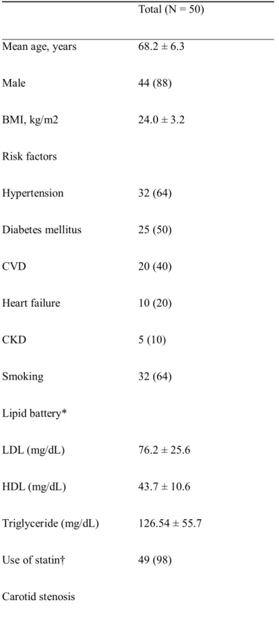
Between July 2019 and April 2020, 50 patients with carotid artery stenosis who underwent CEAs with tissue and blood sampling were consecutively enrolled in this prospective study. The baseline and clinical characteristics of the included patients are presented in Table 1. Their mean age was 68.2 years (median, 67 years; range, 53–79 years), and 88% of the patients were men. The mean degree of carotid
artery stenosis was 75.3% (median, 73.5%; range, 54–90%). Among the study population, 13 symptomatic patients (26%) presented with neurological symptoms (within six months prior to CEA) ipsilateral to the carotid artery stenosis; six had strokes and seven had transient ischemic attacks. All of the 37 asymptomatic patients (74%) presented with carotid artery stenosis with greater than 70%
diameter reduction. Preoperatively, most patients (49/50, 98%) had been taking a statin for different durations. Postoperatively, all patients were given a statin and there were no perioperative neurologic or other cardiovascular events associated with the CEAs.
TABLE 1.Baseline demographic and clinical characteristics of the study population Total (N = 50)
Mean age, years 68.2 ± 6.3
Male 44 (88)
BMI, kg/m2 24.0 ± 3.2
Risk factors
Hypertension 32 (64)
Diabetes mellitus 25 (50)
CVD 20 (40)
Heart failure 10 (20)
CKD 5 (10)
Smoking 32 (64)
Lipid battery*
LDL (mg/dL) 76.2 ± 25.6
HDL (mg/dL) 43.7 ± 10.6
Triglyceride (mg/dL) 126.54 ± 55.7
Use of statin† 49 (98)
Carotid stenosis
Degree of stenosis, %
ICA 75.3 ± 7.9
ECA 27.9 ± 12.5
Symptomatic 13 (26)
Stroke 6 (12)
TIA 7 (14)
The continuous data are expressed as mean ± standard deviation and the categorical data as n (%).
* Preoperative lipid levels, † Preoperative use of statin.
CVD, cardiovascular disease; CKD, chronic kidney disease; ECA, external carotid artery; HDL, high- density lipoprotein; ICA, internal carotid artery; LDL, low-density lipoprotein; TIA, transient ischemic attack.
Selection and Validation of miRNAs Targeting ABCA1
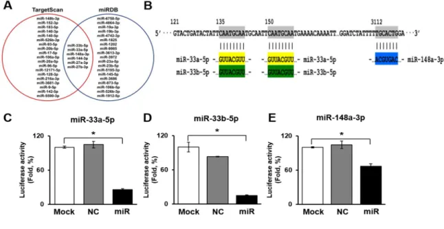
To determine the miRNAs targeting ABCA1, we searched two miRNA search sites (e.g., TargetScan and miRDB). In the sites, six miRNAs (miR-27a-3p, 27b-3p, 33a-5p, 33b-5p, 144-3p, and 148a-3p) were overlapped as target miRNAs of ABCA1. Among the six miRNAs, the target scores of miR-33a- 5p, 33b-5p, and 148a-3p were the highest (Figure 2A). As shown in Figure 2B, miR-33a-5p and 33b- 5p bound to two sites of the ABCA1 3’ untranslated region (UTR) region and miR-148a-3p bound to one site of the ABCA1 3’UTR region (Figure 2B). To determine whether ABCA1 was a direct target of miR-33a-5p, 33b-5p, and 148a-3p, the 3’ UTR reporters for these target genes were cloned into HUVECs. Reporter genes with miRNA mimics were also transfected into HUVECs. Interestingly, when transfected with miRNA mimics, the luciferase activity of the ABCA1 was significantly reduced (Figure 2C-E). These results indicate that these three miRNAs directly targeted ABCA1.
Figure 2. miRNAs targeting ABCA1. (A) Venn diagram of miRNAs targeting ABCA1. (B) The 3’ UTR of the ABCA1 gene containing miRNA binding sites for miR 33a-5p, miR-33b-5p, and miR-148a-3p.
Luciferase assays to validate the interaction of miR-33a-5p (C), miR-33b-5p (D), and miR-148a-3p (E) with ABCA1. Mean ± SD (n = 3 per group), p < 0.05; t-test. NC, negative control; miR, miR transfected group.
Histopathological Transitions in Carotid Stenosis Arteries
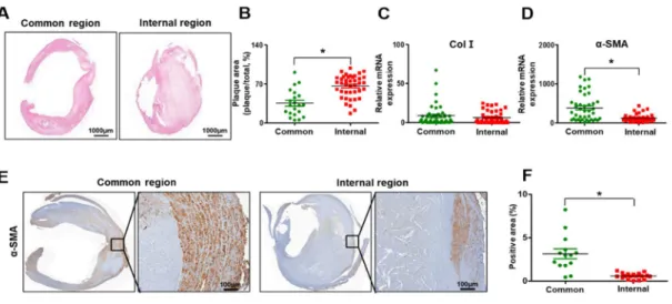
The internal carotid artery exhibited accumulated plaque and shrunken arterial walls compared with the common carotid artery (Figure 3A). To evaluate whether plaque, which is the by-product of inflammation and lipid toxicity, was accumulated in the carotid artery, its area was measured in the common and internal carotid regions. As shown in Figure 3B, the plaque area was significantly increased in the internal carotid artery compared to the common carotid artery (p<0.05, Figure 3B). To evaluate the functioning of the arterial wall in stenotic arteries, Col I and α-SMA were analyzed at the mRNA or protein level. No difference in the mRNA level of Col I was seen between the common and internal carotid artery (Figure 3C). However, the α-SMA expression at the mRNA and protein levels was significantly decreased in the internal carotid artery compared to the common carotid artery due to the decreased thickness of the arterial wall of the internal carotid artery (p<0.05, Figure 3D-F). These data demonstrate that histopathological transitions such as the accumulation of plaque and shrinkage of the arterial wall occur in carotid artery stenosis.
Figure 3. Histopathological transition in the carotid stenosis artery. (A) Hematoxylin and eosin staining in the common region and internal carotid artery. (B) Quantification of plaque area by hematoxylin and eosin staining. mRNA expression of collagen I (C) and alpha-smooth muscle actin (D) by qRT-PCR.
(E) Expression of alpha-smooth muscle actin by immunohistochemical staining. Scale bar: 100μm. (F) Quantification of the alpha-smooth muscle actin positive area by immunohistochemistry staining. Mean
± SD, p < 0.05; t-test. Common, Common carotid artery; Internal, Internal carotid artery; Col I, collagen I; α-SMA, alpha-smooth muscle actin.
Inflammation in Carotid Artery Stenosis
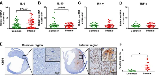
To investigate the degree of the inflammation in the carotid artery, several inflammatory markers (e.g., interleukin-6, IL-6; interferon-gamma, IFN-γ; tumor necrosis factor-alpha, TNF-α) and interleukin-10 (IL-10), which are known as anti-inflammatory factor, were analyzed at the mRNA level by qRT-PCR.
In the carotid arteries, the expression of IL-6 was increased in the internal part versus the common part (p = 0.07, Figure 4A).
The level of IL10 had the slightly decreased tendency in the internal carotid artery compared to the
common carotid artery (p = 0.08, Figure 4B). However, the expression of IFN-γ was similar between the common carotid artery and the internal carotid artery (Figure 4C). Also, a slight increase in TNF-α was seen in the internal carotid artery compared to the common carotid artery although it was not significant (Figure 4D). For histological analysis related to inflammation, the marker of natural killer (NK) cells, CD 56 (neural cell adhesion molecule, NCAM) was analyzed by immunohistochemistry. In the common carotid artery, its expression was barely detected. However, its expression was seen in the peripheral parts of the plaques (Figure 4E). The CD56 positive area in the carotid arteries was increased in the internal carotid artery versus the common carotid artery (p<0.05, Figure 4F). These data indicate that carotid stenosis occurs with inflammation, forming plaques.
Figure 4. Inflammation in carotid artery stenosis. mRNA expression of interleukin-6 (A), interleukin- 10 (B), interferon-gamma (C), and tumor necrosis factor-alpha (D) by qRT-PCR. (E) Expression of CD56 by immunohistochemical staining. Scale bar: 50μm. (F) Quantification of the CD56 positive area.
Mean ± SD, p < 0.05; t-test. Common, common carotid artery; Internal, internal carotid artery; IL-6, interleukin-6; IL-10, interleukin-10; IFN-γ, interferon-gamma; TNF-α, tumor necrosis factor-alpha.
Expression of ABCA1 and its Targeting miRNAs in Carotid Artery Stenosis
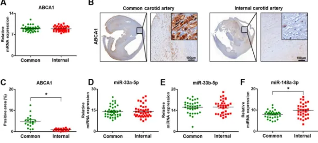
To analyze the expression of ABCA1 in the carotid artery tissues, the mRNA and protein levels were
evaluated by qRT-PCR and immunohistochemistry. The mRNA level of ABCA was not different between the common and internal carotid arteries (Figure 5A). However, the protein expression analyzed by immunohistochemical staining showed a difference between the common and internal regions. ABCA1 was expressed in the cytoplasm of the arterial cells (Figure 5B).
Figure 5. Expression of ABCA1 and its target miRNAs in carotid artery stenosis. (A) mRNA expression of ABCA1 by qRT-PCR. (B) Expression of ABCA1 by immunohistochemical staining. Scale bar:
100μm. (C) Quantification of the ABCA1 positive area by immunohistochemistry staining. Expression of miR-33a-5p (D), miR-33b-5p (E), and miR-148a-3p (F) by qRT-PCR. Mean ± SD, p < 0.05; t-test.
Common, common carotid artery; Internal, internal carotid artery
In particular, ABCA1 was highly expressed in the common carotid artery, whereas its expression was lower in the internal region due to injury by stenosis (p < 0.05, Figure 5C). Also, its target miRNAs, miR-33a-5p, 33b-5p, and 148a-3p, were analyzed by qRT-PCR. In arterial tissues, the expression of miR-33a-5p and 33b-5p was not different, whereas the level of miR-148a-3p was significantly increased (p < 0.05, Figure 5D-F).
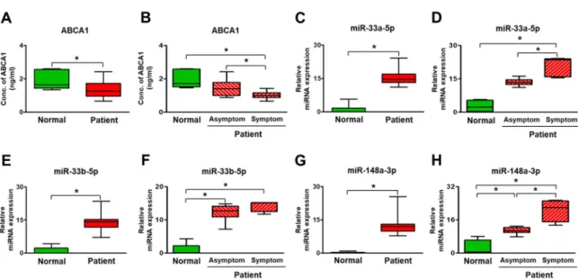
Similar to these analyses, ABCA1 and its target miRNAs were evaluated in plasma. The soluble form
of ABCA1 was significantly decreased in the plasma of stenosis patients compared to the non-stenosis patients (p<0.05, Figure 6A). Also, the expression of the target miRNAs (miR-33a-5p, 33b-5p, and 148a-3p) was significantly increased in the stenosis patients versus the non-stenosis patients showing an inverse correlation between ABCA1 and miRNAs (p<0.05, Figure 6B-D and Figure 7A-C). Further analysis was done between asymptomatic and symptomatic patients to evaluate clinical correlation with ABCA 1 and its target miRNAs. ABCA 1, miR-33a-5a, mi148a-3p show significant difference in plasma between two group (p<0.05, Figure 6B, D, F, and H). These data suggest that ABCA1 was decreased in stenosis, and plaques were formed while its target miRNAs were increased in patient plasma rather than in arterial tissues. Also, this study shows that the increased expression of miRNAs was correlated with carotid stenosis severity and their symptom.
Figure 6. Expression of ABCA1 and its target miRNAs in the plasma of carotid artery stenosis patients.
(A and B) Expression of soluble ABCA1 by ELISA. Expression of miR-33a-5p (C and D), miR-33b- 5p (E and F), and miR-148a-3p (G and H) by qRT-PCR. Mean ± SD, p < 0.05; t-test or One-way ANOVA. Normal, plasma of non-carotid artery stenosis patient; Patient, plasma of carotid artery stenosis patient
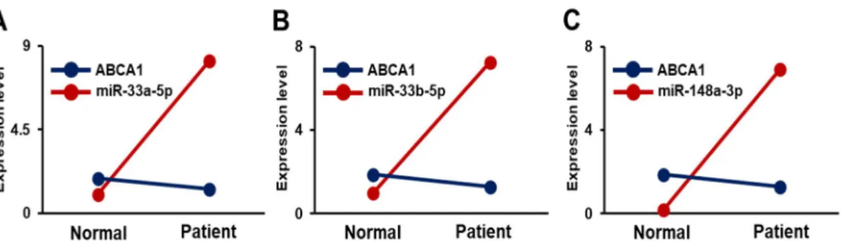
Figure 7. Correlation of ABCA1 and its target miRNAs in the plasma of carotid artery stenosis patients.
Correlation between soluble ABCA1 and (A) miR-33a-5p, (B) miR-33b-5p, and (C) miR-148a-3p.
Normal, plasma of non-carotid artery stenosis patient; Patient, plasma of carotid artery stenosis patient.
Discussion
Cholesterol metabolism is elaborately regulated at the cellular level and homeostasis is essential for this process.24 One of several proteins related to cholesterol homeostasis is ABCA1, a transmembrane protein extensively expressed in many tissues.25ABCA1 functions in the first and crucial step of HDL- C formation.26 Considering that low HDL-C levels are an independent risk factor for coronary heart disease (CHD), genetic variants that are known to increase HDL-C levels would be expected to decrease CHD risk, whereas variants associated with lower HDL-C levels would increase CHD risk.27However, high HDL-C levels are not always protective of CHD, and Mendelian randomization studies suggest that there is no causal inverse relationship between HDL-C levels and the risk of CHD.28,29Deregulation of miRNAs function may be responsible for several pathophysiological disorders, especially in the cardiovascular system.30,31 However, the role of miRNAs in carotid-related stroke remains mostly unknown. Previous studies suggest a potential regulatory function for several miRNAs on dysfunctional endothelium, contributing to the evolution of carotid plaque toward growth, instability, and rupture.
Maitrias et al. evaluated the expression of seven selected miRNAs in human carotid plaques from a small group of patients.32Studies based on larger sample sizes are required to determine the potential use of miRNA as biomarkers in carotid stenosis. In terms of biomarkers, we analyzed miRNAs which are related to carotid plaque and peripheral blood system among patient with carotid stenosis. In our study, the expression and localization of ABCA1 were decreased in patients with atherosclerosis and carotid artery stenosis, as well as their plasma (p < 0.05, Figure 5B-C and 6A).
Generally, miRNA expression shows unique patterns based on developmental stages, specific diseases, specific organs, events, and cancer types. It also regulates the expression of most genes associated with HDL metabolism, including ABCA1. This implies that miRNAs regulate HDL biogenesis, cellular cholesterol efflux and HDL-C hepatic uptake, thereby controlling all steps involved in reverse cholesterol transport (RCT).33A previous study showed that miRNA-33, which is located within the gene encoding sterol-regulatory element-binding factor-2 (SREBF-2), modulates the expression of
genes involved in cellular cholesterol transport and miRNA-33 appears to regulate both HDL biogenesis in the liver and cellular cholesterol efflux.34 Cifani et al. evaluated the expression of miRNA-33 in peripheral blood and carotid specimens of patients with symptomatic and asymptomatic carotid artery stenosis. This study showed a different expression of miRNA-33 in peripheral blood and carotid specimen between symptomatic and asymptomatic patients. In the asymptomatic group, increases in miRNA-33 expression were seen in the peripheral blood and arterial specimens.35 In our data, its expression was evaluated in the plasma and carotid artery (common and internal region) tissues of patients. The expression was significantly increased in plasma of stenosis patients compared to normal patients (p < 0.05, Figure 6B-C). However, in the common and internal carotid artery tissues, its expression was not different (Figure 5D-F).
Considering the role of ABCA1 and miRNA-33 in lipid metabolism, it is not surprising that such miRNA was involved in severe atherosclerotic carotid artery disease.36However, the role of miRNA- 33 in atherosclerosis is complex, and its effects on inflammatory mediators seem to be paradoxical. The expression of miRNA-33 has been inversely correlated to pro-inflammatory cytokines.37Therefore, the inhibition of miRNA-33 promoted macrophage differentiation and pro-inflammatory cytokine release,38 suggesting that miRNA-33 expression can exert pleiotropic action in the different stages of atherosclerotic disease by affecting other risk factors such as obesity, glucose metabolism, and inflammation.39This may explain our results. Dyslipidemia represents a key event in plaque formation and growth whereas inflammation works during all of the phases of atherosclerosis and represents the main factor leading to plaque instability.40
The proinflammatory factors, protein mediators, and other factors such as free fatty acids will aggravate the damage of endothelial cells and then induce or aggravate the process of atherosclerosis.41,42NF-κB, associated with innate and adaptive immunity as well as in inflammatory diseases, regulates the proinflammatory cytokines and NO release.43 Studies have confirmed that NF-κB regulates the development of atherosclerotic disease.44
The use of statin and its effects on the lipid profile are known to be associated with a significant decrease of miRNA-33 and -148a expression and, in recent years, clinically, statin has been used extensively in patients with atherosclerosis risk factors.45,46In our study, most patients (49/50) had been taking statin preoperatively for different durations, and all patients were given a statin after CEA. This can impose bias in our study and is an important limitation that should be acknowledged. Also, our current findings were obtained at a single center, resulting in a small sample size, which limits the overall relevance of our results and some clinical information in normal subjects was not available from the medical records.
The study cohort was entirely Asian; therefore, these results may not be generalizable to other ethnic groups. Further studies are needed for more robust clinical correlation evidence of target mi-RNAs.
Conclusion
In carotid artery stenosis, histopathological transitions such as the accumulation of plaque and shrinkage of the arterial wall occurred as well as inflammatory reactions. ABCA1 was decreased by stenosis and the formation of plaques while its target miRNAs (miRNA-33a-5p, 33b-5p, and 148a-3p) were significantly increased in patient plasma but not in arterial tissues. Therefore, we suggest the potential of miRNA-33a-5p, 33b-5p, and 148a-3p, which directly target ABCA1, as biomarkers of carotid artery stenosis.
References
1. Rundek, T.; Arif, H.; Boden-Albala, B.; Elkind, M.S.; Paik, M.C.; Sacco, R.L. Carotid plaque, a subclinical precursor of vascular events: the Northern Manhattan Study. Neurology 2008, 70, 1200- 1207, doi:10.1212/01.wnl.0000303969.63165.34.
2. Spence, J.D.; Eliasziw, M.; DiCicco, M.; Hackam, D.G.; Galil, R.; Lohmann, T. Carotid plaque area: a tool for targeting and evaluating vascular preventive therapy. Stroke 2002, 33, 2916-2922, doi:10.1161/01.str.0000042207.16156.b9.
3. den Hartog, A.G.; Achterberg, S.; Moll, F.L.; Kappelle, L.J.; Visseren, F.L.; van der Graaf, Y.;
Algra, A.; de Borst, G.J.; Group, S.S. Asymptomatic carotid artery stenosis and the risk of ischemic stroke according to subtype in patients with clinical manifest arterial disease. Stroke 2013, 44, 1002- 1007, doi:10.1161/STROKEAHA.111.669267.
4. Noh, M.; Kwon, H.; Jung, C.H.; Kwon, S.U.; Kim, M.S.; Lee, W.J.; Park, J.Y.; Han, Y.; Kim, H.; Kwon, T.W.; et al. Impact of diabetes duration and degree of carotid artery stenosis on major adverse cardiovascular events: a single-center, retrospective, observational cohort study. Cardiovasc Diabetol 2017, 16, 74, doi:10.1186/s12933-017-0556-0.
5. Saba, L.; Sanfilippo, R.; Sannia, S.; Anzidei, M.; Montisci, R.; Mallarini, G.; Suri, J.S.
Association between carotid artery plaque volume, composition, and ulceration: a retrospective assessment with MDCT. AJR Am J Roentgenol 2012, 199, 151-156, doi:10.2214/AJR.11.6955.
6. Hirt, L.S. Progression rate and ipsilateral neurological events in asymptomatic carotid stenosis.
Stroke 2014, 45, 702-706, doi:10.1161/STROKEAHA.111.613711.
7. Kwee, R.M.; van Oostenbrugge, R.J.; Hofstra, L.; Teule, G.J.; van Engelshoven, J.M.; Mess, W.H.; Kooi, M.E. Identifying vulnerable carotid plaques by noninvasive imaging. Neurology 2008, 70, 2401-2409, doi:10.1212/01.wnl.0000314697.76580.cb.
8. O'Brien, J.; Hayder, H.; Zayed, Y.; Peng, C. Overview of MicroRNA Biogenesis, Mechanisms of Actions, and Circulation. Front Endocrinol (Lausanne) 2018, 9, 402, doi:10.3389/fendo.2018.00402.
9. Kweon, M.; Kim, J.Y.; Jun, J.H.; Kim, G.J. Research Trends in the Efficacy of Stem Cell Therapy for Hepatic Diseases Based on MicroRNA Profiling. Int J Mol Sci 2020, 22, doi:10.3390/ijms22010239.
10. Catalanotto, C.; Cogoni, C.; Zardo, G. MicroRNA in Control of Gene Expression: An Overview of Nuclear Functions. Int J Mol Sci 2016, 17, doi:10.3390/ijms17101712.
11. Cannell, I.G.; Kong, Y.W.; Bushell, M. How do microRNAs regulate gene expression?
Biochem Soc Trans 2008, 36, 1224-1231, doi:10.1042/BST0361224.
12. Zhang, J.; Li, S.; Li, L.; Li, M.; Guo, C.; Yao, J.; Mi, S. Exosome and exosomal microRNA:
trafficking, sorting, and function. Genomics Proteomics Bioinformatics 2015, 13, 17-24, doi:10.1016/j.gpb.2015.02.001.
13. Weber, J.A.; Baxter, D.H.; Zhang, S.; Huang, D.Y.; Huang, K.H.; Lee, M.J.; Galas, D.J.; Wang, K. The microRNA spectrum in 12 body fluids. Clin Chem 2010, 56, 1733-1741, doi:10.1373/clinchem.2010.147405.
14. Wang, J.; Chen, J.; Sen, S. MicroRNA as Biomarkers and Diagnostics. J Cell Physiol 2016, 231, 25-30, doi:10.1002/jcp.25056.
15. Hansson, G.K.; Robertson, A.K.; Soderberg-Naucler, C. Inflammation and atherosclerosis.
Annu Rev Pathol 2006, 1, 297-329, doi:10.1146/annurev.pathol.1.110304.100100.
16. Badimon, L.; Vilahur, G. LDL-cholesterol versus HDL-cholesterol in the atherosclerotic plaque: inflammatory resolution versus thrombotic chaos. Ann N Y Acad Sci 2012, 1254, 18-32, doi:10.1111/j.1749-6632.2012.06480.x.
17. Brunham, L.R.; Kruit, J.K.; Pape, T.D.; Timmins, J.M.; Reuwer, A.Q.; Vasanji, Z.; Marsh, B.J.;
Rodrigues, B.; Johnson, J.D.; Parks, J.S.; et al. Beta-cell ABCA1 influences insulin secretion, glucose homeostasis and response to thiazolidinedione treatment. Nat Med 2007, 13, 340-347, doi:10.1038/nm1546.
18. Liu, Y.; Tang, C. Regulation of ABCA1 functions by signaling pathways. Biochim Biophys Acta 2012, 1821, 522-529, doi:10.1016/j.bbalip.2011.08.015.
19. Singaraja, R.R.; Fievet, C.; Castro, G.; James, E.R.; Hennuyer, N.; Clee, S.M.; Bissada, N.;
Choy, J.C.; Fruchart, J.C.; McManus, B.M.; et al. Increased ABCA1 activity protects against atherosclerosis. J Clin Invest 2002, 110, 35-42, doi:10.1172/JCI15748.
20. Kwon, H.; Moon, D.H.; Han, Y.; Lee, J.Y.; Kwon, S.U.; Kang, D.W.; Choo, S.J.; Kwon, T.W.;
Kim, M.J.; Cho, Y.P. Impact of subclinical coronary artery disease on the clinical outcomes of carotid endarterectomy. J Neurosurg 2017, 126, 1560-1565, doi:10.3171/2016.3.JNS16287.
21. Barkat, M.; Hajibandeh, S.; Hajibandeh, S.; Torella, F.; Antoniou, G.A. Systematic Review and Meta-analysis of Dual Versus Single Antiplatelet Therapy in Carotid Interventions. Eur J Vasc Endovasc Surg 2017, 53, 53-67, doi:10.1016/j.ejvs.2016.10.011.
22. Kwon, H.; Kim, H.K.; Kwon, S.U.; Lee, S.W.; Kim, M.J.; Park, J.W.; Noh, M.; Han, Y.; Kwon, T.W.; Cho, Y.P. Risk of major adverse cardiovascular events in subjects with asymptomatic mild carotid artery stenosis. Sci Rep 2018, 8, 4700, doi:10.1038/s41598-018-23125-8.
23. Betel, D.; Wilson, M.; Gabow, A.; Marks, D.S.; Sander, C. The microRNA.org resource:
targets and expression. Nucleic Acids Res 2008, 36, D149-153, doi:10.1093/nar/gkm995.
24. Goedeke, L.; Fernandez-Hernando, C. Regulation of cholesterol homeostasis. Cell Mol Life Sci 2012, 69, 915-930, doi:10.1007/s00018-011-0857-5.
25. Oram, J.F. Molecular basis of cholesterol homeostasis: lessons from Tangier disease and ABCA1. Trends Mol Med 2002, 8, 168-173, doi:10.1016/s1471-4914(02)02289-x.
26. Jacobo-Albavera, L.; Dominguez-Perez, M.; Medina-Leyte, D.J.; Gonzalez-Garrido, A.;
Villarreal-Molina, T. The Role of the ATP-Binding Cassette A1 (ABCA1) in Human Disease. Int J Mol Sci 2021, 22, doi:10.3390/ijms22041593.
27. Wilkins, J.T.; Ning, H.; Stone, N.J.; Criqui, M.H.; Zhao, L.; Greenland, P.; Lloyd-Jones, D.M.
Coronary heart disease risks associated with high levels of HDL cholesterol. J Am Heart Assoc 2014, 3, e000519, doi:10.1161/JAHA.113.000519.
28. Dumitrescu, L.; Carty, C.L.; Taylor, K.; Schumacher, F.R.; Hindorff, L.A.; Ambite, J.L.;
Anderson, G.; Best, L.G.; Brown-Gentry, K.; Buzkova, P.; et al. Genetic determinants of lipid traits in diverse populations from the population architecture using genomics and epidemiology (PAGE) study.
PLoS Genet 2011, 7, e1002138, doi:10.1371/journal.pgen.1002138.
29. Voight, B.F.; Peloso, G.M.; Orho-Melander, M.; Frikke-Schmidt, R.; Barbalic, M.; Jensen, M.K.; Hindy, G.; Holm, H.; Ding, E.L.; Johnson, T.; et al. Plasma HDL cholesterol and risk of myocardial infarction: a mendelian randomisation study. Lancet 2012, 380, 572-580, doi:10.1016/S0140-6736(12)60312-2.
30. van Rooij, E.; Olson, E.N. MicroRNAs: powerful new regulators of heart disease and provocative therapeutic targets. J Clin Invest 2007, 117, 2369-2376, doi:10.1172/JCI33099.
31. Fichtlscherer, S.; De Rosa, S.; Fox, H.; Schwietz, T.; Fischer, A.; Liebetrau, C.; Weber, M.;
Hamm, C.W.; Roxe, T.; Muller-Ardogan, M.; et al. Circulating microRNAs in patients with coronary artery disease. Circ Res 2010, 107, 677-684, doi:10.1161/CIRCRESAHA.109.215566.
32. Maitrias, P.; Metzinger-Le Meuth, V.; Massy, Z.A.; M'Baya-Moutoula, E.; Reix, T.; Caus, T.;
Metzinger, L. MicroRNA deregulation in symptomatic carotid plaque. J Vasc Surg 2015, 62, 1245-1250
e1241, doi:10.1016/j.jvs.2015.06.136.
33. Canfran-Duque, A.; Ramirez, C.M.; Goedeke, L.; Lin, C.S.; Fernandez-Hernando, C.
microRNAs and HDL life cycle. Cardiovasc Res 2014, 103, 414-422, doi:10.1093/cvr/cvu140.
34. Rayner, K.J.; Suarez, Y.; Davalos, A.; Parathath, S.; Fitzgerald, M.L.; Tamehiro, N.; Fisher, E.A.; Moore, K.J.; Fernandez-Hernando, C. MiR-33 contributes to the regulation of cholesterol homeostasis. Science 2010, 328, 1570-1573, doi:10.1126/science.1189862.
35. Del Porto, F.; Cifani, N.; Proietta, M.; Dezi, T.; Tritapepe, L.; Raffa, S.; Micaloni, A.; Taurino, M. Lag3(+) regulatory T lymphocytes in critical carotid artery stenosis. Clin Exp Med 2019, 19, 463- 468, doi:10.1007/s10238-019-00570-x.
36. Parahuleva, M.S.; Lipps, C.; Parviz, B.; Holschermann, H.; Schieffer, B.; Schulz, R.; Euler, G.
MicroRNA expression profile of human advanced coronary atherosclerotic plaques. Sci Rep 2018, 8, 7823, doi:10.1038/s41598-018-25690-4.
37. Ouimet, M.; Ediriweera, H.N.; Gundra, U.M.; Sheedy, F.J.; Ramkhelawon, B.; Hutchison, S.B.;
Rinehold, K.; van Solingen, C.; Fullerton, M.D.; Cecchini, K.; et al. MicroRNA-33-dependent regulation of macrophage metabolism directs immune cell polarization in atherosclerosis. J Clin Invest 2015, 125, 4334-4348, doi:10.1172/JCI81676.
38. Koyama, S.; Horie, T.; Nishino, T.; Baba, O.; Sowa, N.; Miyasaka, Y.; Kuwabara, Y.; Nakao, T.; Nishiga, M.; Nishi, H.; et al. Identification of Differential Roles of MicroRNA-33a and -33b During Atherosclerosis Progression With Genetically Modified Mice. J Am Heart Assoc 2019, 8, e012609, doi:10.1161/JAHA.119.012609.
39. Ono, K. Functions of microRNA-33a/b and microRNA therapeutics. J Cardiol 2016, 67, 28- 33, doi:10.1016/j.jjcc.2015.10.017.
40. Rayner, K.J.; Moore, K.J. The plaque "micro" environment: microRNAs control the risk and the development of atherosclerosis. Curr Atheroscler Rep 2012, 14, 413-421, doi:10.1007/s11883-012- 0272-x.
41. Numata, T.; Takahashi, K.; Inoue, R. "TRP inflammation" relationship in cardiovascular system. Semin Immunopathol 2016, 38, 339-356, doi:10.1007/s00281-015-0536-y.
42. Lamers, D.; Schlich, R.; Greulich, S.; Sasson, S.; Sell, H.; Eckel, J. Oleic acid and adipokines synergize in inducing proliferation and inflammatory signalling in human vascular smooth muscle cells.
J Cell Mol Med 2011, 15, 1177-1188, doi:10.1111/j.1582-4934.2010.01099.x.
43. Hayden, M.S.; Ghosh, S. NF-kappaB in immunobiology. Cell Res 2011, 21, 223-244, doi:10.1038/cr.2011.13.
44. Niu, N.; Xu, S.; Xu, Y.; Little, P.J.; Jin, Z.G. Targeting Mechanosensitive Transcription Factors in Atherosclerosis. Trends Pharmacol Sci 2019, 40, 253-266, doi:10.1016/j.tips.2019.02.004.
45. Ouimet, M.; Ediriweera, H.; Afonso, M.S.; Ramkhelawon, B.; Singaravelu, R.; Liao, X.;
Bandler, R.C.; Rahman, K.; Fisher, E.A.; Rayner, K.J.; et al. microRNA-33 Regulates Macrophage Autophagy in Atherosclerosis. Arterioscler Thromb Vasc Biol 2017, 37, 1058-1067, doi:10.1161/ATVBAHA.116.308916.
46. Rotllan, N.; Price, N.; Pati, P.; Goedeke, L.; Fernandez-Hernando, C. microRNAs in lipoprotein metabolism and cardiometabolic disorders. Atherosclerosis 2016, 246, 352-360, doi:10.1016/j.atherosclerosis.2016.01.025.
국문요약
배경
경동맥협착증은 심혈관계 질환의 위험을 증가시키는 것과 연관된 역동적인 과정의 하나 이다. 그러나, 뇌혈관질환의 발생률 증가와 관련된 고위험 경동맥 플라크를 식별하고 정 량화 하는 데에 유용한 바이오마커에 대한 지식은 충부하지 않은 상황이다. 따라서 이 연구의 목적은 ABCA1의 발현을 평가하고 인간에서의 경동맥 협착이 발생한 동맥에서 표적 miRNA 후보를 검증하여 바이오마커로서의 잠재력을 확인하는 것이다.
방법
2019년 7월부터 2020년 4월까지 경동맥내막절제술의 시행 받은 환자 50명이 이 전향 적 연구에 등록이 되었고, 각각에서 조직 및 혈액 검체를 추출하였다. 인간의 경동맥 협 착이 발생한 동맥 조직 및 혈장에서 ABCA1 및 표적 miRNA(miRNA-33a-5p, 33b-5p 및 148a-3p)의 발현을 정량적 실시간 중합효소 연쇄반응과 면역조직화학염색법 등으로 평가하였다. 중증 경동맥 협착이 있는 환자의 특정 혈청 바이오마커를 평가하기 위해서 중증 경동맥 협착이 있는 환자 (50명)의 혈청 바이오마커를 경동맥 협착 또는 기타 죽상 경화증의 위험 인자가 없는 정상 대조군 (6명)의 혈청 바이오 마커와 비교 분석하였다.
결과
ABCA1은 총경동맥에서 높게 발현된 반면, 내경동맥에서는 협착에 의한 손상으로 인해 발현이 낮았다(p<0.05). 동맥 조직에서 miR-33a-5p와 33b-5p의 발현은 차이가 없었으
나 miR-148a-3p의 발현은 유의하게 증가하였다(p<0.05). ABCA1의 발현은 경동맥 협 착증 환자의 혈장에서 유의하게 감소하였으나 동맥 조직에서는 차이가 없었다(p<0.05).
그러나 정상 대조군보다 경동맥 협착증 환자에서 훨씬 더 많은 표적 miRNA가 확인되었 다(p<0.05).
결론
경동맥 협착증에서는 염증 반응과 함께 플라크 축적 및 동맥벽 수축과 같은 조직병리학 적 전이가 발생한 것을 연구를 통해 확인할 수 있었다. ABCA1은 경동맥의 협착 및 플라 크 형성에 의해 감소한 반면, 표적 miRNA(miRNA-33a-5p, 33b-5p 및 148a-3p)는 환 자 혈장에서 유의하게 증가했지만 경동맥 조직에서는 증가하지 않았다. 따라서 miRNA- 33a-5p, 33b-5p 및 148a-3p는 경동맥 협착증에서 ABCA1을 직접 표적화 할 수 있는 잠재적인 바이오마커이다.