의학석사 학위논문
Is multiparametric MRI really helpful to predict upgrading and upstaging in active
surveillance?
능동적 감시 대상의 전립선암 환자에서 암의 병기 상승 및 등급 상승을 예측하는 도구로서 다중 자기 공명 영
상의 가치
울 산 대 학 교 대 학 원 의 학 과
김 휘 우
[UCI]I804:48009-200000177342 [UCI]I804:48009-200000177342 [UCI]I804:48009-200000177342
능동적 감시 대상의 전립선암 환자에서 암의 병기 상승 및 등급 상승을 예측하는 도구로서 다중 자기 공명 영
상의 가치
지도교수
안 한 종
이 논문을 의학석사 학위논문으로 제출함
2018 년 12 월
울 산 대 학 교 대 학 원 의 학 과
김 휘 우
김휘우의 의학석사학위 논문을 인준함
심사위원장 홍 준 혁 ( 인 ) 심사위원 정 인 갑 ( 인 ) 심사위원 안 한 종 ( 인 )
울 산 대 학 교 대 학 원
2018 년 12 월
1
ABSTRACT
Background
We aimed to evaluate whether multiparametric MRI using Prostate Imaging Reporting and
Data System (PI-RADS) is helpful to predict upgrading and upstaging in men with
prostate cancers eligible for active surveillance.
Materials and methods
From January 2014 to December 2017, a total of 223 patients eligible for PRIAS active
surveillance criteria (biopsy Gleason score ≤ 6, PSA ≤ 10, PSA density < 0.2, clinical
T1c/T2, and the positive cores ≤ 2) were analyzed. All patients underwent multiparametric
MRI with PI-RADS scoring and radical prostatectomy. PI-RADS scoring was performed
divided into 12 zones (right/left, anterior/posterior, and base/mid/apex). 4 or 5 PI-RADS
score was regarded as clinically significant cancer is likely to be present
Results
Of 223 patients, 25 (11.2%) patients had upstaging (24 patients to T3a, 1 to T3b) and 108
ii
(48.4%) patients had upgrading (104 patients to GS 7 and 4 to GS 8-10). Patients with
upgrading and upstaging tumors had older age, smaller prostate volume, higher PSA
density, higher percent of positive cores, and larger tumor volume, compared to others. A
total of 2676 sites from 223 patients were evaluated to identify diagnostic accuracy of PI-
RADS scoring using whole mount section analysis. Overall sensitivity and specificity of
PI-RADS were 36.8% and 86.5%, respectively. In multivariate analysis, PI-RADS score
4-5 lesions ≥ 2 was the independent predictor for upgrading/upstaging in patients eligible
for active surveillance.
Conclusions
In this study, multiparametric MRI reported on PI-RADS scoring system has low
sensitivity then previously reported, but it can be useful to detect clinically significant
prostate cancer among patients on AS when there are 2 lesion or more PI-RADS 4-5 score
were seen.
Keywords:prostate cancer, active surveillance, multiparametric MRI, PI-RADS
iii
CONTENTS
English Abstract ... i
Contents...iv
List of tables and figures...v
List of Abbreviations...vi
Introduction...1
Material and Methods...4
Results...7
Discussion ...14
Conclusion ...20
References...21
국문요약...24
iv
LIST OF FIGURES
Table 1.Clinical and pathologic characteristics eligible for PRIAS criteria for AS
Table 2.Clinical and pathologic characteristics subdivided by pathologic findings
Table 3. Diagnostic accuracy of PI-RADS system and MR characteristics subdivided by
pathologic findings
Table 4.Univariate and multivariate analysis for prediction of upgrading and upstaging
v
LIST OF ABBREVIATIONS
PSAprostate specific antigen
PI-RADSProstate Imaging Reporting and Data System
mpMRImulti-parametric magnetic resonance imaging
PPVpositive predictive value
NPVnegative predictive value
vi
1
INTRODUCTION
Active surveillance has risen as another option for replace radical treatment of low-risk
prostate cancer over past decade[1]. The new National Comprehensive Cancer Network
(NCCN) guideline recommends active surveillance for very low-risk and low-risk prostate
cancer groups who have 10 years or more life expectancy. The most definitive method for
diagnosing prostate cancer is histological confirmation through needle biopsy. However,
previous studies in this field have reported a significant discrepancy between the needle
biopsy and radical prostatectomy specimens[2]. So, it is important to identify clinically
significant cancer among low-risk prostate cancer.
Currently, multi-parametric magnetic resonance imaging(mpMRI) has been put to
practical use to detect lymph nodes involvement and to guide prostate needle biopsy to
discover significant high-risk cancer inside the prostate which is easy to can be missed
with standard prostate biopsy[3,7]. But there are still controversies that MR imaging can
find clinically significant prostate cancer, although it is widely used for the staging of
prostate cancer.
2
Recent studies say that we can use magnetic-resonance imaging to identify clinically
significant prostate cancer in patients on active surveillance[4], Because the ability to
detect clinically significant prostate cancer has increased because of the improvement on
interpretation and standardization of multi-parametric magnetic resonance imaging over
past years[5,6]. Therefore, now it is established that using magnetic resonance imaging for
risk assessment and magnetic resonance imaging-targeted biopsy is superior to standard
risk assessment with prostate specific antigen and standard trans-rectal prostate biopsy[7].
Multi-parametric magnetic resonance imaging can be used for work-up with prostate-
specific antigen(PSA), clinical staging and can provide valuable information for risk
stratification of patients with localized prostate cancer[8].
In 2012, European Society of Urogenital Radiology (ESUR) designed The Prostate
Imaging Reporting and Data System(PI-RADS) scoring for standardize interpretation of
multiparametric magnetic resonance imaging of prostate[9]. PI-RADS system is regarded
as the method to differentiate prostate cancer from normal prostate tissue. The higher the
score put, the more possibility of prostate cancer present[10]. PI-RADS score is helping
3
clinicians to select the prostate cancer patients who is available for active surveillance,
standardizing the reporting or prostate multiparametric magnetic resonance imaging[11].
In this study, we tried to clarify the ability of multiparametric prostate magnetic
resonance imaging and PI-RADS scoring whether they can predict the cancer upstaging or
upgrading after radical prostatectomy.
4
MATERIALS AND METHODS
Medical records of patients who underwent radical prostatectomy at our center
from January 2014 to December 2017 for prostate cancer by 5 surgeons were
reviewed retrospectively. Accordingly, 223 patients satisfying the Prostate Cancer
Research International Active Surveillance(PRIAS) criteria (biopsy Gleason
score(GS) ≤ 6, PSA ≤ 10, PSA density < 0.2, clinical T1c/T2, and the positive
cores ≤ 2) were included in the study. Patients who received androgen-deprivation
therapy before radical prostatectomy were excluded from the study. We reviewed
basal demographics, and pathologic findings, and interpretation of pre-operation
multiparametric MRI of prostate.
All patients on analysis were taken multiparametric MRI of prostate after
confirmed diagnosis by standard transrectal-ultrasonography guided prostate
needle biopsy and before radical prostatectomy. All multiparametric MRI was
performed with at least 3 sequences, including T2-weighted sequences [T2WI],
5
diffusion-weighted imaging [DWI], dynamic contrast-enhanced imaging [DCE]. 3
Radiologists interpreted MRI findings on PI-RADS version 2 system for 12
sections of prostate and 12 sections of extraprostatic extension. 4 or 5 PI-RADS
score was regarded as clinically significant cancer is likely to be present, and PI-
RADS score less than or equal to 3 was regarded as clinically significant cancer is
unlikely to be present.
We defined cancer upgrading as definitive Gleason score ≥ 7, and cancer
upstaging as pathological staging ≥ pathologic stage T3a. We divided patients in
two groups : Some has pathologic upstaging or upgrading, Others has not. Each
group was analyzed by the difference of clinical and pathologic characteristics, and
whether multiparametric MRI with PI-RADS scoring and pathologic results were
well correlating. Location of index tumor was also analyzed, divided as
anterior/posterior and base/midgland/apex.
All 2,676 PI-RADS scored sections on MRI were evaluated to identify diagnostic
6
accuracy of PI-RADS scoring using whole mount section analysis. MRI
characteristics for each group were examined. Comparison of pathologic findings
on 12 sections and PI-RADS scored section was done for discover pathologic and
imaging correlation. Pathologic results also divided in 12 sections same as PI-
RADS system. We analyzed pathologic results whether a section has tumor or not,
regardless of size. Overall section review, and Separative review for anterior and
posterior section were done. Sensitivity, specificity, positive predictive value(PPV),
negative predictive value(NPV) was considered for measure diagnostic accuracy
of PI-RADS scoring system.
We sorted risk factors for cancer upgrading and upstaging that considered
different by two groups, and calculated odds ratio of these risk factors for
upstaging and upgrading using logistic regression analysis. We analyzed factors by
both univariate and multivariate method. We used IBM SPSS Statistics software
version 25.0 to perform statistical analysis. Results were regarded as significant
when the p-value is under 0.05.
7
RESULTS
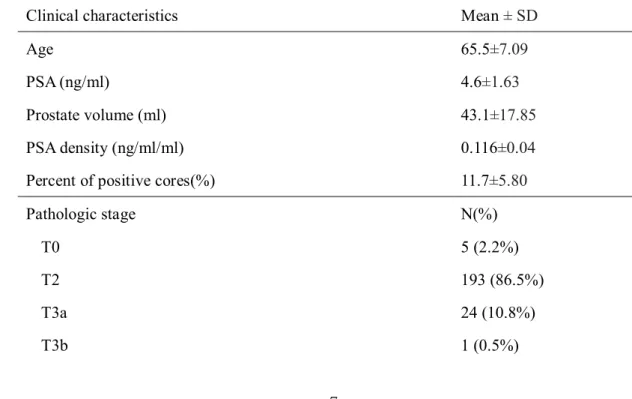
223 people who met the PRIAS criteria were analyzed. The clinical and
pathologic characteristics of these patients are reported in table 1. The mean-age
was 65.5±7.09, mean PSA was 4.6±1.63ng/dl, mean prostate volume was
43.1±17.85cc, mean PSA density was 0.116±0.04 ng/ml/cc, and mean percentage
of positive core was 11.7±5.80%.
Table 1. Clinical and pathologic characteristics eligible for PRIAS criteria for AS
(N=223)
Clinical characteristics Mean ± SD
Age 65.5±7.09
PSA (ng/ml) 4.6±1.63
Prostate volume (ml) 43.1±17.85
PSA density (ng/ml/ml) 0.116±0.04
Percent of positive cores(%) 11.7±5.80
Pathologic stage N(%)
T0 5 (2.2%)
T2 193 (86.5%)
T3a 24 (10.8%)
T3b 1 (0.5%)
8
N1 0
Pathologic GS N(%)
0 5 (2.2%)
6 110 (49.3%)
3+4 82 (36.7%)
4+3 22 (9.8%)
8 3 (1.3%)
9 1 (0.5%)
Tumor volume percentage 4.3%
25 of the 223 patients had T-stage elevation on post-operation pathologic
examination, 24 as T3a(tumor with extracapsular extension), and 1 as T3b(tumor
invading seminal vesicle). 108 of the 223 patients has elevated Gleason score on
pathology, 104 as Gleason score 7(both 3+4 and 4+3), 4 as Gleason score more
than 7. Mean tumor-volume percentage were 4.3%.
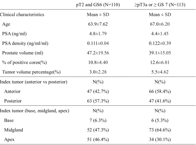
When patients were classified in 2 groups depending on whether up-staging or
up-grading occurred on pathologic finding or not, 110 patients has no upstaging or
upgrading, and other 113 patients has upstaging or upgrading. The clinical and
pathologic characteristics of each group were reported on Table 2. The group with
upstaging or upgrading had relatively older age(63.9±7.62 vs. 67.0±6.20), smaller
9
prostate(47.2±19.56cc vs. 39.1±15.05cc), higher PSA density(0.111±0.04ng/ml/cc
vs. 0.122±0.39ng/ml/cc), more positive biopsy core percentage(10.8±4.40% vs.
12.6±6.81%), higher tumor-volume percentage(3.0±2.28 vs. 5.5±4.62%)then other
group. The PSA has no significant difference in two groups. Also, the ratio of
index tumor on anterior prostate was higher(42.7% vs. 58.4%) in group with up-
staging or up-grading.
Table 2. Clinical and pathologic characteristics subdivided by pathologic findings
pT2 and GS6 (N=110) ≥pT3a or ≥ GS 7 (N=113)
Clinical characteristics Mean ± SD Mean ± SD
Age 63.9±7.62 67.0±6.20
PSA (ng/ml) 4.8±1.79 4.4±1.45
PSA density (ng/ml/ml) 0.111±0.04 0.122±0.39
Prostate volume (ml) 47.2±19.56 39.1±15.05
% of positive cores(%) 10.8±4.40 12.6±6.81
Tumor volume percentage(%) 3.0±2.28 5.5±4.62
Index tumor (anterior vs posterior) N(%) N(%)
Anterior 47 (42.7%) 66 (58.4%)
Posterior 63 (57.3%) 47 (41.6%)
Index tumor (base, midgland, apex) N(%) N(%)
Base 7 (6.3%) 6 (5.3%)
Midgland 52 (47.3%) 73 (64.6%)
Apex 51 (46.4%) 34 (30.1%)
10
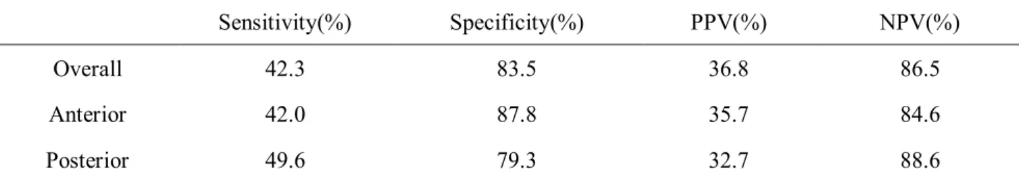
2676 sections of multiparametric-MRI of prostate were reviewed with pathologic
findings to survey diagnostic accuracy of PI-RADS scoring system. Overall
sensitivity was 42.3%, specificity was 83.5%, PPV was 36.5%, and NPV was
86.5%. When analyzed anterior and posterior prostate separately, 42.0% of
sensitivity and 87.8% specificity, 35.7% of PPV and 84.6 NPV for anterior
prostate and 49.6% sensitivity and 79.3% specificity, 32.7% of PPV and 88.6%
NPV for posterior prostate(Table 3).
Table 3. Diagnostic accuracy of PI-RADS system and MR characteristics subdivided by pathologic findings
Diagnostic accuracy of PIRADS score
Sensitivity(%) Specificity(%) PPV(%) NPV(%)
Overall 42.3 83.5 36.8 86.5
Anterior 42.0 87.8 35.7 84.6
Posterior 49.6 79.3 32.7 88.6
MR characteristics subdivided by pathologic findings
pT2 and GS 6 (N=110) ≥pT3a or ≥ GS 7 (N=113)
N(%) N(%)
Presence of PI-RADS 4-5 91 (82.7) 103 (91.2)
No. of PI-RADS 4-5
11
0 16 (14.5) 10 (8.8)
1 28 (25.5) 16 (14.2)
≥2 66 (60) 87 (77.0)
Presence of PI-RADS 5 31 (28.2) 50 (44.2)
No. of PI-RADS 5
0 79 (71.8) 63 (55.8)
1 15 (13.6) 21 (18.6)
≥2 16 (14.5) 29 (25.7)
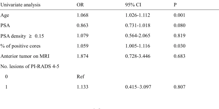
According to results above, we presumed age, PSA, PSA density, positive-biopsy
core percentage, Index tumor at anterior prostate, numbers of lesion with positive
findings PIRADS as the risk factor of upstaging and upgrading for prostate cancer.
Logistic regression analysis was done for reveal these suspected risk factors really
affects upstaging and upgrading statistically. Univariate logistic regression showed
the age(OR 1.068, 95% CI 1.026-1.112, p=0.001), PSA density over 0.1(OR 2.125,
95% CI 1.215-3.715, p=0.008), percentage of positive core(OR 1.059, 95% CI
1.005-1.116, p=0.030), index tumor at anterior prostate(OR 1.882, 95% CI 1.106-
3.203, p=0.020), 2 or more PI-RADS 4 or 5 at multiparametric-MRI(OR 2.143,
95% CI 0.923-4.978, p=0.046) seems to be statistically meaningful to influence
12
cancer upstaging and upgrading. PSA was not statistically intentional to predict
cancer upstaging and upgrading(p=0.080), and 1 lesion of PI-RADS 4-5 at
multiparametric MRI does not affected cancer upstaging and upgrading(p=0.807)
Finally, multivariate logistic regression revealed that the age(OR 1.072, 95% CI
1.028-1.119, p=0.001), PSA density more than 0.10(OR 2.097, 95% CI 1.166-
3.771, p=0.013), Index tumor at anterior prostate(OR 1.921, 95% CI 1.095-3.371,
p=0.023), 2 or more lesions of PIRADS 4-5 score on mpMRI(OR 1.544, 95% CI
0.524-3.817, p=0.034) are the factors that impacts upstaging/upgrading of prostate
cancer. Percentage of positive core was not statistically voluntary(p=0.071)
Table 4. Univariate and multivariate analysis for prediction of upgrading and upstaging
Univariate analysis OR 95% CI P
Age 1.068 1.026-1.112 0.001
PSA 0.863 0.731-1.018 0.080
PSA density ≥ 0.15 1.079 0.564-2.065 0.819
% of positive cores 1.059 1.005-1.116 0.030
Anterior tumor on MRI 1.874 0.728-3.446 0.683
No. lesions of PI-RADS 4-5
0 Ref
1 1.133 0.415–3.097 0.807
13
2 2.143 0.923-4.978 0.046
Multivariate analysis OR 95% CI P
Age 1.069 1.024-1.116 0.003
% of positive cores 1.059 1.001-1.120 0.046
No. lesions of PI-RADS 4-5
0 Ref
1 1.184 0.413-3.398 0.754
2 1.879 0.776-4.552 0.043
14
DISCUSSION
Managing patients of prostate cancer on active surveillance is on the challenge,
which are avoiding delay in providing treatment to higher risk for progression and
avoiding overtreatment[12]. Standard prostate biopsy and PSA has limited role in
pretreatment prostate cancer risk and prognosis evaluation[13]. Comparing contemporary programs of AS, PRIAS and University of Miami protocols shows
the best ability to select as many insignificant prostate cancer patients as possible
with guaranteed safety[13]. The PRIAS study is the largest prospective study about
AS at this time, coming close to representing a real-world situation, and its ability
in selecting low-risk PCa patients is well known[14]. So, our study used PRIAS
criteria for AS classification.
Need for the tool for screening the clinically insignificant prostate cancer and
monitor progression on active surveillance with non-invasive method is still
15
present.[15]. Shoots et al, did systematic review with 19 studies, and revealed that
MRI can be used in prostate cancer on active surveillance to find clinically
significant disease, and it is evident that positive MRI finding can be resulted in
reclassification after MRI-targeted biopsies or radical prostatectomy[4]. More
recently, Klots et al did prospective multicenter trial that results MRI with targeted
biopsies did not significantly increase the upgrading rate and addition of MRI
imaging and targeted biopsy discovers most clinically significant diseases in men
on active surveillance[16].
The American Urological Association(AUA) presents consideration of
multiparametric MRI as a component for AS for localized prostate cancer patients,
and European Association of Urology(EAU) proposed that imaging with
multiparametric MRI has high negative-prediction value for lesion upgrading in
low-risk prostate cancer patients. Siddiqui et al. represents the performance of a
multiparametric MRI based nomogram can reduce repeat biopsy in AS patients[17].
And Park et al. reveals that tumor identification with multiparametric MRI can be
16
used as an initial management strategy because it is predictive of adverse
pathologic features in localized prostate cancer patients who are eligible for
AS[18].
In our study, PI-RADS showed low sensitivity in prostate cancers suitable for
active surveillance criteria, as 0.423 overall, and it showed specificity of 0.835. It
is relatively lower sensitivity and higher specificity, compared with recent meta-
analysis that PI-RADS scoring system shows pooled sensitivity of 0.89 and
specificity of 0.73[19]. The other study also showed similar results, as 0.78 of
sensitivity and 0.79 of specificity[20]. However, these meta-analyzes are aimed at
all of risks of prostate cancer patients, and it is different from those of this study.
Kornberg Z et al. represent 41% of positive predictive value and 85% of negative
predictive value for PI-RADSv2[21], which is similar with our result. And they
also represent PI-RADSv2 score of 4 or 5 is associated with increased risk of
biopsy upgrade.
17
Multiparametric-MRI is believed to be useful to identify high-grade and
significant cancers[22]. Marcus et al studied the leverage of multi-parametric MRI
on risk evaluation of prostate cancer, that 16.9% of patients have re-sorted to a
higher risk due to MRI findings, and 8.5% of patients has been changed the
therapeutic strategy[8]. And recent prospective study says standardized reported
MP-MRI using PI-RADS may be a promising tool for the selection of patients
suitable for AS[23]. Our study supports this concept, as resulted that existence of 2
or more lesions of PI-RADS score of 4 or 5, and 0.15 or more PSA density has
strong relationships with upgrading or upstaging in prostate cancer patients fitting
for AS, confirms the multiparametric MRI has notable capacity to identify
clinically significant prostate cancer in patients satisfying PRIAS criteria for AS.
On the other hand, Ploussard et al. mentioned that multiparametric MRI does not
make better prediction of high-risk disease in patients that are enrolled in an AS
protocol according to rigid criteria[24]. Guzzo et al. demonstrated that MRI was
not a significant predictor of upgrading[25]. This study had no statistical
18
correlation between upgrading and visible disease (PIRADS 4–5) on
multiparametric MRI. We observed the better ability of multiparametric-MRI that
predicts upgrading, upstaging, and unfavorable diseases.
There are studies that anterior prostate cancer has significant nature, and harder to
detect by standard TRUS biopsy[26]. Lawrentschuk et al. insisted that MRI and
further biopsy should be considered, because their pathology could be
aggressive[27]. Song et al. said that anterior site of cancer on MRI can be expected
for Gleason upgrading or unfavorable pathological outcome[28]. But our study
suggests there are no statistically meaningful results that anterior location of
prostate tumor on MRI is associated with upstaging and upgrading. We think that
low sensitivity and PPV of PI-RADS score makes it, because there are definite
evidence between pathologic results of anterior prostate tumor is associate with
cancer upstaging and upgrading(HR 1.885, p<0.001)
Our study has several limitations. First, relatively small number of patients in
19
single center were enrolled; a multicenter-cohort, large pool of patients could be
more probable to define the role of multiparametric MRI to seek clinically
significant disease. Second, this study was not a prospective or screening trial, so
our results may not be generalized to patients that has normal PSA and normal
DRE findings. Third, we did not consider the interval between cancer
diagnosis(biopsy-confirmed) and getting MRI images, so we could not estimate
disease progression in these gaps. Fourth, 3 radiologists reviewed the MRI images,
and there are doubt that reproducibility can be among radiologists.
Despite these limitations, our investigation provided interesting outcomes that
can support the buildup of the role of multiparametric MRI and PI-RADS score
that can be a new criterion to predict upstaging and upgrading disease.
20
CONCLUSION
In conclusion, this study shows multiparametric MRI reported on PI-
RADS scoring system has relatively low sensitivity, but it can be useful to detect
clinically significant prostate cancer among patients on AS according PRIAS
criteria, based on upstaging and upgrading. We believe that multiparametric MRI
has a decisive role to find clinically significant prostate cancer, with combine of
other traditional serum markers and physical findings. Our results can support
establishing the treatment plan of first-diagnosed prostate cancer patients.
21
REFERENCE
1. Bul M, Zhu X, Valdagni R, et al: Active surveillance for low-risk prostate cancer worldwide: the PRIAS study. Eur Urol 2013; 63: 597–603.
2. Kvåle R, Møller B, Wahlqvist R, et al. Concordance between Gleason score s of needle biopsies and radical prostatectomy specimens: a population bas‐ ed study. BJU international. 2009;103:1647-54.
3. Cornud F, Flam T, Chauveinc L, et al. Extraprostatic spread of clinically lo calized prostate cancer: factors predictive of pT3 tumor and of positive end orectal MR imaging examination results. Radiology 2002;224:203–10.
4. Schoots IG, Petrides N, Giganti F,et al: Magnetic Resonance Imaging in Ac tive Surveillance of Prostate Cancer: A Systematic Review. Eur Urol. 2015 Apr;67(4):627-36.
5. Dickinson L, Ahmed HU, Allen C, et al. Magnetic resonance imaging for t he detection, localization, and characterization of prostate cancer: recommen dations from a European consensus meeting. Eur Urol 2011; 59: 477-94.
6. Weinreb JC, Barentsz JO, Choyke PL, et al. PI-RADS Prostate Imaging — Reporting and Data System: 2015, Version 2. Eur Urol 2016; 69: 16-40.
7. Kasivisvanathan V, Rannikko AS, Borghi M, et al, MRI-Targeted or Standar d Biopsy for Prostate-Cancer Diagnosis. N Engl J Med. 2018 May 10;378 (19):1767-1777.
8. Marcus DM, Rossi PJ, Nour SG, et al. The impact of multiparametric pelvi c magnetic resonance imaging on risk stratification in patients with localize d prostate cancer. Urology 2014;84:132–7.
9. Barentsz JO, Richenberg J, Clements R, Choyke P, Verma S, Villeirs G, Ro uviere O, Logager V, Fütterer JJ (2012) ESUR prostate MR guidelines. Eur
Radiol 22(4):746–757.
10. Junker D, Quentin M, Nagele U, et al, Evaluation of the PI-RADS scoring system for mpMRI of the prostate: a whole-mount step-section analysis. W
22 orld J Urol. 2015 Jul;33(7):1023-30.
11. Hoeks CM, Somford DM, van Oort IM, Vergunst H, Oddens JR, Smits GA, Roobol MJ, Bul M, Hambrock T, Witjes JA, Fütterer JJ, Hulsbergen-van d e Kaa CA, Barentsz JO (2014) Value of 3-T multiparametric magnetic reso nance imaging and magnetic resonance-guided biopsy for early risk restratifi cation in active surveillance of low-risk prostate cancer: a prospective multi center cohort study. Invest Radiol 49(3):165–172.
12. Wong LM, Neal DE, Johnston RB, et al: International multicentre study ex amining selection criteria for active surveillance in men undergoing radical prostatectomy. Br J Cancer 2012; 107: 1467–1473.
13. Iremashvili V, Pelaez L, Manoharan M, et al: Pathologic prostate cancer ch aracteristics in patients eligible for active surveillance: a head-to-head comp arison of contemporary protocols. Eur Urol 2012; 62: 462–468.
14. M. Bul, X. Zhu, R. Valdagni, et al. Active surveillance for low-risk prostat e cancer worldwide: the PRIAS study. Eur Urol, 2012; 63: 597-603.
15. Dall’Era MA, Albertsen PC, Bangma C, et al: Active surveillance for prost ate cancer: a systematic review of the literature. Eur Urol 2012; 62: 976–9 83.
16. Klotz L, Loblaw A, Sugar L, et al. Active Surveillance Magnetic Resonanc e Imaging Study (ASIST): Results of a Randomized Multicenter Prospective Trial. Eur Urol. 2018 Jul 13. pii: S0302-2838(18)30450-0. doi: 10.1016/j.e ururo.2018.06.025.:
17. Siddiqui MM, Truong H, Rais-Bahrami S, et al: Clinical implications of a multiparametric magnetic resonance imaging based nomogram applied to pro state cancer active surveillance. J Urol 2015; 193: 1943–1949.
18. Park BH, Jeon HG, Choo SH, et al: Role of multiparametric 3.0-Tesla mag netic resonance imaging in patients with prostate cancer eligible for active s urveillance. BJU Int. 2014 Jun;113(6):864-70.
19. Woo S, Suh CH, Kim SY, et al: Diagnostic Performance of Prostate Imagin g Reporting and Data System Version 2 for Detection of Prostate Cancer:
23
A Systematic Review and Diagnostic Meta-analysis. Eur Urol. 2017 Aug;72 (2):177-188.
20. Hamoen EHJ, de Rooij M, Witjes JA, et al: Use of the Prostate Imaging Reporting and Data System (PI-RADS) for Prostate Cancer Detection with Multiparametric Magnetic Resonance Imaging: A Diagnostic Meta-analysis.
Eur Urol. 2015 Jun;67(6):1112-1121.
21. Kornberg Z, Cowan JE, Westphalen AC, et al: Genomic Prostate Score, PI- RADSv2, and Progression in Men with Prostate Cancer on Active Surveilla nce. J Urol. 2018 Sep 1. pii: S0022-5347(18)43795-0.
22. Wang L, Mazaheri Y, Zhang J, et al. Assessment of biologic aggressiveness of prostate cancer: correlation of MR signal intensity with Gleason grade after radical prostatectomy. Radiology 2008;246:168–76.
23. Hoeks CM, Somford DM, van Oort IM, et al: Value of 3-T multiparametric magnetic resonance imaging and magnetic resonance-guided biopsy for earl y risk restratification in active surveillance of low-risk prostate cancer: a pr ospective multicenter cohort study. Invest Radiol. 2014 Mar;49(3):165-72.
24. Ploussard G, Xylinas E, Durand X, et al: Magnetic resonance imaging does not improve the prediction of misclassification of prostate cancer patients e ligible for active surveillance when the most stringent selection criteria are based on the saturation biopsy scheme. BJU Int 2011; 108: 513–517.
25. Guzzo TJ, Resnick MJ, Canter DJ, et al: Endorectal T2-weighted MRI does not differentiate between favorable and adverse pathologic features in men with prostate cancer who would qualify for active surveillance. Urol Oncol 2012; 30: 301–305.
26. Koppie TM, Bianco FJ, Jr, Kuroiwa K, et al. The clinical features of anterior pros tate cancers. BJU Int. 2006;98:1167–1171.
27. Lawrentschuk N, Haider MA, Daljeet N, et al. Prostatic evasive anterior tumors: th e role of magnetic resonance imaging. BJU Int. 2009;105:1231–1236.
28. Song SH, Pak S, Park S, et al. Predictors of unfavorable disease after radical prost atectomy in patients at low risk by D'Amico criteria: role of multiparametric magn
24
etic resonance imaging. J Urol. 2014 Aug;192(2):402-8.
국문요약
우리는 Prostate Imaging Reporting and Data System (PI-RADS)을 이용한
다중 자기 공명 영상이 능동적 감시 대상의 전립선 암을 가진 남성에서 수술
후 병리 검사상 등급 상승 및 병기 상승을 예측하는 데 도움이 되는지 평가하
고자 하였다
2014 년 1 월부터 2017 년까지 PRIAS 능동형 감시 기준에 해당하는 총
223 명의 환자를 분석하였다. 모든 환자는 전립선 조직 검사 후 다중 자기 공
명 영상을 촬영하였으며, 이를 통해 PI-RADS 점수가 측정되다. 모든 환자는
근치적 전립선 절제술을 시행 받았다. PI-RADS 점수는 전립선 내 12 개의
구역, 전립선 외 12구역으로 나뉘어 측정되었다. 4 또는 5점이 측정된 경우
임상적으로 의미 있는 암으로 간주하였다.
223 명의 환자 중 전립선암의 병기 상승을 보인 환자가 25명이었고 등급 상
승을 보인 환자는 108명이었다. 종양의 병기 상승 및 등급 상승을 보인 환자
는 그렇지 않은 환자에 비해 노령이었으며, 전립선 크기가 작았으며, 전립선
25
특이 항원 밀도가 높았으며, 양성 코어의 비율이 높았으며 종양의 크기가 더
컸다. PI-RADS 채점의 진단 정확도를 확인하기 위해 223 명의 환자로부터
총 2676 개의 병소를 평가했다. PI-RADS 점수 시스템의 민감도와 특이도는
각각 36.8 %와 86.5 %였다. 다변량 분석에서 PI-RADS 점수 4 또는 5점인
병소가 두 군데 이상인 경우 능동적 감시 대상 환자에서 병기 상승, 등급 상승
을 독립적으로 예측할 수 있었다.
본 연구에서 PI-RADS 점수 시스템을 이용한 다중 자기 공명 영상은 이전의
보고들과 같이 낮은 민감도를 가졌지만, 전립선암의 등급 상승 및 병기 상승을
예측할 수 있었다. 이 결과는 능동적 감시 대상의 전립선암 환자에서, 임상적
으로 의미 있는 암을 발견하는 데에 유용하게 이용할 수 있다.