저작자표시-비영리-변경금지 2.0 대한민국 이용자는 아래의 조건을 따르는 경우에 한하여 자유롭게
l 이 저작물을 복제, 배포, 전송, 전시, 공연 및 방송할 수 있습니다. 다음과 같은 조건을 따라야 합니다:
l 귀하는, 이 저작물의 재이용이나 배포의 경우, 이 저작물에 적용된 이용허락조건 을 명확하게 나타내어야 합니다.
l 저작권자로부터 별도의 허가를 받으면 이러한 조건들은 적용되지 않습니다.
저작권법에 따른 이용자의 권리는 위의 내용에 의하여 영향을 받지 않습니다. 이것은 이용허락규약(Legal Code)을 이해하기 쉽게 요약한 것입니다.
Disclaimer
저작자표시. 귀하는 원저작자를 표시하여야 합니다.
비영리. 귀하는 이 저작물을 영리 목적으로 이용할 수 없습니다.
변경금지. 귀하는 이 저작물을 개작, 변형 또는 가공할 수 없습니다.
의학석사 학위논문
성인 털모양 성상세포종의 예후 인자와 성인 털모양 성상세포종 치료에의 수술 후 보조 방사선치료의 효용성
Prognostic Factors of Adult Pilocytic Astrocytoma and Efficacy of Adjuvant Radiotherapy
on Adult Pilocytic Astrocytoma
울 산 대 학 교 대 학 원 의 학 과
박 상 혁
성인 털모양 성상세포종의 예후 인자와 성인 털모양 성상세포종 치료에의 수술 후 보조 방사선치료의 효용성
지 도 교 수 김 영 훈
이 논문을 의학석사 학위 논문으로 제출함
2022년 2월
울 산 대 학 교 대 학 원 의 학 과
박 상 혁
박상혁의 의학석사 학위논문을 인준함
심사위원 김 정 훈 (인) 심사위원 김 영 훈 (인) 심사위원 정 상 준 (인)
울 산 대 학 교 대 학 원
2022년 2월
국문 요약
털모양 성상세포종은 국제 보건 기구 등급 1등급의 종양으로, 양성의 특성을 가지 고 소아 인구에서 흔하다. 성인 인구에서는 비교적 드물기 때문에, 성인 털모양 성 상세포종의 행동 양상과 예후 인자는 비교적 덜 알려져 있다. 기존의 연구들은 성 인 털모양 성상세포종이 좀 더 공격적인 행동을 보인다고 보고하고 있다. 또한, 전 절제술을 시행하지 못한 종양의 보조적 치료는 정립되어 있지 않다. 본 연구는 성 인 털모양 성상세포종의 예후 인자를 찾고, 보조적 방사선치료의 효용성을 규명하 고자 한다.
1999년부터 2018년까지 단일기관에서 수술을 받고 18세 이상의 나이에 털모양 성 상세포종을 처음 진단받은 77명의 환자들의 후향적 데이터 분석을 시행하였다. 저 자는 전체 생존 기간과 무진행 생존 기간을 조사했으며 각 인자들의 예후에의 영향 을 분석하였다. 무진행 생존 기간과 전체 생존 기간에의 예후 인자는 카플란-마이 어 생존 곡선을 이용하여 분석했으며, 다변량 분석은 콕스 비례 위험 모형을 이용 하여 진행하였다.
77명의 성인 환자들이 수술을 받고 털모양 성상세포종으로 진단되었다. 전절제술은 45명 (58%)의 환자들에서 이루어졌다. 중간값 80개월의 관찰 기간동안, 7명 (9%) 의 환자들이 죽었고, 19명 (25%)의 환자들이 종양의 진행을 경험했다. 5년동안의 전체 생존율은 91%이었고, 5년동안의 무진행 생존율은 77%이었다. 단변량 분석에 서는 진단 시점의 나이, 신경학적 증상의 유무, 종양의 위치, 종양 절제 범위가 통
= 0.003). 보조적 방사선치료는 전절제를 시행하지 못한 종양의 예후를 개선하지 못했다.
본 연구에 의하면, 종양의 절제 범위가 성인 털모양 성상세포종의 예후에 유의한 영향을 미친다. 보조적인 방사선치료는 종양의 예후를 개선하지 못했지만, 앞으로 전절제를 시행하지 못한 종양의 보조적 치료 전략을 수립하기 위해 후속연구들이 시행되어야 할 것이다.
Contents
Korean Abstract ···i
List of Tables ···iv
List of Figures ···v
Introduction ···1
Methods ···2
- Patient identification and selection
···2
- Outcome selection and statistical analysis
···3
Results ···3
- Patient clinical and radiological characteristics
···3
- Surgical outcome
···4
-
Overall patient outcome ···4
-
Prognostic factors ···4
-
Adjuvant radiation therapy ···5
Discussion ···6
Conclusion ···10
Reference ···11
Abstract ···20
List of Tables
Table 1.
The baseline clinical and radiological characteristics of the study cohorts (n =
77) ···12
Table 2.The surgical outcome and the parameters of the postoperative treatments of the
study cohorts (n = 77) ···13
Table 3.The surgical outcome by tumor location (n = 77) ···13
Table 4.Favorable prognostic factors of progression-free survival in the adult patients
who underwent surgical treatments for pilocytic astrocytoma (n = 77) ···14
Table 5.Characteristics and overall outcome of the patients who receive adjuvant
radiation therapy (n = 11) ···15
List of Figures
Figure 1.
Overall survival and progression-free survival of adult pilocytic astrocytomas
(n=77) ···16
Figure 2.Progression-free survival curve for prognostic factors ···17
Introduction
Pilocytic astrocytomas (PA) are primary brain tumors classified as World Health Organization (WHO) grade I.1In pathologic evaluation, they classically demonstrate a biphasic architecture of densely packed fibrillary tissue and looser microcystic compartments. They can exhibit eosinophilic granular bodies and Rosenthal fibers, which consist of corkscrew eosinophilic bundles of intermediate glial filaments with intermingled cytosolic proteins. They can also appear aggressive on histopathology with microvascular proliferation and necrosis, which are characteristic feature of high-grade glioma such as glioblastoma multiforme, but not signs of malignancy for pilocytic astrocytomas in contrast to diffuse astrocytomas.2In molecular biology, KIAA1549:BRAFfusion rearrangements or valine-to-glutamate substitution at the BRAF codon 600 may attribute to pilocytic astrocytomas.3
Pilocytic astrocytomas are the most common pediatric brain tumor in age 0 to 14 years and second most common brain tumor in age 15 to 19 years. They consist 15.2% of all primary brain and central nervous system (CNS) tumors in age 0 to 19 years, which is the highest proportion of all primary brain tumors. The incidence of pilocytic astrocytomas in age 0 to 19 years is 0.95 per 100,000 person-years.4 The most common location of pilocytic astrocytomas is cerebellum in pediatric population and cerebral hemisphere followed by cerebellum in adult population.5The 5- year overall survival rate for patients with pilocytic astrocytomas is 100% and the 10-year survival rate is 95.8%.6
However, despite benign characteristics, some reports suggest pilocytic astrocytomas in adult population behave more aggressively than in pediatric population.5,7On the other hand, Paul D.
Brown et al. suggests benign nature in adult population as in pediatric population.8Unfortunately, the behavior of pilocytic astrocytomas in adults is known relatively less due to their rarity in adult population, with incidence less than 0.1 per 100,000 person-years in age over 45 years.4
As for treatment strategy, surgical treatment is treatment of choice and gross-total resection
(GTR) offers the most favorable outcome.6 However, is is not always possible to achieve GTR for especially deep-located tumors such as on thalamus or brainstem. For tumors unamenable to GTR, adjuvant radiation therapy is applied in some centers. However, the efficacy of adjuvant radiation therapy is on controversy.6
The goal of this study is to evaluate the prognosis of adult pilocytic astroctyomas, to find prognostic factors of pilocytic astroctyomas in adult, and to evaluate the efficacy of adjuvant radiation therapy on adult pilocytic astrocytomas.
Methods
Patient identification and selection
We conducted retrospective data analysis of adult pilocytic astrocytomas in single institute, Asan medical center. The patients who underwent operation between 1999 and august 2018, and were diagnosed histopathologically as pilocytic astrocytomas at the age of 18 year-old or over were enrolled in the study. Only newly diagnosed cases were enrolled in the study. Data was based on medical records and images.
In our institute, we tried to achieve gross total resection (GTR) for suspected adult pilocytic astrocytomas. When GTR was achieved, the patients were cautiously observed and followed up without additional treatments. As for the patients who could not undergo GTR, but only subtotal resection (STR) or biopsy, we did careful observation first. However, in case of patients who have deep-seated tumors for which redo operations may be complicated or have severe fear for progression, we considered adjuvant therapy such as radiation therapy.
We collected following data by retrospective review of medical records: gender, age of diagnosis,
magnetic resonance imaging (MRI), perilesional edema, hydrocephalus, pathologic report, extent of removal (GTR / STR or partial resection (PR) / biopsy), residual tumor size, and adjuvant treatment.
We investigated progression-free survival (PFS) and overall survival (OS). Overall survival was defined as the time interval between the date of initial diagnosis and the date of death.
Progression-free survival was defined as the time interval between the date of initial treatment and the date of tumor recurrence or progression based on radiologic findings.
Outcome selection and statistical analysis
Prognostic factors for PFS and OS were analyzed using using Kaplan-Meier survival curve.
Multivariate analysis was conducted using Cox-proportional hazard model.
All statistical analyses were conducted using SPSS ver. 21.0 (SPSS Inc, Chicago, IL). Result of p value ≤ 0.05 was considered statistically significant.
Results
Patient clinical and radiological characteristics
Total 88 adult patients were diagnosed as pilocytic astrocytoma but only 77 patients were enrolled in the study. 3 patients were excluded because of misdiagnosis, 3 were excluded because they were not newly diagnosed but had recurrence, 1 was excluded due to insufficient data, and 4 were excluded due to follow-up loss.
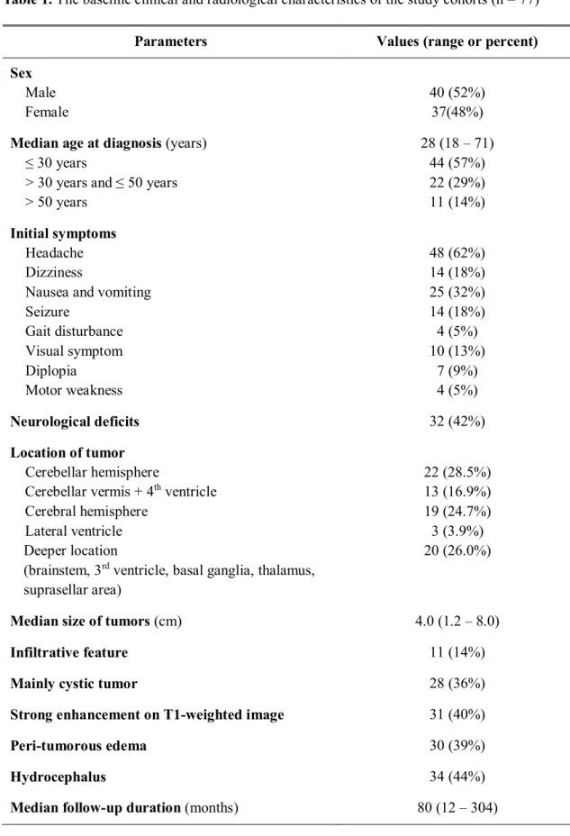
Median age of diagnosis was 28-year old (range, 18-71 years old). Among 77 patients, 44 (57%) patients were diagnosed at 30 years old or younger. Median follow-up period was 80 months (range, 12-304 months). Among 77 patients, male patients were 70 (52%) and female patients were 37 (48%). As for location of tumors, 22 (28.5%) cases were in cerebellar hemisphere, which
is the most common location followed by cerebral hemisphere (19, 24.7%) and cerebellar vermis (13, 16.9%). 20 (26.0%) tumors were in deeper locations such as brainstem, basal ganglia, and thalamus. Other clinical and radiological characteristics are shown in Table 1.
Surgical outcome
Among 77 patients who underwent operation for pilocytic astrocytoma, gross total resection (GTR) was achieved in 45 (58%) patients. Subtotal resection (STR) and biopsy were achieved in 18 (23%) and 14 (18%) patients, respectively. Median size of residual tumors were 2.8 cm (range, 0.4 – 5.6).
After surgery of the tumor, 54 (70%) patients did not receive any form of adjuvant therapy. 11 (14%) patients received adjuvant radiation therapy.
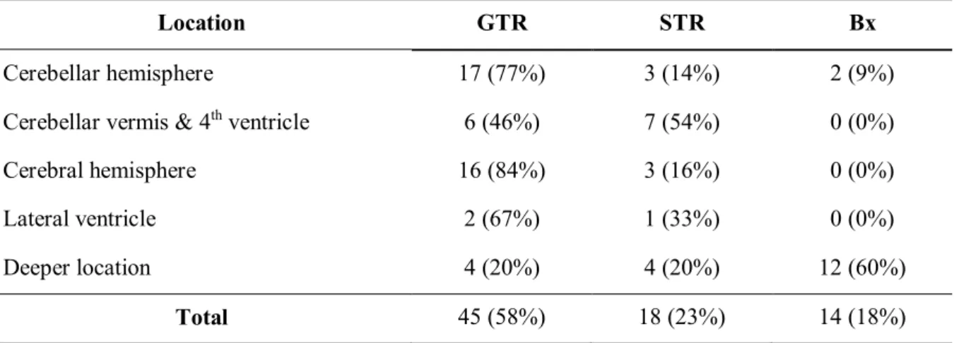
Surgical outcomes and adjuvant treatments are shown in Table 2. In addition, surgical outcomes by the location of tumors are shown in Table 3. GTR was achieved most for tumors in cerebellar hemisphere (17 of 22 patients, 77%) and cerebral hemisphere (16 of 19 patients, 84%).
Overall patient outcome
7 deaths of 77 patients were observed during follow-up period with overall survival rate 91%.
Progression of the disease was the cause of all 7 deaths. Overall survival rates were 96% for 1- year, 91% for 3-year, and 91% for 5-year.
During post-operative follow-up period median of which was 80 months, 19 progressions occurred (25%). Progression free survival rates were 88% for 1-year, 80% for 3-year, and 77%
for 5-year. Overall survival and progression free survival curves are demonstrated on Figure 1.
Prognostic factors
correlated to PFS with statistical significance. Kaplan-Meier survival curves for each factors are shown in Figure 2.
The patients who were diagnosed at 30-year old or younger showed significantly better PFS than older patients (p = 0.009). Progression-free survival rate at 5 years was 80% for the patients at or younger than 30-year old and 49% for the older patients.
When the patients had neurologic deficit, progression-free survival rate was significantly worse (p = 0.005). Progression-free survival rate at 5 years was 80% for the patients without any neurologic deficit and 51% for neurologically compromised patients.
For the location of tumors, the patients with tumor on cerebellum, cerebral hemisphere, and lateral ventricle had better progression-free survival rates than the patients with tumor on deeper location, such as thalamus or brainstem (p = 0.007). 5-year progression-free survival rate was 77%
for the tumors on cerebellum, cerebrum, and lateral ventricles and 29% for deeper tumors.
Extent of resection was strongly correlated to PFS (p < 0.001). 5-year progression-free survival rate was 83% after GTR, 54% after STR, 39% after biopsy, and 45% when combining STR and biopsy.
Other factors including tumor size, gender, tumor infiltration, cyst component, gadolinium enhancement, peri-tumorous edema, and hydrocephalus did not show significant correlation with PFS. P values for each prognostic factors are shown in Table 4.
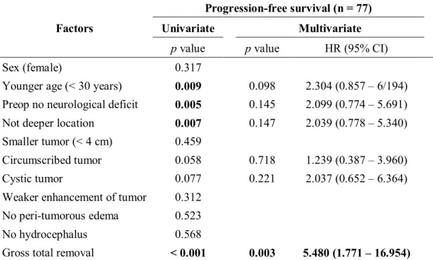
In the multivariate Cox analysis, however, only extent of resection had significant impact for PFS (HR 5.480 [95% CI 1.771-16.954]; p = 0.003). Age of diagnosis, presence of neurologic deficit, and tumor location had no significant effect on PFS in multivariate model. Results of multivariate analysis are demonstrated in Table 4.
Adjuvant radiation therapy
We additionally analyzed efficacy of adjuvant radiation therapy after STR or biopsy. After STR or biopsy for 32 patients, 11 patients were closely observed without adjuvant therapy and 10
patients received adjuvant radiation therapy. 5 of 11 (45%) patients who did not receive adjuvant therapy had recurrence or progression of tumors. 6 of 10 (60%) patients who underwent adjuvant radiation therapy experienced recurrence or progression of residual tumors, which is higher proportion than the group without adjuvant treatment. Median OS of the patients who underwent adjuvant radiation therapy after STR or biopsy was 52.7 months (range, 5.4-158.3) and median PFS of the same group was 25.6 months (range, 2.9-107.2).
Among patients without adjuvant treatment, 9 of 11 (82%) patients underwent STR. In contrast, only 3 of 10 (30%) patients with adjuvant radiation therapy underwent STR and the rest 7 patients underwent only biopsy.
After adjuvant radiation therapy, 6 of 10 (60%) patients had recurrence or progression, and 3 of 6 (50%) patients died.
Characteristics and overall outcome of the adjuvant radiation therapy recipients are demonstrated in Table 5.
Discussion
Pilocytic astrocytomas (PA) are WHO grade I primary brain tumors presenting biphasic architecture of densely packed fibrillary tissue and looser microcystic compartments. They characteristically exhibit Rosenthal fibers and eosinophilic granular bodies.1,2They are the most common primary brain tumor in pediatric age, between 0 and 19 years old, occupying 15.2% of all primary CNS tumors of this age group.4In pediatric population, pilocytic astrocytomas have indolent nature and favorable prognosis, with 100% survival rate for 5 years and 95.8% survival rate for 10 years.6However, adult pilocytic astrocytomas have been reported not to be benign by some authors and their characteristics and prognosis are not well known due to their rarity with incidence less than 0.1 per 100,000 person-years in population over 45 years.4Stüer et al. reported
from pilocytic asstrocytomas.9Theeler et al. also studied 127 adult pilocytic astrcytomas for 22 years. From their report, recurrence rate was 42% cases and death from the tumor was seen in 13 patients during follow-up.7
Our study reports similar distributions of age at diagnosis and tumor location as other studies.
More than half of the adult patients (44 of 77, 57%) were diagnosed at the age of 30 or younger.
22 (28.5%), 19 (24.7%), and 13 (16.9%) of 77 patients had tumors on cerebellar hemisphere, cerebral hemisphere, and cerebellar vermis along with 4thventricle, respectively. According to National Cancer Institute Surveillance from 1973 to 2008, 71.8% (2201 of 3066) patients were diagnosed in pediatric age and 19.2% (590 of 3066) patients were diagnosed at the age from 20 to 39. For tumor location, in adult population, 26.6% (230 of 865) and 29.7% (257 of 865) tumors were found on cerebellum and cerebrum, respectively.5
In our study, recurrence or progression rate was 25% (19 of 77) for follow-up duration median of which is 80 months. For same follow-up period, death occurred in 9% (7 of 77). Progression rate and death rate were both lower in our study than previous long-term studies. This difference might have been attributed to the period of follow-up, which was shorter in our study compared to the previous long-term studies.
45 of 77 (58%) patients received GTR, proportion of which is higher compared to Johnson et al.
studying 965 adult pilocytic astrocytoma cases. Johnson et al. reported 39.8% of GTR, 23.0% of STR, 19.0% of biopsy, 10.4% of unknown treatment, and 8.0% of no surgery. Proportions of STR and biopsy in our study are 23% and 18%, respectively, which are similar to Johnson et al.5For the location of tumor, GTR was achieved most for tumors in cerebellar hemisphere (17 of 22 patients, 77%) and cerebral hemisphere (16 of 19 patients, 84%). It is obvious that deeply located tumors tend not to be amenable for GTR, which is consistent with the results of our study.
In univariate analysis using Kaplan-Meier curves, age at diagnosis, presence of neurologic deficit, tumor location, and extent of resection had statistically significant impact for progression.
These findings are consistent with previous studies. In multivariate analysis, only extent of
resection showed statistic significance in prognostic impact (p = 0.003). Age of diagnosis failed to have significant impact on progression-free survival in multivariate analysis.
For the age of diagnosis, patients diagnosed with pilocytic astrocytomas over the age of 30 had poorer progression-free survival rate in univariate analysis (p = 0.009). Along with the results of previous study stating that pediatric pilocytic astrocytoms have better prognosis than adult pilocytic astrocytomas,5,7pilocytic astroctyomas may present more aggressiveness as increasing age of diagnosis. Johnson et al. concluded that different prognosis for different age groups are attributed to differences in the aggressiveness of pilocytic astrocytomas behavior between age groups.5In these studies, age of diagnosis was a significant prognostic factor also in multivariate analysis, which is different from our study.5This difference of prognostic impact may be attributed to the difference in reference for dividing patients by age. Johnson et al. divided patients into multiple groups, under 5-year-old, under 20-year-old, under 40-year-old, under 60-year-old, and 60-year-old or older,5and proved prognostic impact of age. On the other hand, we divided adult patients with pilocytic astrocytomas into only 2 groups by the age of 30 years old. Because mean age of the patients enrolled in our study was 28 years old, we decided reference point as age of 30-year-old.
Tumor location and presence of neurologic deficit showed significant impact on progression, but these effects were attributed to the fact that extent of resection is strongly related to tumor location and neurologic status. Obviously, these factors had no significant correlation with progression-free survival in the multivariate analysis.
GTR is the only confirmed treatment to have positive impact on survival and prognosis of adult pilocytic astrocytomas to date. Stüer et al. reported 4 times higher recurrence rates of partially resected adult pilocytic astrocytomas than completely resected tumors.9Johnson et al. revealed a significantly low hazard ratio of death of 0.3 for GTR compared to STR or biopsy.5Based on our
patients underwent GTR in our study. Therefore, adjuvant therapy for subtotally resected or biopsied tumors should be established. Then, we analyzed efficacy of adjuvant radiation therapy after STR or biopsy. Post-operative adjuvant radiation therapy remains controversial with both positive and negative reports on prognosis. Ishkanian et al. reported retrospective analysis suggesting superior progression-free survival in adjuvant radiation therapy group, which supports the use of radiation therapy after the operation in adults.10 On the other hand, Theeler et al.
reported significantly reduced progression-free survival with adjuvant radiation therapy.7
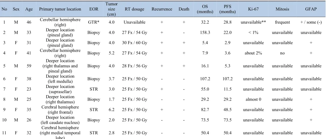
In our study, aside from one patient who received adjuvant radiation therapy after GTR, 10 patients underwent adjuvant radiation therapy after STR and biopsy. After adjuvant radiation therapy, tumor progression rate was higher than no adjuvant treatment group. We analyzed characteristics of these two, observation and adjuvant radiation therapy groups to explain the result. First, extent of resection was different in two groups. In observation after surgery group, 82% (9 of 1) patients underwent STR, whereas only 30% (3 of 10) patients received STR in adjuvant radiation therapy group. As we mentioned before, extent of resection is strong prognostic factor, difference in extent of resection may contribute to negative prognostic impact of adjuvant radiation therapy. We tried to figure out histopathologic characteristics of the tumor with progression after adjuvant radiation therapy, but lack of pathologic report on mitosis or immunohistochemistry such as Ki-67 has hampered analysis. Most importantly, the correlation of adjuvant radiation therapy with higher progression rate is likely a result of patient selection bias.
In practice in our institute, surgeons recommended adjuvant radiotherapy for patients judged to be “high-risk” based on clinical or pathologic factors. Further randomized prospective study with larger numbers should be conducted to find out efficacy of adjuvant radiation therapy for adult pilocytic astrocytomas.
We have some limitations on the study. To compensate the rarity of adult pilocytic astrocytomas, we collected data for long period, 20 years. Through 20 years, surgical technique and equipment that could affect the outcome have evolved. Also, pathologic diagnosis has evolved and there is
possibility of misdiagnosis in earlier period. We reviewed brain magnetic resonance images again, but we were not able to analyze pathologic specimens independently again to confirm the diagnosis of pilocytic astrocytomas. In addition, detailed pathologic information is limited. In the era of the importance of molecular diagnosis of brain tumors, detailed pathologic analysis with immunohistochemistry will be needed in further studies to establish prognostic factors of adult pilocytic astrocytomas.
Conclusion
Adult pilocytic astrocytomas tend to behave more aggressively than pediatric pilocytic astrocytomas. According to our study, extent of tumor resection affects the prognosis of adult pilocytic astrocytomas. Because it is not always possible to achieve GTR, adjuvant treatment strategy should be established. Adjuvant radiation therapy failed to improve the outcome. Further randomized prospective study with larger numbers should be conducted for adjuvant treatment including radiation therapy.
Reference
1. Louis DN, Perry A, Reifenberger G, et al. The 2016 World Health Organization Classification of Tumors of the Central Nervous System: a summary. Acta Neuropathologica 2016;131:803-820.
2. Sadighi Z, Slopis J. Pilocytic Astrocytoma: A Disease With Evolving Molecular Heterogeneity. Journal of Child Neurology 2013;28(5):625-632.
3. Chen Y-H, Gutmann D. The molecular and cell biology of pediatric low-grade gliomas.
Oncogene 2014;33:2019-2026.
4. Ostrom QT, Cioffi G, Waite K, Kruchko C, Barnholtz-Sloan JS. CBTRUS Statistical Report: Primary Brain and Other Central Nervous System Tumors Diagnosed in the United States in 2014–2018. Neuro-Oncology 2021;23(23(S3)):iii1-iii105. DOI:
https://doi.org/10.1093/neuonc/noab200.
5. Johnson DR, Brown PD, Galanis E, Hammack JE. Pilocytic astrocytoma survival in adults: analysis of the Surveillance, Epidemiology, and End Results Program of the National Cancer Institute. Journal of Neuro-oncology 2012;108:187-193. DOI:
10.1007/s11060-012-0829-0.
6. BURKHARD C, PATRE P-LD, SCHÜLER D, et al. A population-based study of the incidence and survival rates in patients with pilocytic astrocytoma. Journal of Neurosurgery 2003;98:1170-1174. DOI: https://doi.org/10.3171/jns.2003.98.6.1170.
7. Theeler BJ, Ellezam B, Sadighi ZS, et al. Adult pilocytic astrocytomas: clinical features and molecular analysis. Neuro-oncology 2014;16(6):841-847. DOI:
10.1093/neuonc/not246.
8. Brown PD, Anderson SK, Carrero XW, et al. Adult patients with supratentorial pilocytic astrocytoma: long-term follow-up of prospective multicenter clinical trial NCCTG- 867251 (Alliance). Neuro-oncology Practice 2015;2(4):199-204.
9. St¨uer C, Vilz B, Majores M, Becker A, Schramm J, Simon M. Frequent Recurrence and Progression in Pilocytic Astrocytoma in Adults. Cancer 2007;110(12):2799-2808.
10. Ishkanian A, Laperriere NJ, Xu W, et al. Upfront Observation Versus Radiation forAdult Pilocytic Astrocytoma. Cancer 2011;117:4070-4079. DOI: 10.1002/cncr.25988.
Tables
Table 1.The baseline clinical and radiological characteristics of the study cohorts (n = 77)
Parameters Values (range or percent)
Sex Male Female
40 (52%) 37(48%) Median age at diagnosis (years)
≤ 30 years
> 30 years and ≤ 50 years
> 50 years
28 (18 – 71) 44 (57%) 22 (29%) 11 (14%) Initial symptoms
Headache Dizziness
Nausea and vomiting Seizure
Gait disturbance Visual symptom Diplopia
Motor weakness
48 (62%) 14 (18%) 25 (32%) 14 (18%) 4 (5%) 10 (13%)
7 (9%) 4 (5%)
Neurological deficits 32 (42%)
Location of tumor Cerebellar hemisphere
Cerebellar vermis + 4thventricle Cerebral hemisphere
Lateral ventricle Deeper location
(brainstem, 3rdventricle, basal ganglia, thalamus, suprasellar area)
22 (28.5%) 13 (16.9%) 19 (24.7%) 3 (3.9%) 20 (26.0%)
Median size of tumors(cm) 4.0 (1.2 – 8.0)
Infiltrative feature 11 (14%)
Mainly cystic tumor 28 (36%)
Strong enhancement on T1-weighted image 31 (40%)
Peri-tumorous edema 30 (39%)
Table 2.The surgical outcome and the parameters of the postoperative treatments of the study cohorts (n = 77)
Parameters Values (range or percent)
Extent of removal Gross total removal Subtotal removal Biopsy
45 (58%) 18 (23%) 14 (18%) Median size of residual tumors(cm) 2.8 (0.4 – 5.6) Adjuvant treatment
Radiation therapy Reoperation
Stereotactic radiosurgery Chemotherapy
23 / 77 (30%) 11 (14%)
7 (9%) 4 (5%) 1 (1%)
Progression at last follow-up 19 (25%)
Table 3.The surgical outcome by tumor location (n = 77)
Location GTR STR Bx
Cerebellar hemisphere 17 (77%) 3 (14%) 2 (9%)
Cerebellar vermis & 4thventricle 6 (46%) 7 (54%) 0 (0%)
Cerebral hemisphere 16 (84%) 3 (16%) 0 (0%)
Lateral ventricle 2 (67%) 1 (33%) 0 (0%)
Deeper location 4 (20%) 4 (20%) 12 (60%)
Total 45 (58%) 18 (23%) 14 (18%)
Abbreviations: GTR, Gross-total resection; STR, Sub-total resection; Bx, Biospy
Table 4. Favorable prognostic factors of progression-free survival in the adult patients who underwent surgical treatments for pilocytic astrocytomas (n = 77)
Factors
Progression-free survival (n = 77)
Univariate Multivariate
pvalue pvalue HR (95% CI)
Sex (female) 0.317
Younger age (< 30 years) 0.009 0.098 2.304 (0.857 – 6/194) Preop no neurological deficit 0.005 0.145 2.099 (0.774 – 5.691)
Not deeper location 0.007 0.147 2.039 (0.778 – 5.340)
Smaller tumor (< 4 cm) 0.459
Circumscribed tumor 0.058 0.718 1.239 (0.387 – 3.960)
Cystic tumor 0.077 0.221 2.037 (0.652 – 6.364)
Weaker enhancement of tumor 0.312 No peri-tumorous edema 0.523
No hydrocephalus 0.568
Gross total removal < 0.001 0.003 5.480 (1.771 – 16.954) Abbreviations: HR, hazards ratio; CI, confidence interval; Preop, preoperative
Table 5.Characteristics and overall outcome of the patients who receive adjuvant radiation therapy (n = 11)
No Sex Age Primary tumor location EOR Tumor size (cm)
RT dosage Recurrence Death OS
(months) PFS
(months) Ki-67 Mitosis GFAP
1 M 46 Cerebellar hemisphere
(right) GTR* 4.0 Unavailable + + 32.2 28.8 unavailable** frequent + / some (-)
2 M 33 Deeper location
(pineal gland) Biopsy 4.0 27 Fx / 54 Gy + - 158.3 22.0 < 1% unavailable unavailable
3 F 31 Deeper location
(pineal gland) Biopsy 4.0 30 Fx / 60 Gy + + 5.4 2.9 unavailable unavailable +
4 F 41 Cerebellar hemisphere
(right) Biopsy 5.2 27 Fx / 54 Gy + + 7.9 3.6 about 2% no +
5 M 59 Deeper location
(right thalamus and
pineal gland) Biopsy 4.0 28 Fx / 56 Gy + + 16.1 5.3 unavailable unavailable unavailable
6 F 38 Deeper location
(left medulla) Biopsy 3.7 25 Fx / 50 Gy - - 107.2 107.2 unavailable unavailable unavailable
7 F 23 Deeper location
(suprasellar) STR 3.0 25 Fx / 50 Gy + - 55.0 11.5 unavailable unavailable unavailable
8 M 25 Deeper location
(right thalamus) Biopsy 1.7 25 Fx / 50 Gy - - 29.2 29.2 almost 0 unavailable +
9 F 35 Cerebral hemisphere
(right frontal) STR 6.2 25 Fx / 50 Gy + - 82.7 48.5 unavailable unavailable +
10 M 26 Deeper location
(left caudate nucleus) Biopsy 2.0 25 Fx / 50 Gy - - 73.5 73.5 unavailable unavailable +
11 F 32 Cerebral hemisphere (right medial temporal
lobe) STR 2.8 25 Fx / 50 Gy - - 50.4 50.4 unavailable unavailable unavailable
Abbreviations: EOR, Extent of resection; RT, Radiation therapy; OS, Overall survival; PFS, Progression-free survival; GFAP, Glial fibrillary acidic protein;
GTR, Gross-total resection; STR, Sub-total resection; Fx, Fraction; Gy, Gray
*The patient who underwent GTR and received adjuvant RT had tumor progression to glioblastoma. There were some cell components without GFAP on pathologic report. Because he received adjuvant RT in other hospital, RT dosage is unavailable to know.
**No information for Ki-67, mitosis, and GFAP is recorded on pathologic reports for some patients. These findings are marked as “unavailable”
Figures
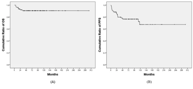
Figure 1.Overall survival and progression-free survival of adult pilocytic astrocytomas (n=77)
(A) Overall survival of adult pilocytic astrocytomas. 96% for 1-year, 91% for 3-year, and 91%
for 5-year. (B) Progression-free survival of adult pilocytic astroctyoma. 88% for 1-year, 80% for 3-year, and 77% for 5-year.
Abbreviations: OS, Overall survival; PFS, Progression-free survival
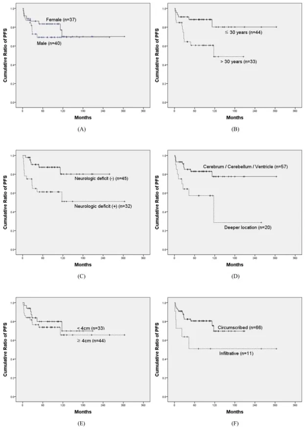
Figure 2.Progression-free survival curve for prognostic factors
years old) (p = 0.009). Younger patients had significantly better progression-free survival. (C) Progression-free survival for the presence of neurologic deficit (p = 0.005). The patients without any neurologic deficit had significantly better prognosis. (D) Progression-free survival for the presence of the location of tumors (p = 0.007). Tumors on deeper location such as thalamus and brainstem had significantly poorer prognosis. (E) Progression-free survival for the size of tumors (p = 0.459). Tumor size had no significant effect on prognosis. (F) Progression-free survival for tumor infiltration (p = 0.058). Infiltrative tumors had more tendency of progression than circumscribed tumor, but there was no statistical significance. (G) Progression-free survival for cystic component (p = 0.077). (H) Progression-free survival for gadolinium enhancement (p = 0.312). (I) Progression-free survival peri-tumorous edema (p = 0.523). (J) Progression-free survival for the presence of hydrocephalus (p = 0.568). (G), (H), (I), (J) Cystic component, gadolinium enhancement, peri-lesional edema, and the presence of hydrocephalus had no significant prognostic impact on progression-free survival. (K) Progression-free survival for extent of resection (GTR vs. STR vs. biopsy) (p < 0.001). (L) Progression-free survival for extent of resection (GTR vs. non-GTR) (p < 0.001). Extent of resection had strong correlation with progression-free survival.
Abstract
Introduction
Pilocytic astrocytomas which are WHO grade I, have benign characteristics and common in pediatric population. Because they are relatively rare in adult population, behavior and prognostic factors of adult pilocytic astrocytomas are known relatively less. Previous studies report adult pilocytic astrocytomas to behave more aggressively. Adjuvant treatment for not gross-totally resected tumor is not established. This study aims to find out prognostic factors and efficacy of adjuvant radiation therapy for adult pilocytic astrocytomas.
Methods
Retrospective data of the 77 patients who underwent surgery and were newly diagnosed with pilocytic astrocytomas at the age of 18-year-old or older between 1999 and august 2018 in single institute was analyzed. We investigated overall survival and progression-free survival, and analyzed prognostic impacts of each factors. Prognostic factors for progression-free survival and overall survival were analyzed using Kaplan-Meier survival curve. Multivariate analysis was conducted using Cox-proportional hazard model.
Results
77 patients underwent operation and were diagnosed with adult pilocytic astrcytomas. Gross total resection was achieved in 45 (58%) patients. During the median follow-up duration of 80 months, 7 (9%) patients died and 19 (25%) patients had disease progression. 5-year overall survival was 91% and 5-year progression-free survival was 77%. In univariate analysis, age of diagnosis, presence of neurologic deficit, tumor location, and extent of tumor resection were significant prognostic factors (p = 0.009, p = 0.005, p = 0.007, p < 0.001, respectively). In
resected tumors.
Conclusion
According to our study, extent of tumor resection affects the prognosis of adult pilocytic astrocytomas. Adjuvant radiation therapy failed to improve the outcome. Further studies should be conducted to establish adjuvant treatment strategy for not gross-totally resected tumors.