Orange and light blue represent Mito-FF and Mito-ff in Mito-rac fibril, respectively. In vitro toxicity analysis of Mito-FF, Mito-ff and Mito-rac in PC-3 cancer cell line.
Research background
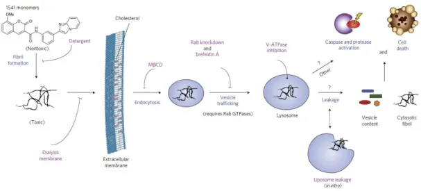
The modification of aligned amphiphilic peptide assembly with IKVAV or RGD motif enhanced neurite growth of neurons encapsulated in the scaffold, while the alignment directed these neurites toward the fiber. Schematic representation of the mechanism of cell death induced by fiber-forming small molecules, “1541”. The small molecules form fibrils in cell culture medium, dialysis or nonionic detergents can block the cellular entry of chemical fibrils (1541).
Enzyme Instructed Intra Cellular Assembly
After cellular uptake, the hydrogelator became two fragments, a hydrogel-forming fragment; Gly-Gly-Gly-His-Gly-Pro-Leu-Gly (G-C16) and a peptide fragment Leu-Ala-Arg-Lys-CONH2. The tetrapeptide segment Gly-Gly-Gly-His acts as both acceptor and donor of hydrogen bonds.
Targeting organelle with enzyme instructed intra cellular assembly
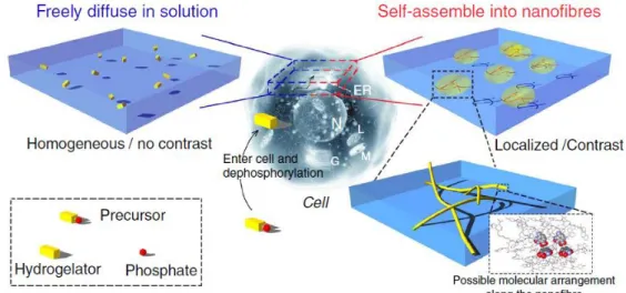
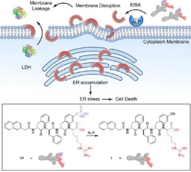
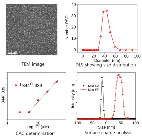
Illustrations of the enzymatically guided self-assembly of 1P to form assemblies of 1 anchored to cell membranes in (b) rod and (c) CPK models (Ref. 45). Self-assembly is an equilibrium process between individual building units and their aggregated state, and the concentration of molecules must be above the critical value to induce assembly (i.e., critical aggregation concentration (CAC)).
Organelle Localization Induced Self-assembly (OLISA)
The self-assembly occurs easily due to the high concentration experienced and undergoes nanofiber formation. The nanofiber formation occurs easily due to the increased CAC which will be characterized by electron microscopy and fluorescence imaging.

Research purpose
More about the enzyme expression level varies from cell to cell, implying that enzyme-responsive composition could not be generalized to all cancer types. In this scenario, there is an urge to find a versatile and general strategy that can be applied to all types of cancer at the lowest possible IC50.
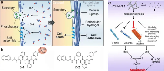
Zhou, J.; Du, X.; Yamagata, N.; Xu, B., Enzyme-directed self-assembly of small D-peptides as a multi-step process for the selective killing of cancer cells. Gao, Y.; Shi, J.; Yuan, D.; Xu, B., Imaging enzyme-activated self-assembly of small molecules inside living cells.
Mitochondria Localization Induced Self-assembly of Peptide Amphiphiles for Cellular
Introduction
Intracellular self-assembly thus requires a higher concentration of the molecules than CAC, which may limit the practical implementations of self-assembling molecules. Transformation of the molecular structure from hydrophilic to hydrophobic entities inside the cell (or pericellular space) through external stimuli (chemical or physical) is a powerful strategy to reduce CAC by increasing the propensity for self-assembly. The small molecules readily diffuse through the cell membrane, reach the target site (organelle or subcellular compartment depending on the targeting moiety), and then undergo self-assembly inside the target organelle as a result of increased local concentration.
The self-assembly process is driven by the increased mitochondrial membrane potential of cancer cells, leading to high mitochondrial accumulation of Mito-FF followed by self-assembly into fibrils.
Results and Discussion
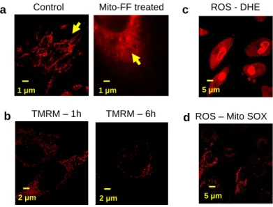
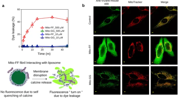
Mitochondrial co-localization of Mito-FF measured with Mito Tracker Red FM shows high localization within mitochondria (a) at 37 oC (scale bar, 2 μm) and (b) 4 oC (scale bar, 5 μm). Blue: nuclei staining, red: Mito Tracker Red FM, green: Mito-FF-NBD (left) Magnified image (middle) Rectangular view (right). Mitochondria showed severe morphological damage with fragmentation after 1 hour of Mito-FF incubation in HeLa cells (Fig. 2-8a).
This mitochondrial fragmentation induced by Mito-FF was also confirmed using MitoTracker (Figure 2-11b, middle panel).

Conclusion
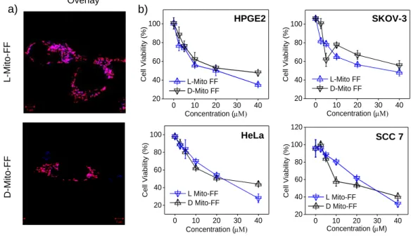
Intra-mitochondrial assembly for cancer therapy. a–d) Analysis of Mito-FF toxicity in different cancer cell lines. e–f) Analysis of mito-peptide toxicity in normal cell lines. g) Cell death induced by Mito-FF was monitored by Annexin V/PI staining, which showed early apoptosis (scale bar, 10 μm). h).
Experimental Section
IMC determination for Mito-FF (a) Emission spectra of Mito-FF in buffer/MeOH mixture (1:1) recorded to generate a calibration plot. Flow cytometric analysis of Mito-FF was obtained with Annexin/PI staining on HeLa cells after treatment with Mito-FF for the desired time point. Determination of the concentration of Mito-FF/Mito-FF-NBD in the mitochondria of HeLa cells.
The sample of Mito-FF fibrils within the isolated mitochondria was observed with a JEM-1400 operating at 120 kV.
Proteomic mapping of the human mitochondrial intermembrane space in living cells via ratiometric APEX labeling.
Mitochondria Localized Self-assembling L-peptides Overwhelm Proteolytic Degradation
Introduction
However, in light of previous reports and the general information that D analog of peptides could show better efficiency than L, we synthesized Mito-ff, the D analog of Mito-FF, under the assumption that we could achieve better tumor growth could achieve. healing efficiency than Mito-FF. Surprisingly, we observed that the efficacy of Mito-FF and Mito-off show no significant difference and show similar performance both in vivo and in vitro. Furthermore, because Mito-FF has a lower molecular weight (1054 g/mol), they enter cells via free diffusion, which is faster than other endocytosis mechanisms.
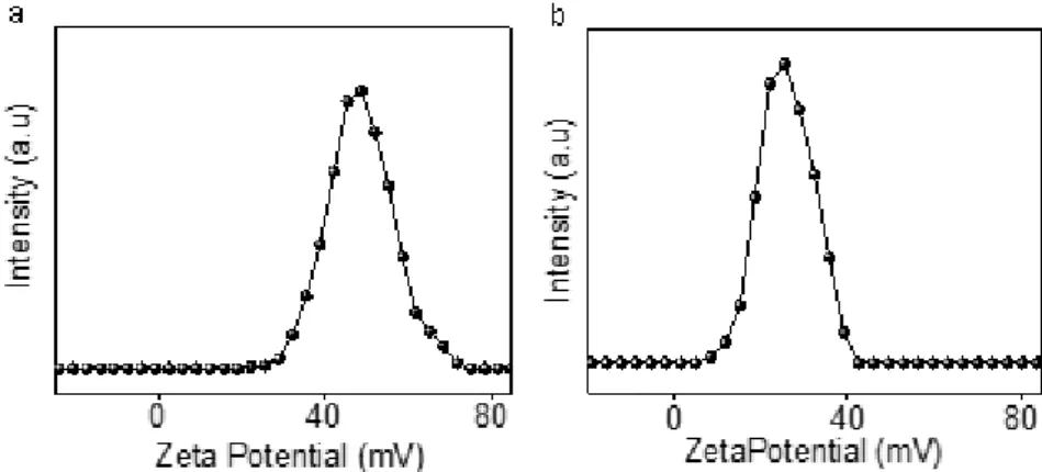
The Mito-FF fiber has a surface charge of approximately + 40 mV, which is quite feasible for specific and non-specific interactions with biomolecules that may reduce the targeting efficiency, but the molecule is not.
Results and Discussion
As we mentioned, we could not observe any difference in the cytotoxicity of Mito-FF and Mito-off; both L and D previously showed similar efficacy. The cellular cytotoxicity of Mito-FF and Mito-off in different cancer cell lines shows similar cytotoxicity after 24 hours of incubation. In vivo therapeutic efficacy of Mito-FF and Mito-off at different doses of (a) 1 mg/kg and (b) 10 mg/kg of mouse, showing similar activity for both Mito-FF and Mito-off.
This finding suggests that Mito-FF and Mito-ff share similar biological properties due to the presence of TPP and consequent mitochondrial localization. a) Molecular design of Cyto-FF and Cyto-ff.
Conclusion
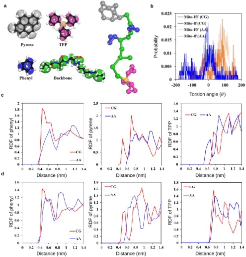
Ⅱ Force field to compare the molecular structure of the D- and L- isomers of the Mito molecule (ie, Mito-FF and Mito-ff). Mito-FF/ff racemic colocalization in the Mito-rac well occurred in the aquatic environment. To analyze the impact of Mitorac inside the mitochondria, compared to Mito-FF/ff we performed several confocal microscopy experiments.
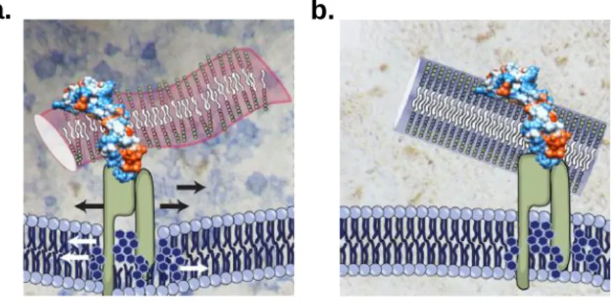
Water permeation at final configuration of penetration of (a) Mito-rac fiber bundle and (b) Mito-FF fibrils on cancer cell membrane. The amidation of Mito-FF with maleic anhydride is found to be unstable (data not shown). The mass analysis of Mito-SA confirming the successful conversion of Mito-FF to Mito-SA.
Abstract
Here, we monitored the altered morphological and biological properties of the short peptide amphiphile, Mito-FF, after their co-assembly with the mirror pair, Mito-ff. Mito-FF is an intra-mitochondrial self-assembling peptide amphiphile that induces cancer cell apoptosis followed by mitochondrial damage after intra-mitochondrial fibrillation. Mito-FF when co-assembled with Mito-ff induced a thick fiber bundle (super fiber) up to 100 nm in diameter, while the enantiomers formed a narrow fiber with a diameter of ~10 nm.
Intracellular co-administration of Mito-FF and Mito-ff resulted in drastic mitochondrial disruption as enantiomers both in vitro and in vivo as a result of intra-mitochondrial racemic co-assembly to form the Mito-rac superfibrillar structure.
Introduction
Mito-rac showed fast and rapid depolarization compared with Mito-FF or Mito-ff. A drop of 100 μM PBS solution of Mito-FF, Mito-ff, and Mito-rac was placed on a formvar/carbon-coated copper grid and allowed to evaporate under ambient conditions. Intra mitochondrial assembly followed by micelle disassembly. a) Schematic representation for the conversion of Mito-FF to Mito-SA.
TEM images after 24 h of pH 6.5 treatment showed fibers, indicating the conversion of Mito-FF to Mito-SA (Fig. 5
Results and Discussion
Conclusion
In conclusion, we have shown for the first time a heterochiral assembly of amphiphilic peptides to form superfibrous structure, which is completely different from the respective enantiomers. The superfibrous structure that is also formed within the mitochondria of cancer cells with faster kinetics leads to drastic mitochondrial damage of the cancer cells and higher in vivo tumor reduction ability.
Experimental Section
The confocal laser scanning microscopy images were acquired using LSM 780. The transmission electron microscopy images were acquired using a BioTEM system (JEM 1400). HeLa cells were seeded in a 25 ml T-flask (Thermo Scientific) in DMEM (Life Technologies) supplemented with 10% FBS and 1% penicillin/streptomycin at 37 °C under 5% CO. According to the manufacturer's protocol (Life Technologies, V13241), the cells were incubated with 5 μl Alexa Fluor 488-conjugated annexin V and 1 μl 100 μgml-1 PI working solution in 100 μl annexin-binding buffer solution for 15 min. room temperature.
The stained cells were then analyzed by flow cytometry (FACS ealibur, BD Bioscience), with the fluorescence emission measured at 530 nm (i.e. FL1) and > 575 nm (i.e. FL3).
Schematic diagram showing the pH-induced disassembly of Mito-SA-like micellar aggregates in the tumor environment. The formation of Mito-SA was confirmed by mass analysis using MALDI-TOF/TOF (Experimental; Fig. 5.6. However, in NIH 3T3 cells, toxicity was not shown suggesting higher tumor specificity of Mito-SA.
In conclusion, we have developed tumor-selective, mitochondrial self-assembling nanostructure aggregates of Mito-SA.
Intra mitochondrial assembly of peptide amphiphile via pH induced disassembly of micelle in
Introduction
As a result, Mito-FF accumulates approximately 10 times more in tumor cells compared to cancer cells and induces tumor-specific killing.7 However, such a system is not sufficient to achieve specificity during in vivo, as it can also accumulate in normal cells at higher doses, furthermore, there is the possibility of specific and non-specific interactions in the bloodstream, as biomolecules such as proteins and enzymes are negatively charged and require a much more tumor-specific design. Mito-FF is a strong candidate for OLISA and is a positively charged molecule, so in order to protect the negative charge to make it more stable during blood circulation, Mito-FF is converted to Mito-SA in which the secondary amine of the lysine side chain is conjugated with parts of succinic anhydride. Succinic anhydride cleaves below tumor pH, thus restoring the original structure of Mito-FF.
However, the micelle cannot be internalized into normal cells because it cannot be cleaved and transformed into Mito-FF under normal conditions (Figure 5-1).
Results and Discussion
However, at pH 7.4 Mio-SA showed no peak shift after 24 hours suggesting higher stability of Mito-SA (Fig. 5-4b). Therefore, we concluded that the presence of TPP facilitates acid hydrolysis of the succinyl amide of Mito-SA. Microscopic analysis showed that mitochondria were severely fragmented with a leakage of Mito Tracker from mitochondria to cytoplasm for the cell treated with Mito-SA in HeLa cells (Fig. 5-6a).
The mitochondria dysfunctional study in HeLa cells (a) Mitochondrial fragmentation monitored with Mito-Tracker Red FM showing drastic fragmentation after treatment with Mito-SA.
Conclusion
Experimental Section
Following the manufacturer's protocol (Life Technologies, V13241), the cells were incubated with 5 µL Alexa Fluor 488-conjugated annexin V and 1 µL 100 µg/mL PI working solution in 100 µL annexin-binding buffer solution for 5 min. room temperature. HeLa cells were seeded on a Lab Tek II slide chamber at 80% confluence in DMEM (Life Technologies) supplemented with 10% FBS and 1% penicillin/streptomycin at 37°C under 5% CO2. The cells were then incubated with the peptides at 10 μM for various intervals and gently washed.
The cells were then gently washed, the medium was replaced with DMEM medium, and the cells were analyzed with an FV1000 laser confocal scanning microscope.
Future Possibilities of Organelle Localization Induced Self Assembly of Peptide