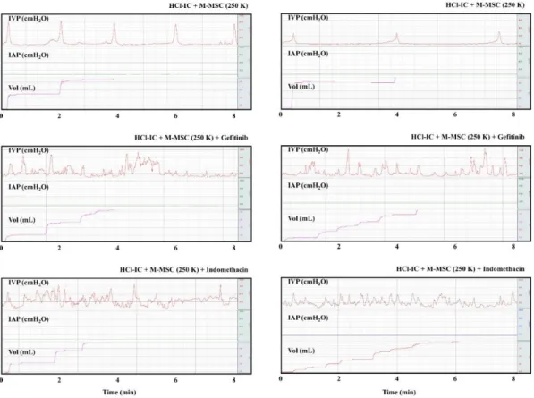
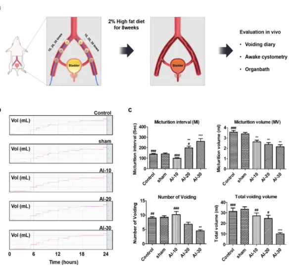
Results: Analysis of the 24-hour voiding pattern showed that in the AI-30 group the voiding interval was significantly extended (p <0.001), while the number of voids and volume of voiding significantly decreased (p <0.01 and p <0.001). . The cystometrogram showed that the frequency of micturition contractions and voiding pressure were significantly lower in the AI-30 group (p <0.01). In the organic bath study, contractile responses to various stimuli were significantly lower in the AI-20 and AI-30 groups (all p <0.001).
Histological examination showed that in the AI-20 and AI-30 group, atherosclerotic occlusion in the iliac arteries followed by tissue inflammation, fibrosis, denervation and apoptosis of the bladder muscle tissue was more prominently observed than in the sham group.
LIST OF ABBREVIATIONS
Improved efficacy and in vivo cellular properties of human embryonic stem cell derivative in a
INTRODUCTION
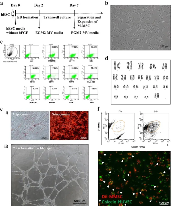
Embryonic SCs (ESCs) are embryos of embryonic stem cells that are capable of differentiating into all cell types in the human body and are able to expand into immortalized cell lines in vitro20, 21. Based on this versatility and infinite scalability, ESCs are considered a promising source for regenerative medicine22. However, safety issues based on hESC treatment will still need to be addressed, including the risk of malformations and other species capable of forming tumors, differentiation as a potential immune response, and unwanted cell types.
In this study, recovery of hESCs derived from multipotent stem cells (M-MSCs) has been demonstrated to more effectively improve the pathological characteristics of the functions of urination and IC/BPS than adult bone marrow, bladder (BM) are the cells that is induced in animal models IC/BPS is induced by instillation of hydrochloric acid (HCl).
MATERIALS AND METHODS
Briefly, after induction of anesthesia, a caged polyethylene container (PE-50, Becton-Dickinson, Parsippany, NJ, USA) was implanted into the dome of the bladder through an abdominal incision. An abdominal balloon (Latex, Daewoo Medical, Incheon, Korea) around the cuff of the catheter tip was placed proximally in the bladder to connect to the other catheter using a silk seal to capture the IAP. Static PET images were acquired over a 15 minute period from 1-5 matches in one view of the MRI range.
A small incision (less than 5 mm) was made in the upper abdominal skin, and the outer surface of the bladder was slightly exposed to any contact with the objective lens.
RESULTS
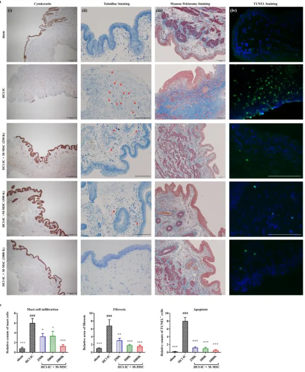
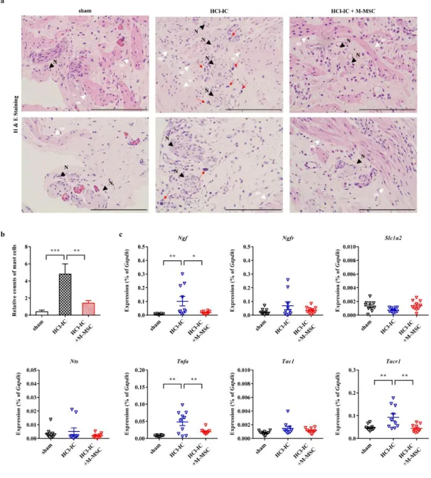
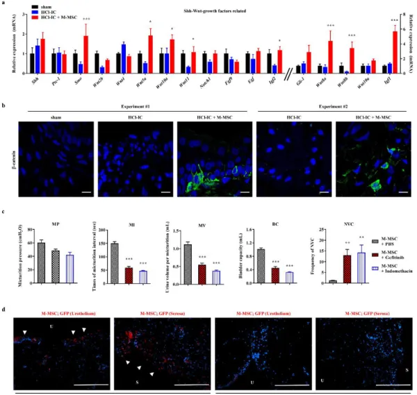
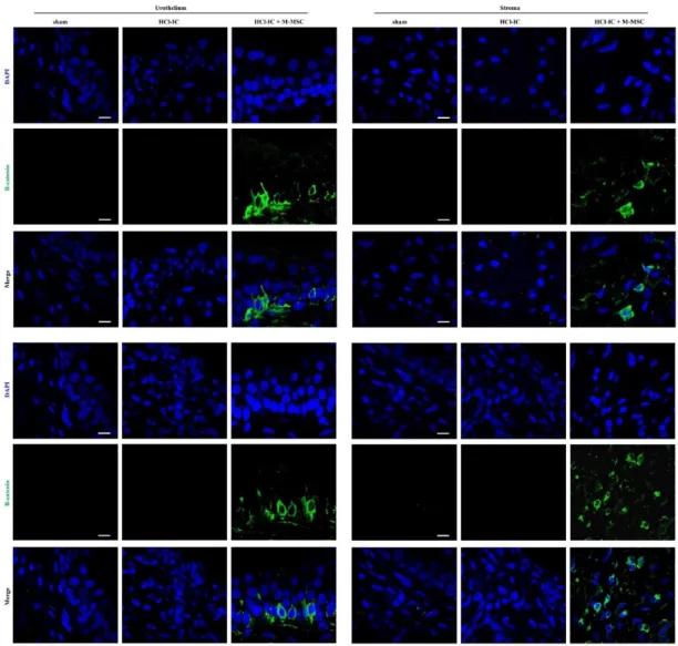
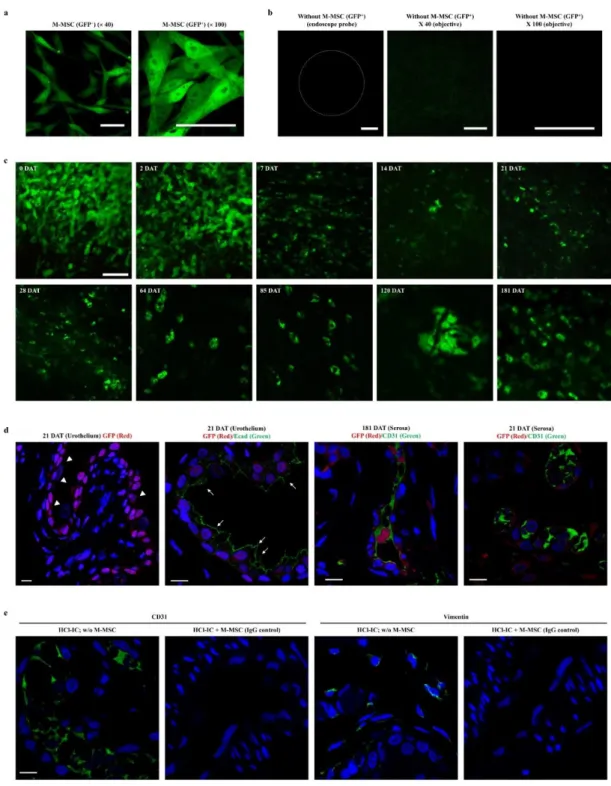
However, the anatomical interactions of mast cells and nerve fibers were significantly reduced by M-MSC treatment (Figs. 1-4a and 1-4b). Furthermore, gene expression analysis showed that bladder tissues in IC-HCl mice were characterized by increased expression of Ngf and other visceral hypersensitivity-related genes such as tumor necrosis factor and tachykinin receptor-1; however, administration of M-MSCs significantly restored their induction in bladder tissue (Fig. 1-4c). In immunofluorescence analysis of M-MSCs stably expressing green fluorescent protein (GFP), the majority of GFP+ cells in HCl-IC animals at 7 DAT were localized to the injection site between muscle and bladder serosa, and some GFP+ cells were observed in the lamina propria and urothelium, but few were detected in the muscular layer (Fig. 1-8a).
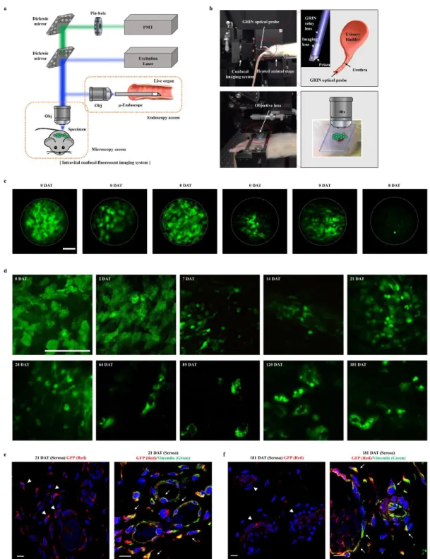
GFP+ cells found in the urothelium expressed E-cadherin in membranes showing differentiation into epithelial cells (Figs. 1-8b and 1-8c). In particular, some GFP+ cells under the serum and urinary tract are distributed in the same vascular structure (Fig. 1-8a). In order to investigate the in vivo characteristics of M-MSCs transplanted at the cellular level in living animals (Fig. 1-9a), intravital fluorescence microscopy, which allows the study of cellular processes in vivo, such as cell traffic, intercellular interactions and vascular changes36 .
Using a front view endoscopic optical probe for urothelium (Fig. 1-9b), longitudinal imaging of GFP+ M-MSC fluorescence injected over 6 months was performed. Subsequently, in vivo confocal microscopy was performed at high resolution using an objective lens aimed at the outer layer of the bladder via minimal incision of the abdominal surface (Fig. 1-9b). During the total observation period (Fig. 1–9d), some autofluorescence was observed in animals injected into the vehicle (Fig.
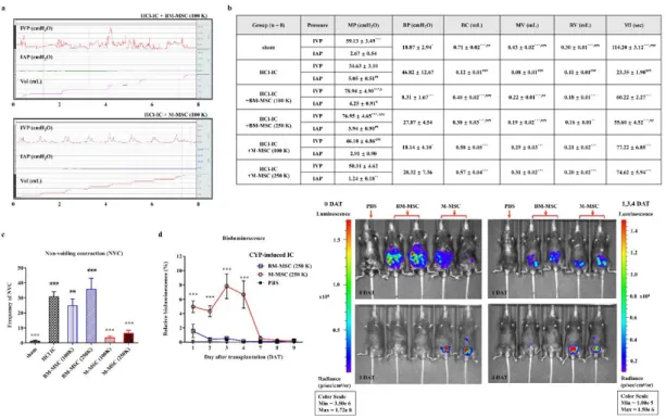
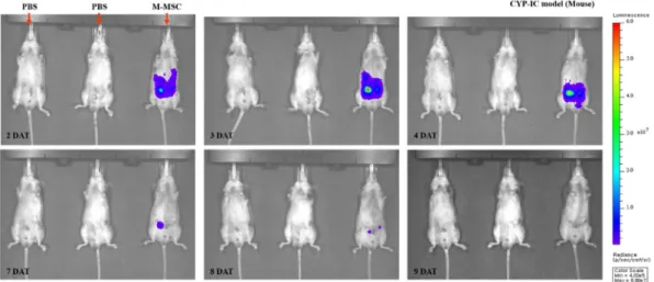
Compared to BM-MSCs, the NVC showing urinary incontinence symptoms were significantly improved in the clinical setting of low dose) (1'105) M-MSC regimens of M-MSCs (Fig. 1-11c). The bioluminescence intensities of the implanted M-MSCs were observed by an optical imaging system up to 8 days post-transplantation (DAT) (Fig. 1-12). After a rapid decrease (~5% compared to when cells were injected) in 1 DAT, the bioluminescent activity of the grafted M-MSC persisted until 4 DAT and significantly decreased after 7 DAT (Fig. 1-11d).
Compared to animals treated with BM-MSCs, animals transplanted with M-MSCs showed significant bright bioluminescence throughout the experimental period (Fig. 1-11d), indicating excellent engraftment ability of M-MSCs.
DISCUSSION
Therefore, the evaluation of pain after treatment of M-MSCs in the intestinal inflammatory condition should be performed at a later time. This critical strength has been shown to increase viability in vivo (Fig. 11d) and is an important challenge for MSCc-based therapies in adult tissue42. Similarly, about 5% of locally injected M-MSCs were detected in the bladder tissue at 1 DAT (Fig. 1-11d).
Despite these advantages, a risk of the treatment of hESC derivatives is the possibility of tumorigenesis. Similarly, long-term longitudinal nanoScanPET/MRI monitoring and thorough testing in tumor and organ transplanted M-MSCs did not reveal abnormal growth (Figs. 1-14). Taken together, clinical data from this study demonstrate that hESCs-derived M-MSCs can overcome the limitations of MSCs therapy known to date, without the deleterious consequences of hESCs derivatives.
In a recent study, several priming strategies have been developed to improve the function of MSCs derived from adult tissues48-50. The present study longitudinally investigated the cellular processes of M-MSCs transplanted into tumors for 6 months (Figs. 1–9). To overcome this drawback, endoscopy was used with a small diameter refractive index (GRIN) lens probe, which is a noninvasive method for visualizing intact tissue (Fig. 1-9b).
It is important to note that the majority of transplanted cells were observed as multiple cells on the bladder surface up to 1 month after injection, but were distributed locally up to 2 months after transplantation (Fig. 1-9d). With the rapid development of SC research, successful clinical application of SC treatment in urology is expected in the near future12, 52, 53.
Development of reliable chronic bladder ischemia rat model for reproducing the
The presence of peripheral nerves in the muscle layer of the bladder was assessed by staining with N,N-dipropyl-2-[4-methoxy-3-(2-phenylethoxy)phenyl] ethylamine (SML0631; NE-100, Sigma-Aldrich). In the AI-10 group, micturition interval (MI) tended to be shorter than in the sham group, but the difference was not statistically significant (Figure 2-1C). More importantly, in the AI-30-treated group, the number of urinations and the total volume of urination were significantly reduced compared to those in the sham group (p <0.01 and p <0.001; respectively).
In the AI-10 group, voiding contractions were more frequently observed (Fig. 2-2a), and the MI was significantly shorter than that of the sham group (p < 0.001; especially in the AI-30 group, both maximal pressures and voiding pressures were significantly reduced (p. The mean contractile responses to KCl in the AI-10, AI-20 and AI-30 groups decreased in accordance with the degree of vascular injury and were significantly lower than in the sham group (all p < 0.001 Fig. 2- 3a).
The contractile responses to 1 mM ATP in the AI-10, AI-20 and AI-30 groups were significantly lower than in the sham group (all p.. lt;0.001) and were lower according to the degree of vascular injury (Fig .The mean iliac artery wall thickness in the AI-10, AI-20, and AI-30 groups were significantly greater than in the sham group (all p < 0.001) and showed a trend toward greater thickness with higher degrees of high vascular damage (Fig. 2-4f). Compared with the sham group, bladder tissues in the AI-10, AI-20, and AI-30 groups demonstrated significantly increased mast cell infiltration stained with toluidine blue (Fig. .
Masson's trichrome staining of bladder tissue revealed an increased percentage of collagen in the muscle layer in the AI-10, AI-20, and AI-30 groups compared to the sham group (Figure 2-4c). In the AI-10 group, micturition contractions were observed more frequently (Figure 2. 2a) and MI was significantly shorter than in the sham group (Figure 2-2b). In particular, in the AI-30 group, micturition pressure was significantly reduced and RV was significantly increased compared to the sham group (Figure 2-2b).
In addition, significant atrophy of the bladder detrusor muscle was also observed in the AI groups in this study (Figs 2-4e and 2-4j).
CONCLUSIONS
Mesenchymal stem cells protect against ketamine-induced cystitis tissue fibrosis in the rat bladder. Kim, H.-S.et al.Clinical trial of umbilical cord blood-derived stem cells for the treatment of moderate to severe atopic dermatitis: Phase I/IIa studies. Zhu, H. et al. Therapeutic effects of human umbilical cord-derived mesenchymal stem cells in mice with acute lung injury.
He, Z. et al. Intravenous hMSCs ameliorate acute pancreatitis in mice via secretion of tumor necrosis factor-α stimulated gene/protein 6. Stem cells display functional characteristics of pericytes and improve retinal vasculature in a rodent model of diabetic retinopathy. Maeda, D. et al. Hunner-type (classical) interstitial cystitis: a distinct inflammatory disorder characterized by pancystitis, with frequent expansion of clonal B cells and epithelial denudation.
Human embryonic stem cells and their differentiated derivatives are less susceptible to immune rejection than adult cells. Kang, H. et al. The therapeutic effects of human mesenchymal stem cells primed with sphingosine-1-phosphate on pulmonary artery hypertension. The paracrine effects of mesenchymal stem cells stimulate the regenerative capacity of endogenous stem cells in the repair of a bladder outlet obstruction-induced overactive bladder.
화학적으로 유발된 쥐의 방광염에 대한 인간배아줄기세포유래 다능성 중간엽줄기세포의 치료효과. 방광통 증후군의 전임상 모델에서 인간 배아 줄기 세포 유도체의 생체 내 특성 조사 및 치료 효과 및 효능 연구.