To examine the conditions under which proliferation was enhanced, corneal endothelial cells were tested using a ROCK inhibitor and hMSC-derived conditioned medium. TGF-β inhibitor and BMP-7 were also used to maintain the morphology of corneal endothelial cells. Finally, spheroids were prepared using microwells and cultured to confirm the morphology of corneal endothelial cells.
As a result, it was confirmed that rabbit corneal endothelial cells maintained their proliferative capacity and cell morphology when cultured in combination with ROCK-inhibitor and hMSC-. Based on these results, a culture method capable of maintaining corneal endothelial cell function during in vitro proliferation was established.
Research background
A corneal transplant can only be performed if the donated cornea is secured by an organ donation, as with other organ transplants. The donor cornea is always underdeveloped in the current situation, when organ donation is not active, and patients with diseases that can only be treated with corneal transplantation, unless they receive some donated cornea. As a result of investigations by the Korean Network for Organ Exchange (KONOS), about 600 corneal transplant operations were performed per year, but 2,000 people waiting for corneal transplants were investigated, which tripled.
In addition, it is cornea donated by domestic donors, approximately 50% of the donated cornea is used for corneal transplantation, and the remaining 50% is cornea donated from abroad.
![Figure 1. Structure of Cornea[8]](https://thumb-ap.123doks.com/thumbv2/123dokinfo/10443223.0/15.892.208.711.757.1088/figure-1-structure-of-cornea-8.webp)
Tissue engineering approach for corneal endothelial regeneration 3
Rho-associated kinase (ROCK) inhibitor
Y27632 is a ROCK inhibitor to fall into the eye of the rabbit, a study of the initial damage to the corneal endothelium has been recovered.[20] And there are studies that the number of proliferating cells increases when Y27632 is treated in CEC injury model and corneal transparency increases to restore function.[26] Further, in case of treatment of Y27632 in vitro culture conditions rabbit CECs, while the process continues to cell cycle G1 phase in S phase (DNA synthesis), adhesion of cells has been improved.[27]. It has also been reported that Y27632 increases proliferation and adhesion in human corneal endothelial cell in vitro culture [28].
Mesenchymal stem cell (MSC)-derived conditioned medium (CM) 4
Similarly, umbilical cord blood (UCB) MSCs are able to “home” to areas of damaged corneal endothelium and can be redifferentiated towards HCEC-like cells. CECs often adopt a fibroblast-like morphology in culture, a process that has been attributed to endothelial-to-mesenchymal transition (EndMT).[33]. Transforming growth factor-beta (TGF-β) and FGF-2 have shown that it is possible to block many pro-EndMT signaling pathways to improve the morphology, phenotype and function of cultured CECs.
The relevant activation of the Wnt/β-catenin pathway appears to be a good candidate to explain many of the phenotypic events observed in in vitro CECs.[33, 34] EndMT can occur postnatally in a variety of pathological settings Endothelial-to-mesenchymal transition-derived cells are thought to function as fibroblasts in damaged tissue and may therefore play an important role in tissue remodeling and fibrosis.[35] The application of SB431542, a selective inhibitor of the TGF-β receptor, reversed the fibroblastic phenotypes to the normal contact-inhibited monolayer, and these polygonal cells maintained endothelial physiological functions. BMP-7 also reversed the fibroblastic phenotypes to the normal endothelial cells with contact-inhibited monolayer and characteristic endothelial adhesion.[36].
Corneal Endothelial Sphere-Forming assay
Thus, the sphere formation assay may contribute to obtaining the new cells needed for regenerative medicine.[38]
Objective of this study
Materials
Methods
- Effect of ROCK inhibitor and mesenchymal stem cell-
- Primary and culture of rabbit corneal endothelial cells · 9
- ROCK inhibitor and MSC-conditioned medium effect
- Effect of medium exchange after RCECs expansion
- RNA sequencing
- Human corneal endothelial cells culture and maintenance
- Effect of hMSC-conditioned medium on HCECs
- Optimization of HCECs culture medium
- Effect of TGF-β inhibitor and BMP-7 treat on HCECs
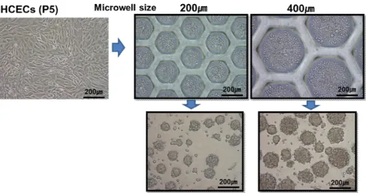
- Corneal endothelial cell spheroids culture using microwell · 13
- Cell density vs spheroids culture
- Different size spheroids formations
- Analysis
- β-galactosidase cell senescence staining
- Immunofluorescent analyses
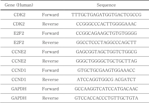
- RT-PCR
- Statistical analysis
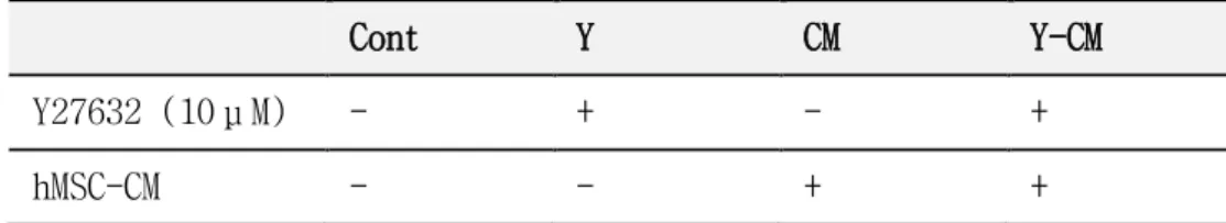
After RCECs were cultured to the 8th passage under Y-CM group conditions, the cells were replaced with the medium of the above four conditions (Cont, Y, CM, Y-CM). Under four conditions of the previous experiment, Cont and Y were prepared as passage 3 with reduced proliferation, and CM and Y-CM were prepared as passage 5. Primary and culture of human corneal endothelial cells (HCECs) HCECs were isolated from human corneal tissue, Descemet's membrane including CECs was removed and digested at 37℃ overnight with 0.02%
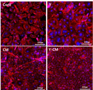
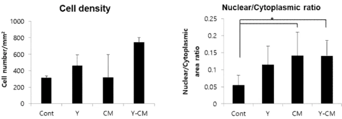
The HCECs were grown in a humidified atmosphere at 37℃ in 5% CO2, and the culture medium was changed every 2 days. To test the MSC-CM effect, the cultured HCECs were passaged with medium supplemented with or without hMSC-CM (50%) and then evaluated after 1 week. In each medium, the effect of the effect of Y27632 hMSC-CM was tested.
To test the anti-fibroblastic effect, the cultured HCECs were passaged with medium supplemented with or without 1μM Repsox (selective TGF-β inhibitor) and 0.1μg/ml BMP-7 and then evaluated after 1 week. StemFIT 3D was purchased from Microfit (Seoul, Korea), which is a full size of 15X15mm inside the well is a concave structure for 400um. Cells were examined microscopically on days 1, 3, and 7, and actin staining, β-galactosidase staining, and immunofluorescence were performed on day 7 of culture.
2X10⁴ cells were seeded on 12-well plates of RCECs, cultured for 7 days under each condition and stained with Senescence β-Galactosidase staining kit (Cell Signaling Technology). All the statistical analyzes were performed with OriginPro 8.0 software using one-way ANOVA analysis, a probability value of 0.05 was considered statistically significant (*, P < 0.05). Effect of ROCK inhibitor and mesenchymal stem cell-derived conditioned medium on rabbit corneal endothelial cells.
Effect of ROCK inhibitor and mesenchymal stem cell-
- Cell morphology
- Cell proliferation
- Cell Functions
- Medium exchange effect
- RNA sequencing
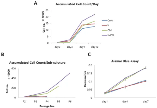
Cell density and size graph of RCECs cultured with or without ROCK inhibitor and conditioned medium. In the early stage of culture (passage 2), cell proliferation was confirmed by cell counting, and Y-CM group showed the highest proliferation (Fig 6A). In the AlamarBlue test, the CM and Y-CM group showed more than twice as much proliferation as the Cont and Y groups.
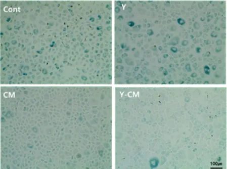
On the other hand, there was no significant difference between the groups with and without Y27632 treatment (Fig. 6C). As a result of confirming the expression of the marker of apoptosis and cell proliferation through immunofluorescent staining, caspae-3 was not expressed in all groups and Ki67 showed high expression in the CM and Y-CM group (Fig. 7). The number of stained cells was counted, and the largest number of cells in the Cont group were stained, which stained more than 10 times more than Y-CM (Fig. 9).
As a result, ZO-1, a tight junction marker, was not expressed at the plasma membrane in all groups (Figure 10). However, it was confirmed that ZO-1 was well expressed on the plasma membrane when the cells were cultured under the same condition they were cultured on the collagen film (Figure 11). ZO-1 was clearly expressed in the Cont group and was not clearly expressed in CM and Y-CM. On the other hand, the expression of Na+K+-ATPase, indicating pump function, was confirmed in only a few cells in the CM and Y-CM groups in plate culture.
Expression of the function-related marker, ZO-1(green), Na+K+- ATPase(red) by immunofluorescence staining in exchanged culture medium after proliferation. When examining the similarity of gene expression between corneal endothelial cells and subcultured cells, the most similarities were observed when cultured in Y-CM (figs. 15, 16). CM and Y-CM, Cont and Y showed more similar results, respectively. The expression of PCNA and Ki67, the genes associated with cell proliferation, was the highest in Y-CM conditions.
Tight junction gene expression was also higher in CM and Y-CM, while ATP1B2, a gene for pump functions, showed a lower difference but higher expression in Y27632-treated group (Fig. 17). In addition, Y-CM showed the highest expression in the gene expression analysis associated with the cell cycle pathway (Fig. 18).
Human corneal endothelial cells culture and maintenance
- Effect of hMSC-CM on HCECs
- Effect of TGF-βinhibitor on HCECs
- Optimization of HCECs culture medium
- Effect of BMP-7 on HCECs
TGF-β inhibitor, Repsox was treated on HCECs already fully fibrotic at passage 6 to confirm whether morphological change was inhibited. However, with the morphology having already changed, it did not return to the original polygonal shape. In order to confirm whether the shape is maintained when subcultured before changing the shape, an experiment was performed in which Repsox was treated from passage 1 (Fig. 21A). In this case, the morphological changes of the cells were not inhibited.
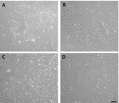
DMEM/F12 and OptiMEM-I (Opti) were compared to optimize media conditions in which HCECs maintain morphology and proliferation is increased. At passage 2, the initial cell culture stage, no significant difference in morphology was observed (Fig. 22A). There was no significant difference in cell proliferation between groups, but DMEM/12 was found to be slightly lower in proliferation (Fig. 22B).
In the DMEM/F12 medium, the cells showed fibrotic morphology and proliferation was also reduced (Fig. 23). Due to genes related to the cell cycle, there was no significant trend of relationship between types of medium and presence or absence of CM treatment (Fig. 24). There was no significant difference between the groups in the cell morphology, and there was no significant difference in proliferation (Fig. 25).

Corneal endothelial cell spheroids culture using
Optimization of CEC cell number on Microwell
Comparison of spheroid and single cell cultures
Different size spheroids culture effect
Because of rabbit-derived cells, it is necessary to apply it to human corneal endothelial cells. Rabbit and human corneal endothelial cells showed different proliferative effects on hMSC-CM in vitro. Human corneal endothelial cell sheets for transplantation: thermo-responsive cell culture carriers to meet cell-specific requirements.
Thermo-responsive poly(NiPAAm-co-DEGMA) substrates for gentle harvesting of human corneal endothelial cell plates. The effects of Rho-associated kinase inhibitor Y-27632 on primary human corneal endothelial cells propagated using a dual media approach. Promoting the expansion and function of human corneal endothelial cells with an orbital adipose tissue-derived stem cell conditioned medium.
Effects of corneal stromal cell and bone marrow-derived endothelial progenitor conditioned media on corneal endothelial cell proliferation. Inhibition of TGF-beta signaling allows for in vitro expansion of human corneal endothelial cells for use in regenerative medicine. Human corneal endothelial cells utilize p27 (Kip1) phosphorylation at both Ser10 and Thr187 sites for FGF-2-mediated cell proliferation via PI 3-kinase.
Effect of overexpression of the transcription factor E2F2 on cell cycle progression in rabbit corneal endothelial cells. In vitro evaluation of the interactions between human corneal endothelial cells and extracellular matrix proteins. Effect of shear stress on corneal endothelial cell attachment in association with corneal endothelial cell loss after laser iridotomy.