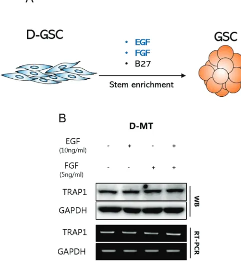
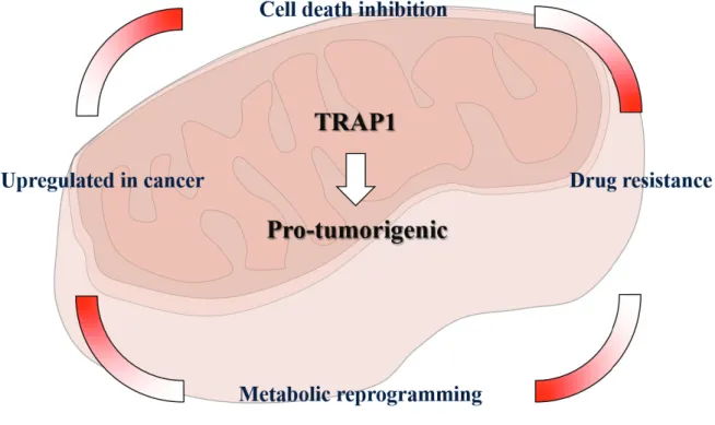
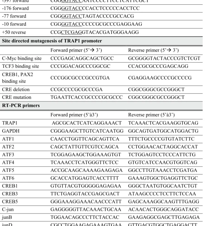
Protective functions of TRAP1 against cancer cells include regulation of reactive oxidative species, influence on energy metabolism, and prevention of cell death leading to drug resistance. Subsequently, specific TRAP1 inhibitors were introduced with high cancer targeting while exhibiting low cytotoxic effect on normal cells, indicating TRAP1 as a novel cancer drug target protein. Previously, TRAP1 has been found to be overexpressed in glioblastoma cancer stem cells (GSCs) compared with differentiated glioblastoma.
In study 1, we investigated the transcriptional mechanism of TRAP1 in cancer, based on the fact that TRAP1 is upregulated in the transcriptional level of GSC. Using the luciferase reporter assay, we specified the TRAP1 promoter and identified that the c-AMP-responsive element (CRE) is an essential regulatory element for TRAP1 expression. Since TRAP1 is a key mitochondrial monitor of homeostasis, we can understand communication between cellular transcription signal and mitochondrial stress response by finding out TRAP1's regulatory mechanism.
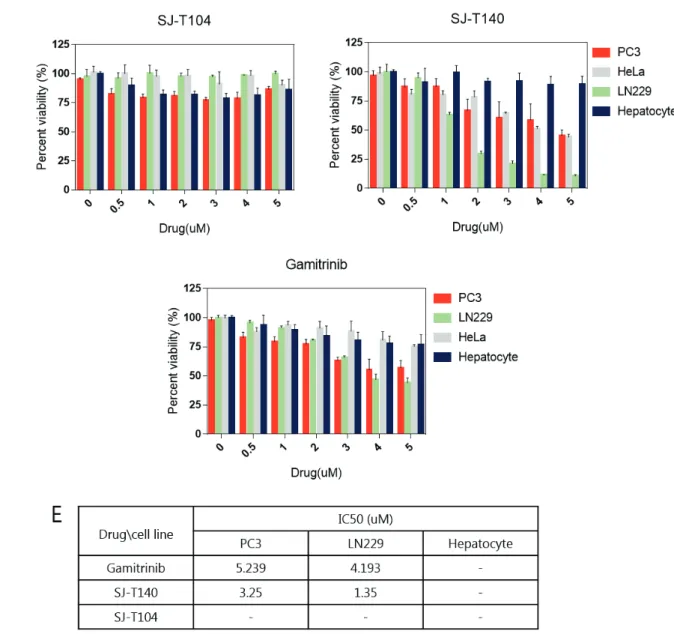
We analyzed specific binding to TRAP1 in vitro by the fluorescence polarization assay and the ability to inhibit TRAP1 function in cancer cells by observing client protein stability. Through the xenograft assay, we recognized that SJ-T140 had enhanced in vivo activity compared to the positive control, gamitrinib.
List of tables
Chapter1. Transcription regulation of mitochondrial chaperone TRAP1]
Introduction
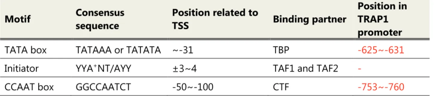
Promoter is the genomic region consisting of core promoter and proximal promoter that has the ability to transcribe certain genes. & Stark, 2018) Core promoter can be categorized into canonical core promoter and non-canonical core promoter. Canonical promoter contains general transcription factor binding motifs such as TATA box, initiator and TFIIB recognition element (BRE). Ponjavic et al., 2006 ) Despite the TATA box largely retaining its consensus, few eukaryotic promoters contain it.
An initiator considered a TFIID binding element, such as the TATA box (Roeder, 1996), has the consensus YYA(+1)NT / AYY. The downstream promoter element (DPE), one of the core promoter elements, is located with the initiator located at +28 to +32 from the TSS and supports the binding of TFIID to the initiator. The University of California Santa Cruz (UCSC) Genome Browser is an online database where researchers can access the results of the ENCODE project.
The UCSC Genome Browser provides transcription factor binding sites, histone marks, chromatin accessibility, DNA methylation, RNA expression, RNA binding, and indicators of cellular state ( Raney et al., 2010 ). Although the exact transcription factors and regulatory elements may be different depending on the cell types, the information provided by the genome browser is very useful for determining gene regulatory elements and putative transcription factors.
Material and method
- Cell culture
- Total RNA extraction and Reverse Transcriptional – PCR (RT-PCR)
- promoter construct cloning
- Immunoblotting analysis
- Promoter reporter assay
- siRNA treatment
- Software for analyzing TRAP1 promoter
Whole lysate from cells was resuspended in RIPA with protease inhibitor cocktail and phosphatase inhibitor cocktail (R&D system). All luciferase reporter constructs were generated by PCR using the primer sets described in Table 1. Cells were harvested after 36 h with passive lysis buffer in the Dual Luciferase Ratio Assay System (Promega).
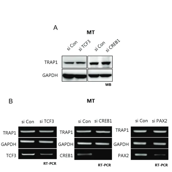
Luciferase assays were performed with the Dual-Luciferase Reporter Assay System (Promega) according to the manufacturer's instructions. Promoter activity is measured by the Relative Light Unit (RLU); Firefly (Promoter construct) / Renilla (pRL-SV40). For the RNA interference, the following siRNA is applied: TCF3 5'- GCGGAGAAAUGGAAACAUA -3', CREB1 5'- AACAGTATTCTGTAGGATCTA -3', CREB5 5'- ACGGTGCCAACTCCAATTTAC - 3', PAX2 5'- GAAGUCAAGUCGACUGACUGACUGACUGACUGACUGA, AAGUCAACUGACUGA -3' Cells are plated in 6mm2 dish and incubated for 12 hours until si RNA treatment, G-fectin (Genolution) mixed with siRNA 1:1, injected into the cell.
Result
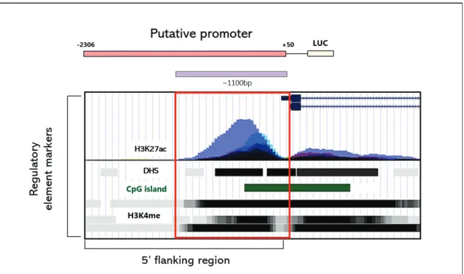
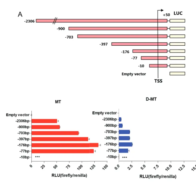
To understand the transcriptional regulation of TRAP1, we first studied the regulatory elements of the TRAP1 gene near the TSS. According to the H3K4me, H3K27ac and DNase I hypersensitivity site (DHS), the 5' flanking region of the TRAP1 gene regulatory element adjacent to the transcription start site (TSS) has putative regulatory elements. We decided to start searching the TRAP1 promoter from -2306 to fully encompass the desired region.
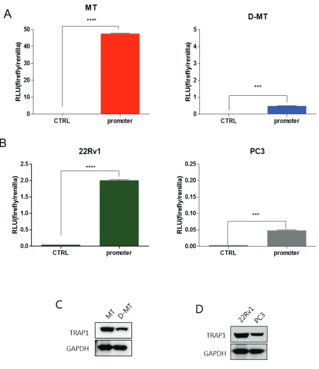
However, the TRAP1 promoter lacks the consensus or predicted consensus is located at irrelevant locus. Below the TRAP1 gene and 5' flanking region, partial TRAP1 5' flanking region, exon 1 and partial intron 1 appear in the enlarged image. To confirm that putative TRAP1 promoter is functioning properly, we performed luciferase reporter assay with various cancer cell lines.
In cell MT, deletion of 1406 base pairs between -2306/+50 and -900/+50 did not alter TRAP1 promoter activity, indicating that this region does not function as a promoter. However, by removing an additional 200 base pairs, the promoter activity increased, suggesting the possibility of the presence of a negative regulatory element. Subsequent deletion showed a weak decrease in promoter activity, but still the -77/+50 construct retained 76% of the maximum promoter activity.
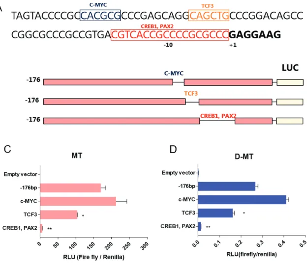
However, quantitative promoter activity was much higher in MT, proportional to TRAP1 expression in both cells. Deletion of promoter region including putative transcription factor binding sites showed a significant reduction in promoter activity. Deletion of the C-Myc binding site showed 25% higher promoter activity compared to -176/+50 construct, while deletion of the TCF3 binding site was reduced to 61% of the original construct.
Promoter activity almost disappeared in the third construct, with only 3.7% of the original construct displayed. Of the three candidates, CREB1 and PAX2 binding sites had the greatest effect on promoter activity. Deletion of the C-Myc binding site increased transcriptional activity, whereas deletion of the TCF3 binding site and the CREB1 and PAX2 binding sites decreased promoter activity.
To specify the most crucial element of the TRAP1 promoter, we compared the homology of the TRAP1 promoter across the species. Figure 5B,C) However, MT showed much higher promoter activity quantitatively, suggesting that the overall trans-acting element responsible for TRAP1 expression would be activated in GSC.
Discussion
Chapter 2. Development of TRAP1 inhibitor for future cancer drug]
- Immunoblotting analysis
- MTT assay
- Fluorescence polarization
- Analysis of mitochondrial ROS and mitochondrial membrane potential
- Tumor Xenograft Experiments
Cytotoxic effect of the anti-cancer drug was determined using MTT tests and was calculated by measuring the absorbance of the tetrazolium at 595 nm. To measure mitochondrial membrane potential and mitochondrial ROS induced by drugs, PC3 cells were treated with SJ-T140 and gamitrinib 10 μmol for 6 h and incubated with 100nM Mito-Sox (Invitrogen, M36008) or 100nM of TM50 mint at 50 . °C. To accumulate drugs in mitochondria, we conjugate SJ-T104 with mitochondrial targeting molecule, triphenylphosphonium (TPP), and this compound is SJ-T140.
Interestingly, SJ-T140 had much higher cancer cytotoxicity than SJ-T104, because TRAP1-specific binding moiety SJ-T140 directly inhibits TRAP1 upon drug accumulation in mitochondria. -T140 showed relatively higher anticancer effect than gamitrinib in both prostate cancer and glioblastoma cancer cell lines. Three chemicals showed innocuous toxicity to hepatocytes at the dose shown, while SJ-T140 and gamitrinib showed a cytotoxic effect on cancer cells.
Compared to DMSO, both gamitrinib and SJ-T140 reduce membrane potential as severe energy depletion occurs due to mitochondrial dysfunction. We had discovered TRAP1 inhibitor SJ-T140 that specifically binds to TRAP1 in vitro and inhibits TRAP1 function in cancer cells. We performed a xenograft assay to find out the inhibition ability of SJ-T140 on tumor progression in vivo.
Accordingly, SJ-T140-treated mouse groups showed dramatic growth inhibition, but gamitrinib, which showed similar cell toxicity and mitochondrial dysfunction, was unable to suppress tumor growth. Mitochondrial membrane potential and mitochondrial ROS were measured in PC3 cell line after 6 h exposure to 10țmol of TRAP1 inhibitor SJ-T140 and positive control gamibrinib. Conjugating Hsp90 inhibitors with mitochondrial targeting chemicals, TPP, these inhibitors showed a higher anti-cancer effect.
In this study, SJ-T104, the precursor of SJ-T140, was discovered as TRAP1 inhibitor by chemical screening and by adding TPP, SJ-T140 obtained cancer cell toxicity. Compared with gamitrinib, mitochondrial Hsp90 inhibitor, SJ-T140 has a slightly enhanced cytotoxic effect with similar mitochondrial dysfunction. Thus, we identified novel TRAP1 inhibitor SJ-T140 as improved in vivo activity against cancer drug candidate.
HH&3DUN+.-HRQJ+/LP-/HH$-&KHRQ.<.LP1''HYHORSPHQWRID PLWRFKRQGULDWDUJHWHG +VS LQKLELWRU EDVHG RQ WKH FU\VWDO VWUXFWXPDQWHVR$5 &KHPLFDO6RFLHW\. 3RQMDYLF - /HQKDUG % .DL & .DZDL - &DUQLQFL 3 +D\DVKL]DNL < 6DQGHOLQ $ 7UDQVFULSWLRQDO DQG VWUXFWXUDO LPSDFW RI 7$WLDWHLQDQDL 7$7LQDQ RUH SURPRWHUV*HQRPHELRORJ\5. 6DQGHOLQ$&DUQLQFL3/HQKDUG%3RQMDYLF-+D\DVKL]DNL< +XPH'$0DPPDOLDQ 51$ SRO\PHUDVH , FRUH SURPRWHUV LQVLJKWV IURP JHQWRPHWHVHGL.
7DOGRQH 7 3DWHO 3 ' 3DWHO 0 3DWHO + - (YDQV & ( 5RGLQD $ *HZLUWK ' ([SHULPHQWDODQGVWUXFWXUDOWHVWLQJPRGXOHWRDQDO\]HSDUDORJXHVSHFLILFLW\DQGDIILQLW\LQ WKH+VSLQKLELWRU VVHULHV-RXUQDORIPHGLFLQDOFKHPLVWU\.