To our knowledge, this is the first study of SARS-CoV-2 whole-genome sequencing by the ONT approach in Kazakhstan. It was then renamed SARS-CoV-2 by the International Committee on Taxonomy of Viruses (ICTV) due to its genetic relevance to previously verified coronaviruses (Lu et al., 2020). The genomic surveillance is critical in the representation of the genetic diversity of circulating SARS-CoV-2, which in form reflects the clinical and epidemiological situation in the country (Yegorov et al., 2021).
The SARS-CoV-2 whole-genome sequencing by Oxford Nanopore Technology was performed for the first time in Kazakhstan by the National Laboratory of Astana (NLA) at Nazarbayev University (Nur-Sultan, Kazakhstan) and aimed to perform further monitoring of viral circulation in the country. The complete genome sequencing facilitated the advancement of RT-PCR assays for SARS-CoV-2 investigation to standardize the diagnostics of the COVID-19 outbreak (Lu et al., 2020). More comprehensive studies of spike protein are expected to expand the current understanding of viral infection and immune escape of SARS-CoV-2.
Taxonomy
Epidemiology and surveillance
SARS-CoV-2 genome sequencing provided important data on the viral mutation rate, transmission dynamics and its taxonomic origin. Taken together, cumulative studies have facilitated the establishment of nomenclature systems for different SARS-CoV-2 lineages (Rambaut et al., 2020). The evidence confirming the reinfections suggests that different SARS-CoV-2 strains are capable of infecting the same person (Tillett et al., 2021) (To et al., 2020).
The main advantages of this platform are long genome reads, an optimized analysis pipeline, rapid sequencing and data collection (Li et al., 2020).
Viral genome sequencing
This method also has a high false-negative rate in clinical settings, which may lead to the spread of disease through delayed patient isolation and treatment, facilitating further transmission of the virus (Wang et al., 2020). In conjunction with various sequencing techniques, the current third-generation sequencing of the entire SARS-CoV-2 genome using Oxford nanopore technology is one of the main approaches. Advances in next-generation sequencing (NGS) offer opportunities for the ubiquitous detection of pathogens directly from clinical samples (Goldberg et al., 2015; Forbes et al., 2017).
The sequencing step itself usually takes from 1 to 6 days, depending on the length of the reads, the platform and the amount of data obtained (Petersen et al., 2019). Viral genome surveillance is essential to monitor disease transmission during major outbreaks (Gardy et al., 2015). Real-time gene sequencing is a key parameter for managing viral outbreaks because it expands our understanding of virus propagation, spread and evolution (Dudas et al., 2017) (Gardy et al., 2015).
Rapid and reliable sequencing techniques in clinical application are crucial for epidemiological surveillance (González-recio et al., 2021). In conjunction with different sequencing techniques, third-generation sequencing of the SARS-CoV-2 whole genome by Oxford nanopore technology is currently one of the prominent approaches. Oxford nanopore technology (ONT) is based on nanopore channels for sequencing, the ionic current passes across the flow cell, so as the nanoscale genetic polymer passes through the nanopore, each nucleotide base is distinguished according to the changes in current (Laver et al. , 2015).
Moreover, ONT sequencing could detect an unexpected diversity of structural variation of viral samples that was supported by verification from short-read sequencing on corresponding samples (Bull et al., 2020). Further improvements simplified library preparation steps, reduced hands-on time and extended sample multiplexing up to 96, reducing reagent cost to almost £10 per sample, which increased the availability of this technique for epidemiological surveillance and further studies (Brejová et al. , 2021).
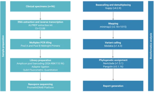
2 MATERIALS & METHODS
- Sample collection
- ONT Library Preparation and Sequencing
- Software setup and installation
- Demultiplexing of multiple barcoded samples
- Variant calling and phylogenetic profiling
- SDM analysis
- Molecular dynamics simulation analysis
Two midnight primer pools were used to anneal to 4.5% of the genome and produce 1200 bp amplicons overlapping by approx. 20 bp. The ARTIC sequencing data obtained by the Midnight protocol were analyzed by the wf-artic bioinformatics pipeline (https://artic.network/ncov-2019/ncov2019-bioinformatics-sop.html. The installation of the software on the Linux operating system is straightforward and supported on GirdION and PromethION devices.
Additionally, the ARTIC pipeline has been adapted to use a primer scheme that specifies the sequencing primers used in Midnight protocol and their genomic localization on the SARS-CoV-2 genome. The wf-artic pipeline classifies the sequenced samples by Nexclade clastid analysis and Pangolin lineage assignment. To prevent rebasing, the software copies the reads pertaining to each barcode to the corresponding label output directory.
Since the Midnight protocol uses a fast barcode kit, the demultiplexing step does not need barcodes at both ends of the sequence. The pipeline output includes NextClade (https://clades.nextstrain.org/ ) and Pangolin analysis that includes the clade designation according to GISIAD (https://www.gisaid.org/ ) and Pangolin. To run the Site Directed Mutator, the wild-type structure in PDB format must be submitted to the http://marid.bioc.cam.ac.uk/sdm2/ web server.
To run the Desmond simulation (https://www.schrodinger.com/), the structure file in PDB format was first imported. The mutations of the sequenced samples were entered into the wild-type PDB file in PyMOL (https://pymol.org/2/) and loaded into the software.
AIMS OF THE THESIS PROJECT
RESULTS
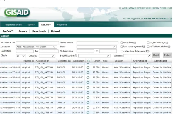
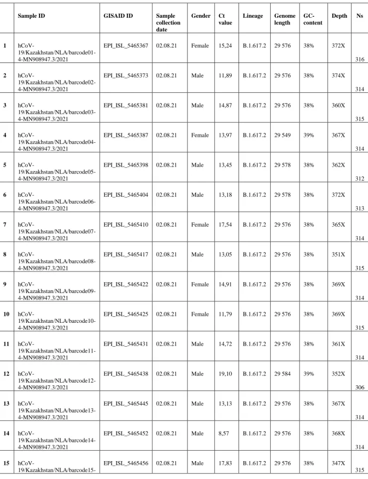
- The 77 genetic sequences upload on GISAID A)
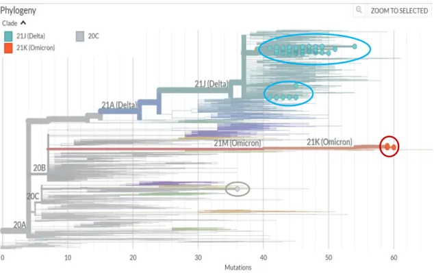
- Phylogenetic tree
- Lineage distribution
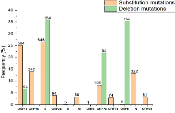
- Frequency of mutations in SARS-CoV-2
- Spike glycoprotein of EPI_ISL_5465615
- Protein stability analysis by SDM upon mutation A)
- Protein-Ligand Molecular Dynamics analysis A)
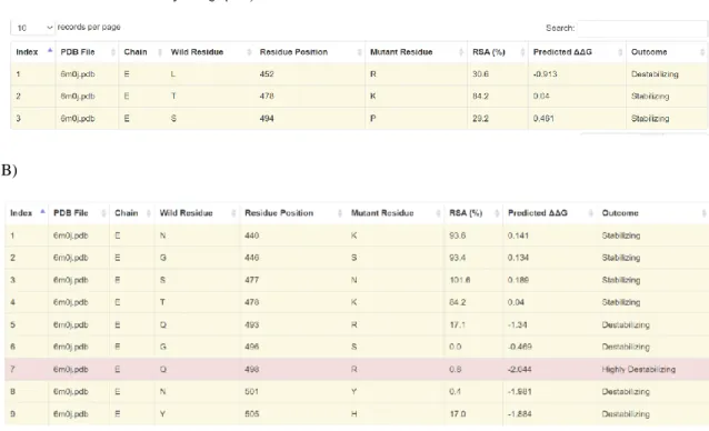
- Protein-Protein Affinity Change Upon Mutation A)
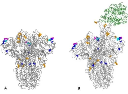
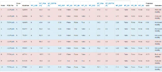
Amino acid changes in the peak glycoprotein of query sequences EPI_ISL_5465615 are distinguished by distinct colors, as black font represents amino acid changes with unknown effects, blue font represents frequent and epidemiologically relevant amino acid changes, orange fonts cell-associated host illustrates receptor binding or antigenicity, and magenta font indicates addition or deletion of glycosylation sites. The results suggest that the rare mutation Q271E slightly increases the stability of spike protein by ΔΔG=0.27 kcal/mol, and generally most mutations contribute to increased protein stability. Barcode 18 (omicron) protein stability analysis suggests that the overall DΔG of peak protein stability increased to 3 kcal/mol.
The rare mutation in genome EPI_ISL_5465615, specifically, the Q271E mutation, was detected in the peak region. The amino acid changes in peak glycoprotein of EPI_ISL_5465615 query sequences are distinguished by colors. Specifically, the blue color is associated with amino acid changes found more than 100 times, while the orange color represents the changes that affect the phenotype in terms of host cell receptor.
Magenta-colored amino acid mutations account for the addition or elimination of the glycosylation sites, whereas cyan-colored amino acid mutations facilitate the insertion or deletion of amino acid residues. While the effect of black-colored amino acid changes is not known, they occurred only once in the current set of sequences. The protein stability analysis after insertion of specific mutations was performed by a site-directed mutator.
The results tables in figure 7 demonstrate that the rare mutation Q271E of EPI_ISL_5465615 (delta) facilitates a slight increase in the stability of the peak protein up to ΔΔG=0.27 kcal/mol and, in general, most mutations contribute to an increase in protein stability (figure 7A). The protein stability analysis of barcode 18 (omicron) indicates that the overall ΔΔG of peak protein stability increased to 3 kcal/mol (figure 7B).
DISCUSSION
In general, viral whole-genome sequencing confirmed that the SARS-CoV-2 variants in the region predominantly belong to the delta strain, which corresponds to a global trend. Since the emergence of SARS-CoV-2 in December 2019, it has undergone various mutations that alter its properties, such as transmissibility, virulence, antigenicity, and vaccine efficacy (Cosar et al., 2021). Notably, according to CoVsurver enabled by GISAID research tool analysis, a Q271E rare substitution mutation in the spike region was detected in sample EPI_ISL_5465615.
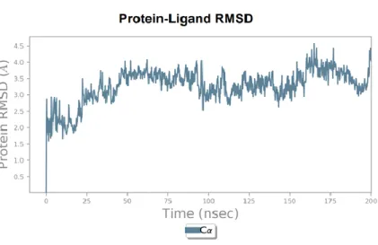
The result of the run suggests that the net increase of predicted ΔΔG was 1.5 kcal/mol, whereas the rare mutation Q271E slightly increased the stability of the spike protein by ΔΔG=0.27 kcal/mol (Figure 7A). In other words, the predicted ΔΔG value specifies whether the indicated mutations stabilize the mutated spike protein in its folded conformation or in its correct native structure to be functionally active state, and therefore the higher ΔΔG of the spike should facilitate higher virulence of SARS-CoV -2. Since RMSD measures the change in position of carbon backbones from their initial to final conformation, the stability of the protein is found according to its conformation by changes in
According to the manual, fluctuations of around 1-3 Å are expected for small globular proteins, while values in excess of this indicate that the protein is substantial. In addition to being responsible for the interaction with the ACE2 receptor, the RBD considered in this analysis is expected to facilitate the pre-fusion state, so the molecular stability of the protein-ligand complex is considered in this study. In addition, the RMSD molecular dynamics plot indicates that the barcode 18 (omicron) RBD with the ACE2 receptor has a higher RMSD value and weaker plot stabilization compared to the interaction of the EPI_ISL_5465615 RBD with the receptor.
In other words, mutations in the omicron spike RBD region facilitate destabilization of binding to the ACE2 receptor. Although the omicron strain has a weaker binding affinity for the ACE2 receptor, it has a higher entry rate than that of delta.
CONCLUSION
GC usage of SARS-CoV-2 genes can adapt to the environment of human lung-expressed genes. Multiplex PCR method for MinION and Illumina sequencing of Zika and other virus genomes directly from clinical samples. Strain wars 3: Differences in infectivity and pathogenicity between Delta and Omicron strains of SARS-CoV-2 can be explained by thermodynamic and kinetic parameters of binding and growth.
Nanopore targeted sequencing for accurate and comprehensive detection of SARS-CoV-2 and other respiratory viruses. Role of structural and non-structural proteins and therapeutic targets of SARS-CoV-2 for COVID-19. The COVID-19 Resource Center is hosted on Elsevier Connect, public news and company information.
APPENDICES