The current study demonstrates the green synthesis of silver nanoparticles from leaf extract of Artemisiae scopariae. Therefore, the preliminary screening of the green-synthesized silver nanoparticles showed potent cytotoxicity and antioxidant activity.
Silver Nanoparticles
However, prolonged exposure to higher doses of silver nanoparticles will cause toxicity such as argyria (Lansdown, 2010). Green synthesis of silver nanoparticles involves mixing natural products such as plant extracts with metal salts.
Cancer
- Geographical Distribution
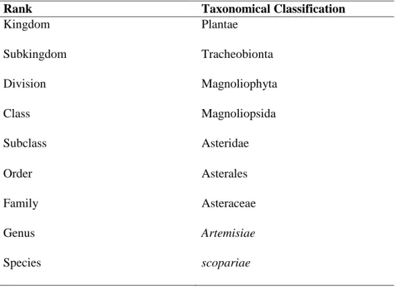
- Taxonomical Classification
- Antinociceptive Activity
- Antipyretic Activity
- Antioxidant Activity
- Anticancer Activity
- Chemical Method
- Biological Method
The present study focused on the cytotoxicity and antioxidant activity of silver nanoparticles synthesized from A. Silver nanoparticles are silver nanoparticles with sizes ranging from 1 to 100 nm (Prabhu and Paulosee, 2012).
Characterization of Silver Nanoparticles
19 researchers to synthesize nanoparticles due to the non-pathogenic, availability of secondary metabolites and the removal of the extra step needed to avoid particle aggregation. The other advantages of biological method are ease in controlling shape, size and distribution of the synthesized nanoparticles, high density and ready solubility of synthesized nanoparticles in water (Ahmed et al., 2016; . Zhang et al., 2016).
Antioxidant Assay
Cytotoxic Assay
21 Figure 2.3: The chemical structure of DPPH (purple color) (left) and reduced form of DPPH (colorless) (right) (Adapted from Kedara and Singh, 2011). 22 Figure 2.4: The formation of formazan by NAD(P)H-dependent cellular oxidoreductase (Adapted from Bahuguna et al., 2017).
Materials .1 Plant Source
Cancer Cell Lines
The reagents and chemicals used in this research are listed in Table 3.1, and the equipment used is listed in Table 3.2. 1 L of deionized water was added to the powdered sample and boiled on a hot plate for 30 minutes. Stir the mixture constantly while boiling and let it cool to room temperature.
The frozen sample was freeze-dried using a freeze-dryer, and the lyophilized sample was stored in a −20°C freezer until further use.
Synthesis of Silver Nanoparticles
27 silver nanoparticles was repeated with different ratios of aqueous extract to AgNO3 which were 1:1 and 9:1. The absorption spectrum optimization of all solutions was performed at different time intervals which were 0.5 hours, 4 hours and 24 hours.
Characterization of Silver Nanoparticles .1 Color Changes
- UV-Vis Spectrophotometer
- Preparation of Reagents and Samples
- DPPH Assay
- Preparation of Reagents, Medium and Chemicals
- Cell Counting
A concentration of 0.2 mM DPPH reagent was prepared by dissolving 1.56 mg of DPPH in 20 mL of methanol and was incubated for 30 minutes in the dark at room temperature. Ascorbic acid of 5 mg/ml was prepared by dissolving 5 mg of ascorbic acid in 1 ml of deionized water. Complete medium was prepared by adding 10 ml of fetal bovine serum to 90 ml of basic DMEM.
Phosphate buffer was prepared by adding two tablets of phosphate-buffered saline (PBS) to 200 mL of deionized water. A concentration of 5 mg/ml of MTT reagent was prepared by adding 100 mg of MTT powder to 20 ml of phosphate buffer solution. Doxorubicin hydrochloride was used as a positive control and 1 mg/mL working solution was prepared by adding 100 µL of doxorubicin hydrochloride to 900 µL of basic DMEM.
The cell suspension was cultured in a new 25 cm3 culture flask containing 5 mL of complete medium. A stock concentration of AgNPs at a ratio of 1:9 was prepared by adding 10 mg of freeze-dried sample to 2 mL of sterile deionized water to obtain a concentration of 5 mg/mL.
Statistical Analysis
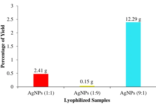
Percentage of Lyophilized Yield
Characterization of Silver Nanoparticles .1 Color Changes
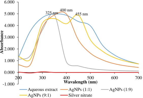
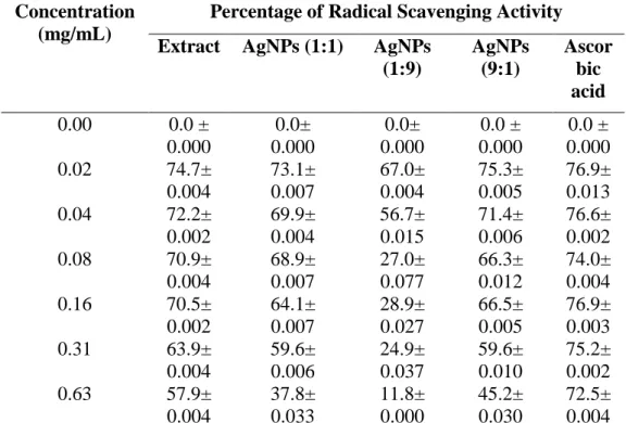
The color changes observed before (top) and after (bottom) the synthesis of silver nanoparticles. The absorption spectrum of the aqueous extract, silver nitrate and different ratios of AgNPs were measured at wavelengths ranging from 300 to 700 nm using UV-Vis spectrophotometer. Among all the samples, AgNPs at a ratio of 9:1 exhibited the highest radical scavenging activity of 75.3% at 0.02 mg/mL.
The antioxidant activity of the aqueous extract and silver nanoparticles decreased slightly as the concentration increased. In AgNPs at ratio 1:9, a sharp drop of radical scavenging activity was observed from concentration of 0.04 to 0.08 mg/ml. Meanwhile, a steady trend of higher radical scavenging activity was observed in ascorbic acid at different concentrations.
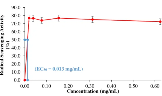
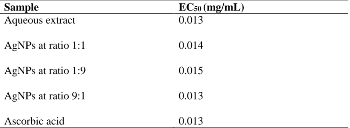
39 Table 4.1: The percentage radical scavenging activity of aqueous extract, silver nanoparticles and ascorbic acid. 40 The EC50 values of the aqueous extract, different proportions of silver nanoparticles and the ascorbic acid were interpolated from graphs as shown in Appendix A (Figures A and B).
MTT Assay .1 HeLa Cells
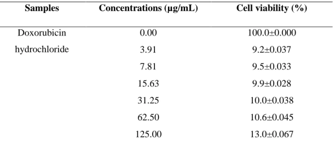
41 Table 4.3: The percentage of cell viability of HeLa cells after 24 hours of treatment with samples and doxorubicin hydrochloride. In general, the percentage of cell viability of HeLa cells decreases sharply after treatment and further becomes constant with increasing concentration. 43 Figure 4.5: The percentage of cell viability of HeLa cells after 24 hours of treatment with aqueous extract, different ratios of AgNPs and doxorubicin hydrochloride at different concentrations.
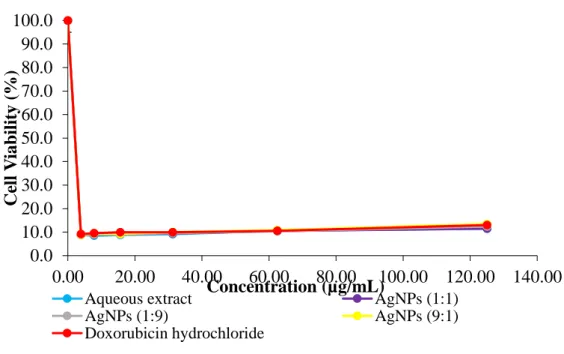
The percentage of viability of HT-29 cells after 24 and 48 hours of treatment with aqueous extract, different proportions of silver nanoparticles and doxorubicin chloride is shown in Table 4.5. All treatments showed a decrease and then an increase in the percentage of cell viability as the concentration increased. All treatments showed a slight increase in the percentage of cell viability as the concentration increased.
45 Table 4.5: The percentage of cell viability of HT-29 cells after 24 and 48 hours of treatment with samples and doxorubicin hydrochloride. 47 Figure 4.6: The percentage of cell viability of HT-29 cells treated with aqueous extract, different ratios of AgNPs and doxorubicin hydrochloride at 24 hours.
Lyophilization Yield
UV-Vis Spectrophotometry .1 Color Changes
Absorption Spectrum
This is due to the presence of phytochemicals in the plant extract and was detected in the range of spectrophotometry (Ramteke, 2013; Aziz et al., 2017). This is due to the absence of the reduction process of Ag+ to Ag atom, which will give rise to the SPR band. The SPR phenomenon occurs due to collective oscillations of conduction electrons when nanoparticles are irradiated with visible light (Amin et al., 2012).
A broad peak at a longer wavelength reflects increased particle size, while a sharper band at a shorter wavelength indicates smaller particle size (Shaik et al., 2018). The increase in particle size, peak broadening and peak absorption are due to the poly-dispersive nature of the particles without sedimentation (Moosa et al., 2015). The presence of more functional groups in the plant extract facilitates the formation of more AgNPs due to the availability of more reduction sites.
This ultimately results in the large shift of the SPR band to a longer wavelength and indicates the formation of large AgNPs. This further concludes that among the AgNPs synthesized, the ratio 9:1 was the largest, followed by 1:1, and the smallest nanoparticles were the AgNPs with a ratio of 1:9 (Moosa et al., 2015; Shaik et al., 2018).
DPPH Assay
Herbal plants are a good source of antioxidants for humans, as plants contain many phytochemicals that can act as antioxidants (Singh et al., 2009; Mahdi-Pour et al., 2012). The higher the amount of antioxidant, the higher the radical scavenging activity (Marxen et al., 2007). Phytochemicals such as polyphenols have strong antioxidant activity and the antioxidant activity was further enhanced by conversion to AgNPs (Otunola et al., 2017). The cessation of radical scavenging activity may be due to the antagonistic activity of antioxidants (Nambikkairaj and Thanighararassu, 2018).
These functional groups serve as capping and stabilizing agents for silver nanoparticles to exert their effects (Keshari et al., 2018). By scavenging free radicals, flavonoids are able to prevent LDL oxidation (Nijveldt et al., 2001). Phenol consists of a benzene ring with one of its hydroxyl groups replaced by a hydrogen atom (Adams et al., 2014).
The ability to chelate metal ions that participate in the formation of free radicals also contributes to the antioxidant properties of phenol (Pereira et al., 2009). The presence of antioxidant in both the aqueous extract and silver nanoparticles contributes to the significant antioxidant activity (Chen et al., 2012).
MTT Assay
57 The signal generated from the MTT assay used in this study is highly dependent on the MTT concentration, the number of viable cells, the metabolic activity of the cells, and the duration of incubation. With longer incubation time, the accumulation of dye will occur, thus increasing the sensitivity of the MTT assay on the HT-29 cells (Riss et al., 2013). HT-29 cells were incubated for a longer time due to the fact that the silver nanoparticles may take time to diffuse into the cells to exert cytotoxic effect.
The result of this study showed that the treatments were highly cytotoxic to HeLa and HT-29 cells due to IC50 values falling within the NCI recommended criteria. Cell death occurs to eliminate damaged or unwanted cells (Chahar et al., 2011). CDKs are one of the key regulators of cell cycle progression and by suppressing the activity of CDKs, the cell cycle will be arrested (Chahar et al., 2011).
Based on the results, HT-29 cells were resistant to doxorubicin hydrochloride as the IC50 slightly increases with longer incubation Multidrug resistant protein recognizes doxorubicin as one of the substrate and thus causes inactivation of doxorubicin. 59 dependent flux pump reduces the concentrations of doxorubicin in the cancer cells (Melguizo et al., 2015).
Limitation of Study
Future Studies
Green synthesis of silver nanoparticles using leaf extracts from the endemic Buddleja globose Hope. Green synthesis of silver nanoparticles using Ganoderma neojaponicum Imazeki: a potential cytotoxic agent against breast cancer cells. Green synthesis of silver nanoparticles using seed extract of Alpinia katsumadai and their antioxidant, cytotoxicity and antibacterial activities.
Green synthesis of silver nanoparticles using oak fruit peel extract (Jaft): synthesis and in vitro cytotoxic effect on MCF-7 cells. Rapid green synthesis of silver nanoparticles using (Prunus persica) plant extract: exploration of its antimicrobial and catalytic activities. Process parameter for green synthesis of silver nanoparticles using leaf extract of Aloe vera plant.
Green production of silver nanoparticles and characterization with plant leaf essential oil compound Geraniol and their antifungal activity against human pathogenic fungi. Evaluation of antioxidant, antibacterial and cytotoxic effects of green synthesized silver nanoparticles by Piper longum fruits.