In addition, these selected characteristics are used to develop a liver disease index (LDI) to differentiate normal, FLD and cirrhosis classes using a single number. It has been reported that 9 to 37% of people are affected with NAFLD in different parts of the world [52, 68]. These studies reported accuracies of 82.2% and 82.6% in identifying normal, FLD, and cirrhosis ultrasound images, respectively.
Few studies [36, 55] have used second-order statistics to examine the texture features of ultrasound images from normal, FLD, and cirrhosis ultrasounds. The overview table shows that second order statistics [33], HOS [1], wavelet transform [1, 76] and discrete cosine transform (DCT) [10] are used in the characterization of the liver diseases. Cropped images are then scaled down to 500 × 500 using bicubic interpolation [28] and subjected to contrast-limited adaptive histogram smoothing (CLAHE) [56] to improve the contrast of the ultrasound images.
CLAHE was developed for medical imaging [74] and reduces noise in the homogeneous area of the images. One of the properties of the LSDA algorithm is that it preserves the discriminative and local geometric structure in the data [17]. It is a supervised learning that uses neighborhood classification to predict the class of the test data.
In order to identify the class of the new test data, k number of training data neighbors to the test data will be assessed.
Results
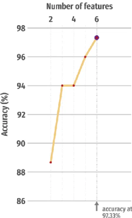
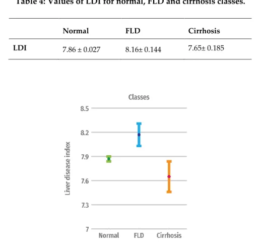
An integrated index, the liver disease index (LDI), is developed in this work using two LSDA coefficients (LSDA7 and LSDA4) to distinguish three classes. This concept was first proposed by Ghista [27] and later used by Acharya for various applications for the identification of medical diseases. The selection of LSDA7 and LSDA4 was made purely based on the empirical study that was conducted on the listed features.
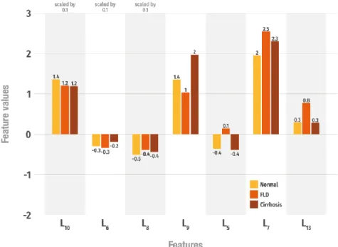
These two features are combined in such a way that the range of values is unique for normal, FLD and cirrhosis classes. It is clear from Table A1 and Figure 3 that LSDA coefficients of entropy features show clear distinction between the three classes. This implies that approximately 4% of the images were incorrectly classified as normal and 100% normal images were correctly classified as normal cases.
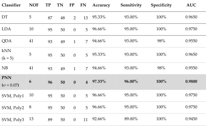
NOF: number of functions; TP: true positive; FP: false positive; TN: true negative; FN: false negative. The LDI ranges for subjects with FLD, cirrhosis and normal people are shown in Table 4. It can be seen from Figure 5 that the LDI helps to clearly separate the three classes using a single number.
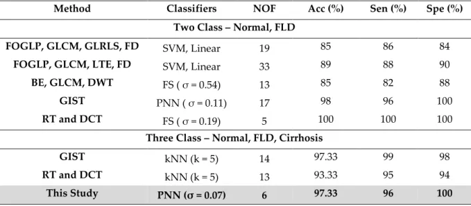
In addition, we compared our results with other techniques using our set of ultrasound images. It can be seen from Table 5 that our proposed method achieved the highest efficiency with the minimum number of features.
Discussion
In this work, we used CT and extracted different types of entropy features from ultrasound images to evaluate normal patients, patients with FLD and cirrhosis. In addition, LDI is developed using only two features to classify three classes. The main novelty of our proposed method is that it estimates inherent signatures present in images [4].
Fuzzy entropy quantifies uncertainties of the highly irregular signals and is not affected by noise [53]. Thus, the algorithm developed using these significant nonlinear features can effectively classify the normal, FLD, and cirrhosis subjects using ultrasound images. But our proposed method uses entropy features which can identify normal, FLD and cirrhosis images with accuracy of 97.33%, specificity of 100.00% and sensitivity of 96.00% using only six features.
In addition, ultrasonography is a relatively inexpensive and widely available imaging technique that provides important information on liver architecture. It is often the first imaging modality that can be used to detect FLD and liver cirrhosis, which are major risk factors for hepatocellular carcinoma (HCC). However, the sensitivity and specificity of gray-scale ultrasound for the detection of FLD and cirrhosis are less than computed tomography or MRI [64].
Using our proposed CAD algorithm, the liver can be automatically classified into normal, FLD, and cirrhosis using the highest performance gray-scale sonographic images. This is achieved without the need for additional computed tomography or MRI scans, potentially replacing the relatively more expensive shear wave elastography (commonly known as fibroscan). Early detection of FLD and cirrhosis is an essential approach for the clinicians to advise on necessary treatments to prevent the onset of HCC and the related complications.
Automated characterization of three (normal, FLD and cirrhosis) classes without the use of image segmentation techniques. LDI is able to differentiate the three classes using a number, and therefore the diagnosis is quick. The performance of the proposed method needs to be tested with a large diverse database for diversity and applicability.
Conclusion
Appendix
Acharya UR, Vidya K., Ghista DN., Eugen LWJ, Molinari F, Sankaranarayanan M, Computer-aided diagnosis of diabetic subjects by heart rate variability signals using discrete wavelet transform method. Acharya UR, Fujita H, Sudarshan VK, Mookiah MRK, Koh EWJ, Tan JH, Hagiwara Y, Chua CK, Junnarkar SP, Vijayanathan A, Ng KH. An integrated index for identification of fatty liver disease using radon transform and discrete cosine transform features in ultrasound images.
Classification of thyroid lesions in 242 patient populations using Gabor transform features from high-resolution ultrasound images. Acharya UR, Mookiah MRK, Koh JEW, Tan JH, Noronha K, Bhandary SV, Rao AK, Hagiwara Y, Chua CK, Laude A. New risk index for identification of age-related macular degeneration using radon transform and DWT functions.
Practices of liver biopsy in France: results of a prospective nationwide survey, for the Group of Epidemiology of the French Association for the Study of the Liver (AFEF). Salehi, Using morphological transformations to enhance the contrast of medical images, The Egyptian Journal of Radiology and Nuclear Medicine. Computer-aided characterization of diffuse liver disease using image texture analysis techniques on B-scan images.
Benjamin EJ, Bennett D, Bhalla K, Bikbov B, Bin Abdulhak A, Birbeck G, Blyth F, Bolliger I, Boufous S, Bucello C, Burch M, et al: Global and regional mortality from 235 causes of death for 20 age groups in 1990 and 2010: a systematic review for the Global Burden of Disease Study 2010. Noninvasive steatosis assessment through computerized processing of ultrasound images: attenuation versus first-order texture parameters. Mendler MH, Bouillet P, Le Sidaner A, et al.: Dual-energy CT in the diagnosis and quantification of fatty liver: limited clinical value compared to ultrasound scanning and single-energy CT, with special reference to iron overload.
Mookiah MRK, Acharya UR, Fujita H, Koha JEW, Tan JH, Chua CK, Bhandary SV, Noronha K, Laude A, Tong L. Murray CJ, Vos T, Lozano R, Naghavi M, Lino A. D., Michaud M. C., Ezzati , Shibuya K, Solomon JA, Abdalla S, Aboyans V, Abraham J, Ackerman I, Aggarwal R, Ahn SY, Ali MK, Alvarado M, Anderson HR, Anderson LM, Andrews KG, Atkinson C, Baddour LM, Bahalim AN -Collo S , Barrero LH, Bartels DH, Basanez MG, Baxter A, Bell ML, Benjamin EJ, et al: Disability-adjusted life years (DALYs) para kadagiti 291 a sakit ken pannakadangran iti 21 a rehion Panagadal 2010. Oliva A, Torralba A, Panagmodelo ti sukog ti eksena: maysa a holistiko a pannakabagi ti espasial a sobre, Internasional.
An integrated index for focal electroencephalogram signal identification using discrete wavelet transform and entropy measures. SPIE 5852, Third International Conference on Experimental Mechanics and Third Conference of the Asian Committee on Experimental Mechanics.