Bioactive compounds from the cultured broth of the fungus Polycephalomyces nipponicus and active extracts from the root. Mahasarakham University has given approval to accept this thesis in partial fulfillment of the requirements for the Master of Science Chemistry. TITLE Bioactive compounds from the cultured broth of the fungus Polycephalomyces nipponicus and active extracts from the root of Smilax verticalis.
INTRODUCTION
Meanwhile, various pharmacological activities of the isolated compounds from Cordyceps have also been reported. The bioassay shows that this compound is the first effective treatment for malaria, which appeared in therapeutic form in the 17th century [7]. Search for bioactive natural products from the culture broth of the fungus Polycephalomyces nipponicus and the root of Smilax verticalis.
LITERATURE REVIEW
C. brunnearubra
C. cicadae
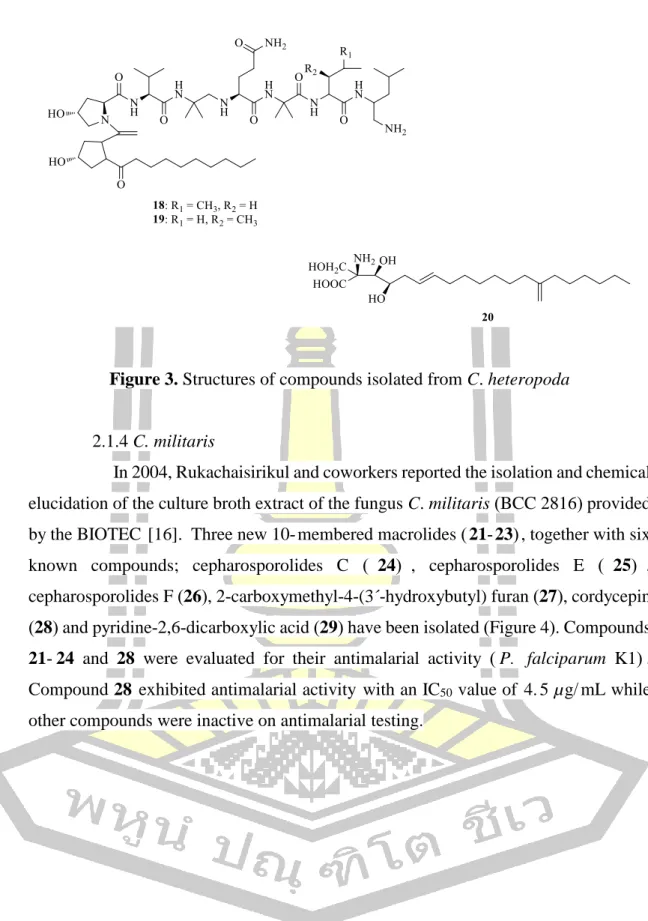
C. heteropoda
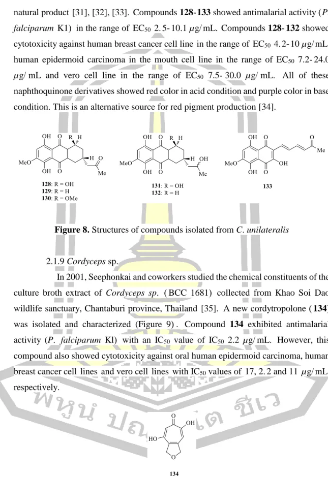
In 2004, Rukachaisirikul and colleagues reported the isolation and chemical elucidation of the culture broth extract of the fungus C. In 2014, Kim and colleagues investigated the characterization of active constituents from the fruiting body extract of the fungus C. In 2017, Sun and colleagues isolated the chemical constituents from the culture of the fungus C.militaris obtained from Yanbian Foresty Science Institute, Yanji, China [20].
C. nipponica
C. pseudomilitaris
In 2010, Wang and colleagues investigated the chemical constituents from the broth extract of mushroom culture C. The results revealed that this polysaccharide had a significant protective effect of chronic renal failure at doses of 40 mg/kg and 80 mg/kg. In 2011, Yang and colleagues investigated the chemical constituents from the mycelium of the mushroom culture C.

C. unilateralis
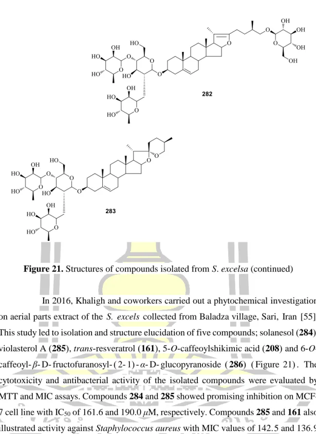
In 2001, Seephonkai and colleagues investigated the chemical constituents of the culture broth extract of Cordyceps sp. In 2007, Isaka and colleagues investigated the chemical constituents of the cultured mycelium extract of the fungus Cordyceps sp. In 2006, Xu and colleagues reported on the chemical constituents from the root extract of S.
S. china
Compound 213 showed little tyrosinase inhibitory activity, while compound 209, a known tyrosinase inhibitor, showed strong tyrosinase inhibitory activity. Interestingly, the mixture of compound 213 and compound 209 (ratio 1:1) showed greater inhibition of tyrosinase activity by L-tyrosine with an IC50 value of 5. The inhibitory activities of PTP1B, α-glucosidase and DPP-IV of all the isolated compounds were evaluated by molecular levels.
In the kinetic study for the PTP1B enzyme, compounds 220, 214 and 223 showed competitive inhibition with Ki values of 3. Molecular docking study for the competitive inhibitors (220, 214 and 223) radically confirms the binding affinities and the inhibition of PTP1B enzymes . All isolated compounds were tested for their inhibitory effects against advanced glycation end products, as well as aldose reductase, α-gucosidase and lipase assays were also performed.
S. corbularia
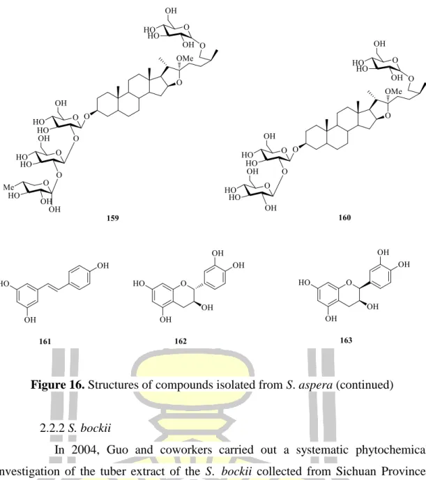
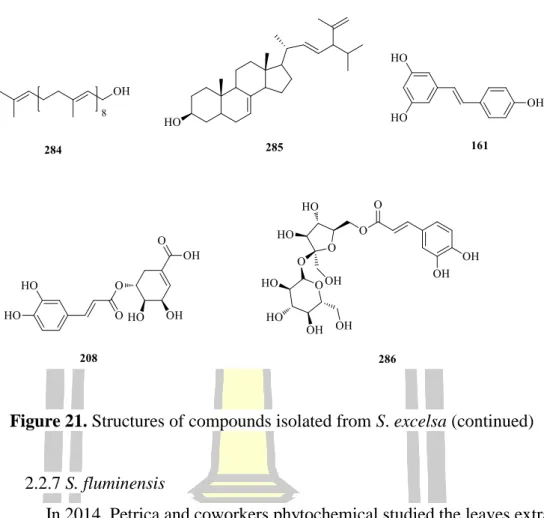
S. excelsa
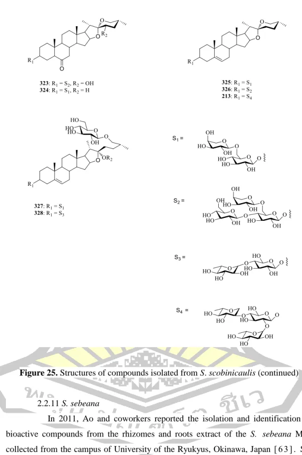
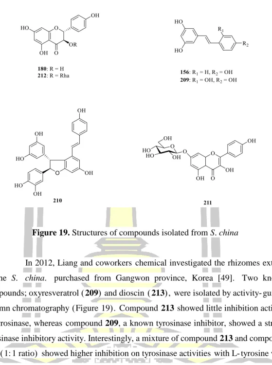
Cytotoxicity and antibacterial activity of the isolated compounds were evaluated by MTT and MIC tests. The results led to the isolation and elucidation of the structure of two flavonoids; quercetin-3-O-β-L-rhamnopyranoside (1-6)-O-β-D-glucopyranoside (287) and quercetin-3-O-β-L-galactopyranoside (288) (Figure 22). Two new steroidal saponins of the spirostan type; smilskobinosides A (294) and smilskobinosides B (295) together with the known congener (296) were isolated and reported (Figure 25). Compounds 294-296 were tested in vitro for their cytotoxicity against A549, Hela and LAC human cancer cell lines.
In 2014, Xu and colleagues reported the isolation of the rhizomes and root extract of S. The isolated saponins were evaluated for cytotoxic activity against two human cancer cell lines, including Hela and SMMC-7221. The in vitro cytotoxicity evaluation of the new compounds showed that compound 304 exhibited weak activity for the tested MCF-7 and H520 cancer cell lines with IC50 values of 65.
The isolated compounds were evaluated for their cytotoxicity against four human tumor cell lines (SH-SY5Y, SGC-7901, HCT-116 and Lovo). In 2011, Ao et al reported the isolation and identification of bioactive compounds from the root extract and roots of S. In 2015, Shu et al reported the isolation and identification of bioactive compounds from the root extract of S.
METHODOLOGY
- Crystal data of compound A
- Antifungal activity assay (Pore plate technique)
- Biological assay
- Small scale extraction
- Large scale extraction
- Antifungal activity assay (Pore plate technique)
- Biological assay
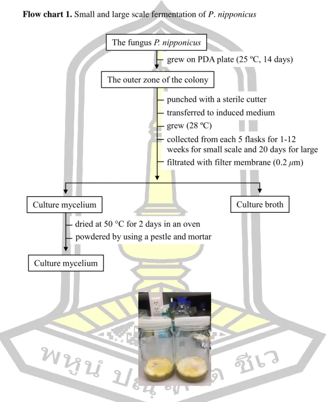
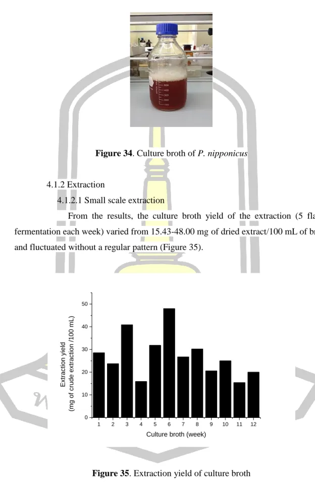
Culture broth and mycelia were collected from every 5 flasks for 1–12 weeks. The mycelium on the surface of the induced medium was collected and dried at 50 °C for 2 days in an oven. The mycelium on the surface of the induced medium was collected and dried at 50 °C for 2 days in an oven. The collected ethyl acetate layer was concentrated under reduced pressure at 45 ºC to obtain the crude ethyl acetate layer extract from the culture broth (Flow Chart 2).
The hexane and ethyl acetate layers were concentrated under reduced pressure at 45 ºC to obtain crude ethyl acetate and hexane extracts (Flow diagram 3). The collected ethyl acetate layer was combined and concentrated under reduced pressure at 45 ºC to obtain crude ethyl acetate extract from the culture broth (Flow diagram 4). The ethyl acetate-soluble fraction was subjected to purification over silica gel column chromatography to obtain six fractions (Fractions 1-6).
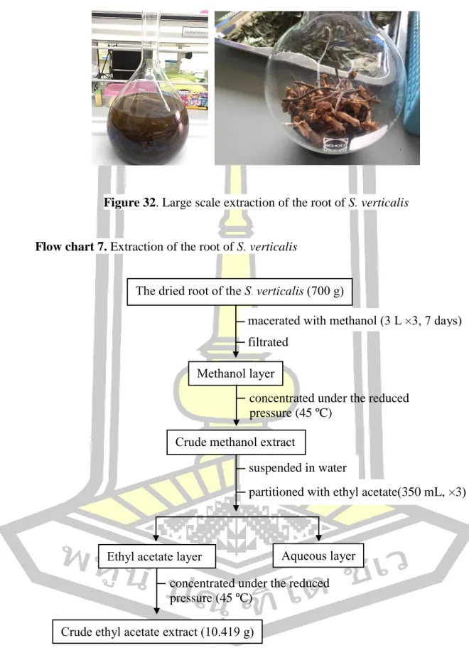
The concentration of the sample required to scavenge 50% of the DPPH free radicals (IC50) was determined from the curve of percentage DPPH radical scavenging activity plotted against the respective concentration. The filtrate was concentrated under the reduced pressure at 45 ºC to get crude methanol extract of the root (SV-R), stem (SV-S) and leaf (SV-L) of the S. The crude methanol extract was suspended in water and partitioned with ethyl acetate (350 ml, . × 3) to obtain crude ethyl acetate extract of the root of the S.
The concentration of sample required to scavenge 50% of the DPPH free radical (IC50) was determined from the percentage of DPPH radical scavenging activity plotted against the respective curve.
RESULTS AND DISCUSSION
Large scale fermentation
According to the results, the extraction yield in the culture broth (5 fermentation flasks every week) varied from mg dried extract/100 ml broth and fluctuated without a regular pattern (Figure 35). The mycelial yield of the extraction culture (5 fermentation flasks each week) varied from mg extract/g dried mycelium and fluctuated without a regular pattern (Figure 36). The crude extract from the bulk extraction in the first bath was 8.95 g, while the bulk extraction in the second bath was 1.38 g.
These results confirmed that the amount of extract from this mushroom is not stable under the same fermentation conditions.
Isolation of the crude extract
Structural elucidation
- Structural elucidation of A
- Structural elucidation of B
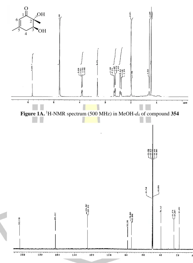
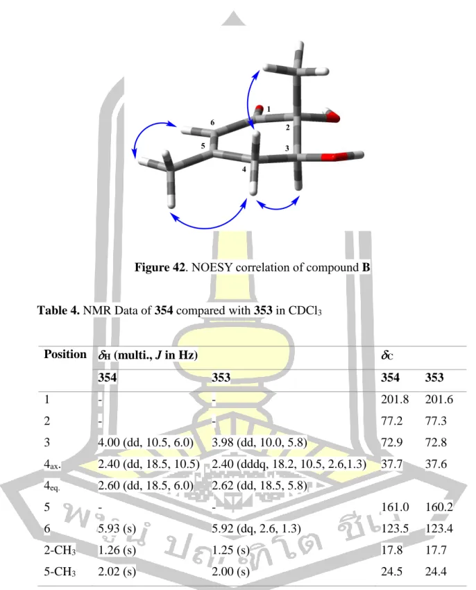
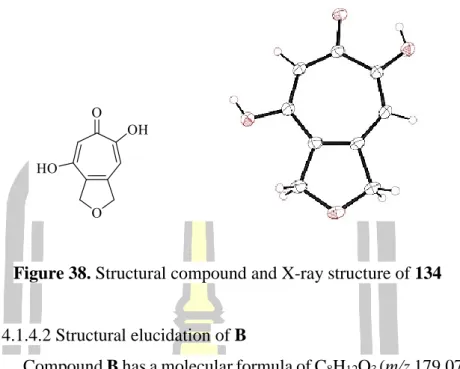
Based on spectroscopic data, the structure of B was elucidated as the same structure of leptosphaerones A and B which are isolated from the fungus Leptosphaeria herpotrichoides [73]. Comparison of the NMR spectroscopic data of B in CDCl3 (Table 4) with those reported in the literature for leptosphaerone A (353) (Figure 41) in CDCl3 were identical, except for their optical rotation values. The amount of 134 in the extracts was determined by its peak area calculated based on a standard linear equation of pure compound 134 (r2 0.988) which was extremely high at weeks 11 and 12 (1 mg/mg dried extract ).
Surprisingly, the production of 134 in week 3 (21 days) was very low compared to most other weeks. Therefore, a second batch was subjected to large-scale fermentation and the isolation of 134 was repeated under the same procedure. These results confirmed that 134 is produced by this fungus, but the amount of 134 is not stable under the same fermentation conditions. mg/mg dried extract).
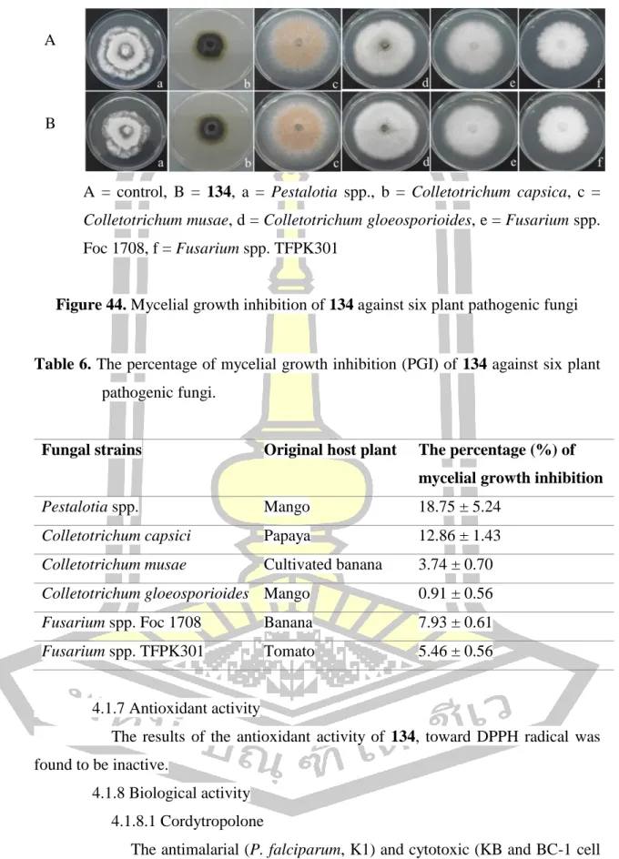
The results revealed that 134 had a slight inhibitory effect against fungal mycelium growth, as shown in Figure 44 and Table 6. The results of the antioxidant activity of 134, against DPPH radical, was found to be inactive.
Biological activity
- Cordytropolone
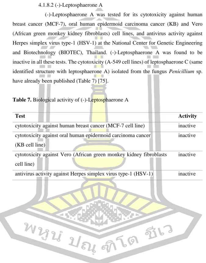
Leptosphaerone A was tested for its cytotoxicity against human breast cancer cell lines (MCF-7), human oral squamous cell carcinoma (KB) and Vero (African green monkey kidney fibroblasts) and antiviral activity against Herpes simplex virus type-1 (HSV-1) at the National Center for Genetic Engineering and Biotechnology (BIOTEC), Thailand.
Extraction
- Small scale extraction
- Large scale extraction
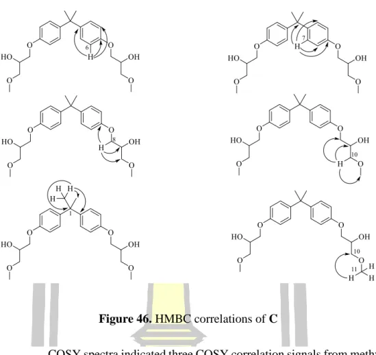
- Structural elucidation of C
- Structural elucidation of D
- Spectoscopic data of E
Based on 1H and 13C NMR spectral data and comparison of 1H and 13C with NMR spectral data with 355, the structure of compound D was proposed (Figure 50).
Proof of contamination by thin layer chromatography (TLC)
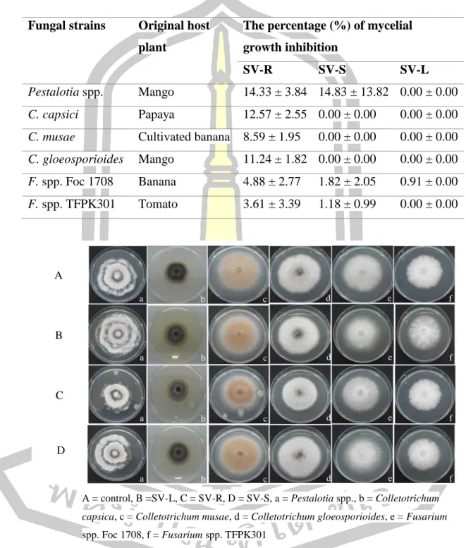
Antifungal activity
The percent mycelial growth inhibition (BGI) of crude methanol extract of the root, stem and leaf against six plant pathogenic fungi. Mycelial growth inhibition of crude methanolic extract of the root, stem and leaf against six plant pathogenic fungi. The scavenging activity of standard ascorbic acid to DPPH radical is shown in Figure 54.
Based on the calculation of %RSA to IC50, ascorbic acid showed scavenging activity of to DPPH radical at IC50 value of. The DPPH scavenging activity of the crude methanol extract from the root, leaf and stem is shown in Table 12. The IC50 values of all crude methanol extract were found significant (P < 0.05) compared to ascorbic acid.
All the data were expressed as mean ± SD and analyzed by One-way ANOVA with the post-hoc Tukey's test and values were considered significant at P < 0.05. The total phenolic content was calculated using the following linear equation based on the calibration curve of gallic acid; Y = 0. The content of total phenolic compounds expressed as mg/100 mg gallic acid equivalent (GAE) of dry extract.
The content of total phenolic compounds of crude methanol extract from the root was detected at mg/100 mg GAE, while the content of total phenolic compounds of crude methanol extract from the leaf and stem was not detected with this test at the concentration of 50 µg/ml ( Table 9).
Biological activity
CONCLUSION
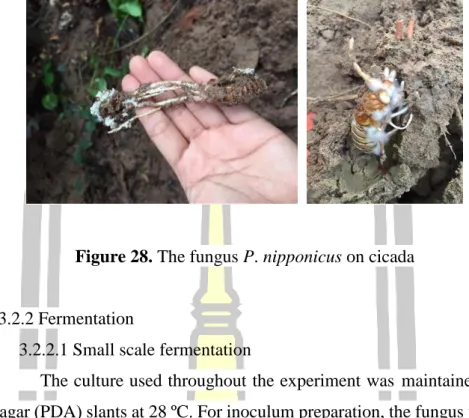
The fungus P. nipponicus (Cod - MK1201)
The Smilax verticalis
The TLC chromatogram of 355 compared to the methanol, ethyl acetate and ethanol extracts of the root of S. Proteases released by entomopathogenic fungi impair phagocytic activity, attachment and spreading of plasmatocytes isolated from haemolymph of the greater wax moth Galleria mellonella. Effects of the peptide mycotoxin destruxin E on insect hemocytes and on dynamics and efficiency of the multicellular immune response.
9] Flora editorial board of the Chinese Academy of Sciences, Flora reipublicae popularis sinicae. Structures of cordypyridones A-D, antimalarial N-hydroxy- and N-methoxy-2-pyridones from the insect pathogenic fungus Cordyceps nipponica. PTP1B, α-glucosidase and DPP-IV inhibitory effects of chromene derivatives from Smilax china L leaves.
Four new furostanol saponins from the rhizomes and roots of Smilax scobinicaulis and their cytotoxicity.