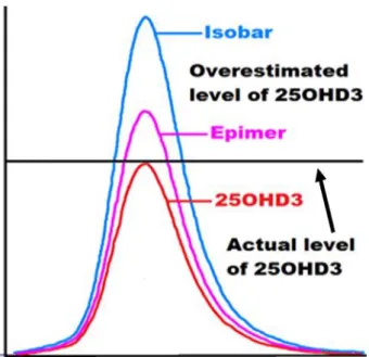
The aim of this study was to develop and validate a UHPLC-MS/MS method for the determination of vitamin D metabolites and their epimers and then apply the new method to the determination of vitamin D levels in the obese Emirati population. Epimers of vitamin D metabolites were found to cause an overestimation of 25OHD and false positive results in the obese Emirati population.
Introduction
Overview
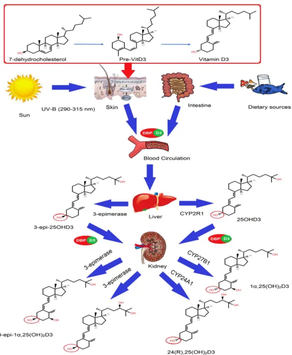
The second step is driven by the conversion of vitamin D3 to 25OHD3 in the liver via the enzyme 25-hydroxylase (CYP2R1), as shown in Figure 1 [27]. It was recently discovered that the C3 epimers of vitamin D may play an important role in the body.
C3-Epimerization of Vitamin D
This was done using keywords such as vitamin D epimers, C3 epimers, metabolism of vitamin D epimers, epimerization pathway for standard vitamin D metabolism, function of 1α,25(OH)2D epimer, detection and quantitative analysis of vitamin D epimers, and the effects of epimers on the routine assay of circulating total vitamin D (25OHD) and its determination in serum. Because of a growing interest in vitamin D epimers and their potential relationship to health and disease, it was decided to conduct this subjective literature review. Cell lines respond to PTH, prostaglandins and bone-resorbing steroids such as vitamin D).
Role of Epimers in Calcium, Phosphorus and PTH
Similarly, 3-epi-1α,25(OH)2D3 has been shown to have more differentiation or antiproliferative activities (about 30% and 10%) than the non-epimeric compound. Moreover, 3-epi-1α,25(OH)2D3 has been suggested to have higher metabolic stability than 1α,25(OH)2D3, despite having non-equivalent VDR binding [24].
The Potency of Epimerization in Microsomal Fractions
The Role of DBP, VDR, and Genetics in Epimerization
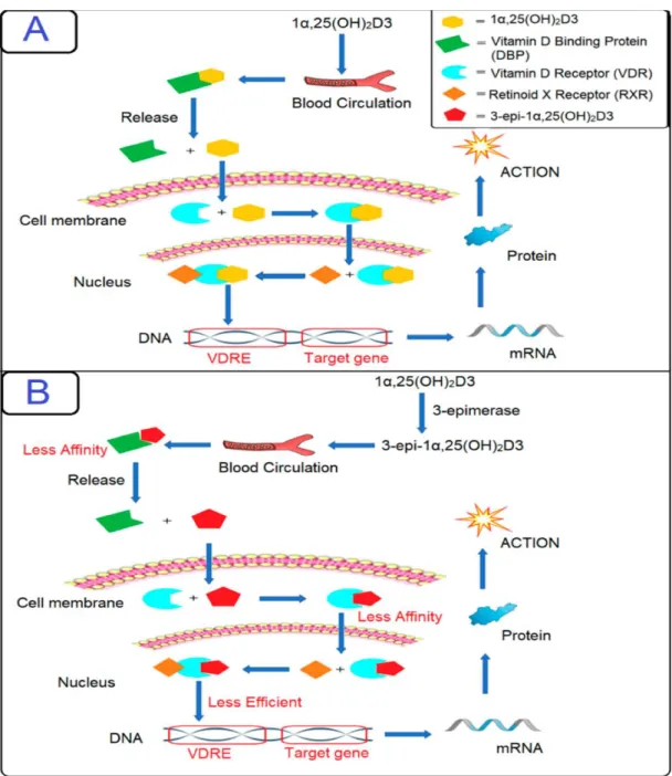
It is then attached to vitamin D receptors on the cells, and the target cells will take up this free active metabolite 1α,25(OH)2D3 within the cell; the metabolite will then either be rapidly metabolized by the 1α,25(OH)2D-24-hydroxylase (CYP24A1) enzyme through the C-24/23 oxidation pathway leading to the formation of other metabolites, or it will be reattached to VDR . Therefore, it was concluded that 3-epi-1α,25(OH)2D3 exerts gene regulation in the same manner as its non-epimeric analogue, but less efficiently. The 1α,25(OH)2D3-VDR complex undergoes translocation to the nucleus and performs conformational changes to associate with other transcription factors and heterodimerize with RXR.
The RXR complex binds to the vitamin D response element (VDRE); then the transcription of RNA for specific genes is initiated, which are then expressed as various proteins responsible for vitamin D homeostasis. A recent study hypothesized that genetic polymorphisms in the vitamin D-related gene pathway cause changes in C3-epimer levels.
Vitamin D Epimer Levels in Humans
Further experiments showed that sun exposure causes a decline in epimer levels in an animal model; however, the mechanism for this process has not been identified. Epimerization can also occur in the skin and cause epimerization of previtamin D3, so the expression of 25-hydroxylase (CYP2R1) will be less efficient, leading to a decrease in the production of 3-epi-25OHD3 [31] . One study found a significant increase in CYP24A1 gene expression in kidney cells of sunlight-exposed (UV-irradiated) mice compared to those receiving vitamin D supplementation [63].
If true, CYP11A1 may be activated by UVB exposure, and the availability of the vitamin D3 substrate for classical hydroxylation to form 25OHD3 and its epimer may be lower in the absence of sunlight [31]. For example, melanocytes in human skin are present in the basal layer of the epidermis, while in mice, melanocytes are present in the hair follicle and dermis.
C-3 Epimer Levels in Newborn and Adults
The activity of the CYP11A1 enzyme was observed in epidermal keratinocytes, where 20, 22(OH)2D3 and 22OHD3 were formed in higher proportions [66]. Mice also have a higher pelage density compared to humans; furthermore, the permeability and fragility of the stratum corneum is higher for rodents than for humans [67-69]. 70] determined that the percentage of the C3 epimer of 25OHD was up to 60% higher in infants compared to only 22% in adults.
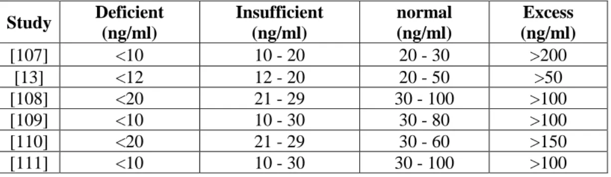
However, with the epimer included in the assessment of insufficiency, 33% of women and 73% of newborns were found to have sufficient levels of vitamin D. 74], insufficient levels of vitamin D are estimated below 50 nmol/L, while the Concentration of the epimer can be equal to 2.5 nmol/L.
Techniques for Measurement of Vitamin D Metabolites
74], concluded that the 25OHD3 epimer plays an important role in clinical applications; however, it could have minimal effect on routine LC-MS/MS measurements. Furthermore, mass differentiation is not possible using most standard LC-MS techniques, as these epimers require chromatographic separation [24, 88]. LC-MS/MS is known to be the most recommended method for vitamin D analysis, especially in young infants, due to its ability to detect and quantify epimeric and non-epimeric metabolites separately in plasma [ 28 , 97 ].
In addition, this technique has advantages over HPLC, which uses CO2 as the supercritical fluid; therefore, UPSFC –MS/MS is more cost-effective than HPLC [79]. 78] used ultra-performance liquid chromatography–tandem mass spectrometry (UPLC–MS/MS) coupled with a pentafluorophenyl (PFP) column to determine epimers.
Quality Assurance in Vitamin D Metabolites and C-3
SRM 972 contains four different concentration levels of vitamin D serum samples, and each level has different ratios of vitamin D metabolites (25OHD3, 25OHD2, 3-epi-25OHD3). The standard measurement for the SRM was performed by three methods using isotope dilution (ID) mass spectrometry at the Centers for Disease Control and Prevention (CDC) and the NIST [52]. The Vitamin D External Quality Assessment Scheme (DEQAS) also uses NIST standards to ensure the analytical reliability of 25OHD, 1,25(OH)2D, and 3-epi-25OHD3 assays [98].
These studies emphasize the fact that the epimers of vitamin D must be separated from the non-epimeric components of the vitamin in order to properly assess circulating vitamin D levels in humans. It is also very important that current methods are standardized in laboratories around the world to account for the interference caused by epimers and isobars.
Objectives of this Study
LC–MS/MS techniques should be implemented in all laboratories worldwide, especially in Middle Eastern countries where these techniques do not exist. To develop, validate and improve the UHPLC-MS/MS method that can accurately detect and quantify the various vitamin D metabolites and separate epimers and isobars. To apply the developed and validated UHPLC-MS/MS method for accurate measurement of blood vitamin D levels in the obese Emirati population.
To determine whether supplementation could increase levels of major vitamin D metabolites or their epimers or both in follow-up serum samples.
Materials and Methods
Materials
Methods
- Preparation of Standards Solution
- Collection of Blood Samples
- Plasma Samples Extraction Method
- Method Development and Validation
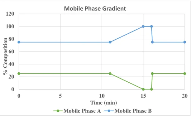
The column temperature was kept at 30℃, and the flow rate of the mobile phase was set at 0.5 ml/min. A separation of vitamin D metabolites and their epimers used two mobile phases or solvents: Mobile Phase A contained 0.1% formic acid or 5mM ammonium formate, and Mobile Phase B contained 0.1% formic acid or 5mM ammonium formate. In the second set, the internal standard was added to all QC samples; then the extraction method was carried out while the rest two sets were returned to the refrigerator and stored at -20℃.
In the third set, the internal standard was added to all QC samples; then the extraction method was carried out while the remaining set was returned to the refrigerator and stored at -20℃. In the fourth set, the internal standard was added to all QC samples; then the extraction method was carried out.

Results and Discussion
Chromatogram of Major Vitamin D Metabolites and
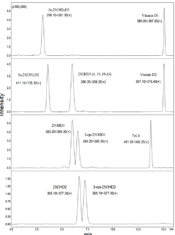
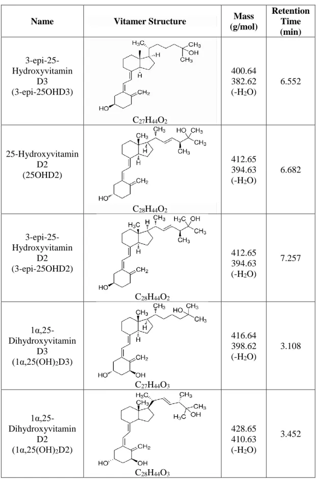
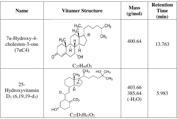
According to Figure 7, the universally measured vitamin D metabolites (25OHD3 and 25OHD2) were separated from their epimers. Vitamin D metabolites with epimers and isobars were separated using a UHPLC-MS/MS instrument. Although some metabolites have similar structure and mass, the retention time of each metabolite is different.
Although some metabolites have similar structure and mass, the retention time of each metabolite is different (continued). Although some metabolites have similar structure and mass, the retention time of each metabolite is different (continued).
Determination of Method Validation Parameters
The instrument's sensitivity to vitamin D metabolites was measured using the Lower Limit of Detection (LOD). For example, the percentage of the coefficient of variations and precision indicates that the method used to analyze vitamin D metabolites and their epimers has good precision and accuracy for intra- and inter-day. The specificity experiment results illustrate that the interfering or co-eluting peaks did not exist at the respective retention times of vitamin D metabolites.
The stability of the vitamin D metabolites and their epimers was assessed by analyzing six QC samples of each concentration level (QCH, QCM and QCL). Ultimately, the stability or intactness results of vitamin D metabolites and their epimers showed that these metabolites are not very stable with successive cycles of freezing and thawing.

Identification of Precursor and Product Ions (Multiple
Analysis of Human Serum Samples (Baseline, Follow-up
The vitamin D3 concentration in the follow-up samples from both men and women is high; The 3-epi-25OHD2 concentration was not detected in baseline samples, while 1α,25(OH)2D was not detected in the baseline, follow-up, and healthy samples. The standard errors of the mean were represented by the error bars in the graph.
3-epi-25OHD2 concentration was not detected in baseline samples while 1α,25(OH)2D was not detected in baseline, follow-up and healthy samples. Standard errors of the mean were represented by error bars in the graph.
Conclusion
Torugsa et al., "Genetic determinants of circulating C3 epimers of 25-hydroxyvitamin D," Journal of Clinical & Translational Endocrinology, vol. 34; Clinical Diagnostic Tools for Vitamin D Assessment," The Journal of Steroid Biochemistry and Molecular Biology, vol. Kamao et al., "Cell Specificity and C-3 Epimerization Properties of Vitamin D3 Metabolites," The Journal of Steroid Biochemistry and Molecular Biology, vol.
Slominski et al., “New activities of CYP11A1 and their potential physiological significance,” The Journal of Steroid Biochemistry and Molecular Biology , vol. Carter et al., "25-Hydroxyvitamin D Assays: Potential Interference from Other Circulating Vitamin D Metabolites," The Journal of Steroid Biochemistry and Molecular Biology, vol.
![Figure 2: The C-23 and C-24 oxidation pathways for 1α,25(OH) 2 D3 and its epimer [30]](https://thumb-ap.123doks.com/thumbv2/azpdfco/10576114.0/25.892.174.788.108.1032/figure-c-oxidation-pathways-1α-oh-d3-epimer.webp)