If the work of others has been used, this is duly acknowledged in the text. Goldring for his excellent supervision, continued encouragement, support, patience, understanding, humor and all help with this thesis.
Introduction
- Overview of malaria
- Life cycle
- Malaria control
- Immune response to malaria
- Immune evasion by the parasite
- Antigenic variation
- Alternative erythrocyte invasion pathways
- Cryptic epitopes
- Vaccines
- Attenuated sporozoite vaccine
- Pre-erythrocytic vaccines
- Asexual erythrocytic stage vaccines
- Transmission blocking vaccines
- Alternative vaccine approaches
- Objective of the present study
The sporozoites do not develop into blood stage forms, regardless of the number of inoculated sporozoites (Labaied et al., 2007). NYVAC-Pf7 was shown to be safe in Rhesus monkeys and to produce antibodies against antigens in all stages of the parasite's life cycle (Tine et al., 1996).
General materials and methods
Materials
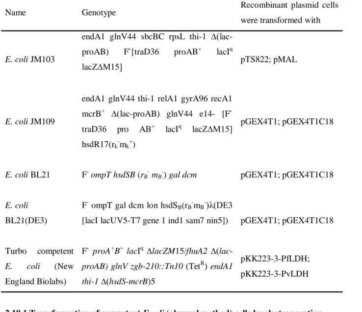
Amylose resin, rabbit anti-MBP antiserum, pMAL-c2X, SacI, BamHI, AccI, and Factor Xa were purchased from New England Biolabs (USA). Syringes (2 ml) were from Promex (South Africa) and 20G x 1½” hypodermic needles were from Tae-Chang Industrial Company (Korea).
Bradford protein assay
- Reagents
- Procedure
Images of western blots, SDS-PAGE gels and agarose gels were captured using the VersaDocTM imaging system and Quantity One software from Bio-Rad (USA). Agarose gels were run using GES and mini-GES electrophoresis equipment from Wealtec Corporation (USA).
Concentration of protein samples
- Dialysis against PEG 20 000
- Materials
- Procedure
- SDS-KCl precipitation of proteins
- Reagents
- Procedure
The protein sample to be concentrated was placed in dialysis tubing (10,000 MW cutoff) and sealed with dialysis clamps. The dialysis bag was removed from the container and rinsed with dH2O to remove any PEG 20,000 that had adhered to the dialysis tubing, before the concentrated protein sample was removed.
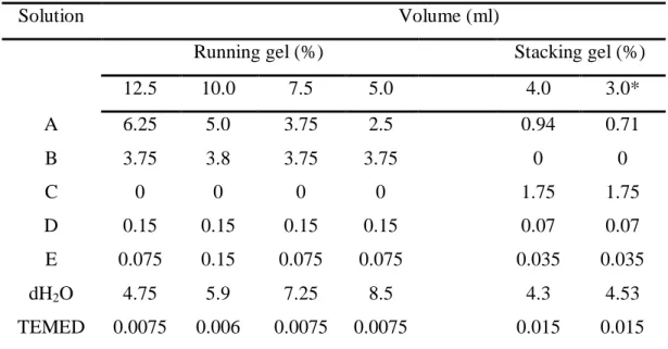
Sodium dodecyl sulfate polyacrylamide gel electrophoresis (SDS-PAGE)
- Reagents
- Procedure
Gloves were worn during the concentration process to prevent contamination of the protein sample with skin keratins. β-Mercaptoethanol reduces the disulfide bonds of proteins and thus disrupts the protein structure for easier separation on SDS-PAGE gel.
Staining protein gels
- Coomassie staining
- Reagents
- Procedure
- Silver staining
- Reagents
- Procedure
Destaining of the gel was achieved by soaking the gel in destain I (overnight or until the gel had a fairly clear background). The gel was then washed with 50% (v/v) methanol (approximately 20 min), photographed and then stored in a Ziploc® plastic bag.
Western blotting
- Reagents
- Procedure
The nitrocellulose was placed on top of a foam pad and three sheets of filter paper. Once the color was removed, the nitrocellulose was washed several times with dH2O and allowed to air dry.
Agarose gel electrophoresis of DNA
- Reagents
- Procedure
After the agarose gel solidified, the comb and seals were removed from the casting tray and the tray was placed in The electrical cables were disconnected and the gel holding the casting tray was removed from the apparatus.
Preparation of protease inhibitors
The lid of the electrophoresis apparatus was firmly attached to the tank and the electrical leads were connected to a power pack so that the DNA would migrate from the negative cathode (black) to the positive anode (red). A voltage of 60-80 V was applied until the loading dye migrated about 1 cm from the end of the gel.
Polymerase chain reaction
- Reagents
- Procedure
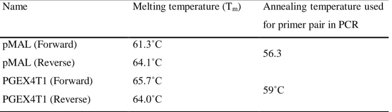
The primer annealing temperature (χ˚C) was set at 5˚C lower than the lowest melting temperature (Tm) of the primer pair (see Table 2.4 for the temperatures used for each primer pair used). Annealing temperatures used for each primer pair used in screening for recombinant bacterial clones by PCR.
Sequencing
Isolation of plasmid DNA
- Midi-preparation of plasmid DNA by alkaline lysis with SDS
- Reagents
- Procedure
- Wizard Plus SV Minipreps DNA Purification System
The solution was vortexed vigorously to ensure that the bacterial cells were completely suspended in solution and then transferred to a 1.5 ml microcentrifuge tube. The microcentrifuge tube was inverted several times to ensure mixing of the solution before being left on ice for 5 min.
Restriction enzyme digestion of DNA
- Reagents
- Procedure
Plasmid DNA can be used immediately for other molecular biology applications such as restriction enzyme digestion, cloning and sequencing, without further purification. Omitting the restriction enzyme and adjusting the volume of water accordingly provided a non-digested control.
Dephosphorylation
- Reagents
- Procedure
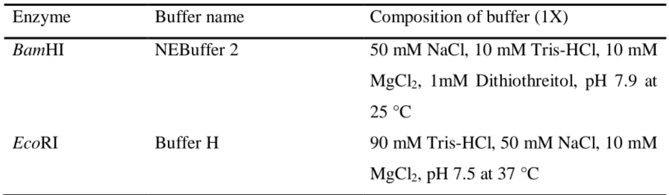
The volumes of reagents used were dependent on the amount of digested DNA required. The following procedure was typically used, although scaled up as needed: 10X restriction enzyme buffer (4μl), dH2O (14μl), plasmid DNA from miniprep (20μl) and restriction enzyme (2μl) were pipetted into a sterile 1.5ml microcentrifuge tube and incubated at 37C.
Purification of plasmid DNA
- Wizard ® DNA Clean-up System
- Materials and reagents
- Procedure
- Low melting point (LMP) agarose purification of DNA
- Materials and reagents
- Procedure
The minicolumn was disconnected from the syringe and the plunger was removed from the syringe barrel. The plunger was inserted into the syringe barrel and the propan-2-ol was pushed through the minicolumn.
Ligation of plasmid DNA and insert DNA
- Reagents
- Procedure
The plunger was inserted into the barrel of the syringe and used to slowly push the resin-DNA slurry into the minicolumns. The plunger was inserted into the barrel of the syringe and used to gently push the propan-2-ol through the minicolumn.
Concentration and purification of DNA
- Reagents
- Procedure
Sodium acetate, pH 5.5 was added to the DNA sample to 1/10th of the sample volume and mixed by gentle pipetting. The DNA pellet was allowed to air dry for 10 min before being dissolved in an appropriate volume of sterile dH2O.
Transformation of bacterial cells with DNA
- Transformation of competent E.coli (glycerol method) cells by electroporation
- Reagents
- Procedure
- Transformation of competent E. coli cells (calcium chloride method) using heat
- Reagents
- Procedure
Cells were aliquoted (20 μl) into sterile 1.5 ml microcentrifuge tubes and stored on ice until use. LB agar plates containing the appropriate antibiotics were then plated with cells (50 μl) and then incubated overnight inverted at 37°C.
Preparation of glycerol stocks
- Reagents
- Procedure
Super coiled recombinant plasmid DNA (1 μl) was incubated with freshly prepared competent cells (20 μl) on ice for 30 min.
Recombinant expression and purification of Pf33-MBP and MBP from pMAL-c2X
- Confirmation of the identity of the pMAL-c2X and pTS822 plasmids
- Expression of Pf33-MBP or MBP alone
- Reagents
- Procedure
- Affinity purification of fusion protein and MBP on amylose resin
- Reagents
- Procedure
- Factor Xa cleavage of fusion protein Pf33-MBP
- Reagents
- Procedure
- DEAE-Sepharose ion exchange separation of cleaved Pf33-MBP
- Reagents
- Procedure
- Reagents
- Procedure
The column was then washed with column buffer until the absorbance of a fraction (1 ml) was approximately 0.010. Factor Xa (20 μg) was added and the column was allowed to gently rock overnight at room temperature.
- Subcloning 822 bp PFC0760c gene sequence into pGEX4T1
- Expression of Pf33-GST
- Glutathione agarose purification of Pf33-GST
- Reagents
- Procedure
This resulted in approximately 1 mL of resin, which was loaded onto a Poly-Prep® chromatographic column and equilibrated with 20 column volumes of PBS. Column regeneration was achieved by washing with 5 column volumes of purification buffer 1, 5 column volumes of dH2O, 5 column volumes of purification buffer 2, followed by 5 column volumes of dH2O.
Intraperitioneal (I. P.) injection of mice
- Materials
- Procedure
Without releasing the tail, the mouse was placed on top of the wire cage and allowed to pull away. The needle was inserted at an angle of approximately 30 degrees to the skin surface and approximately 45 degrees to the midline of the mouse.
Preparation of blood films for determining parasitemia in mice
- Thick blood film
- Materials
- Procedure
- Thin blood film
- Reagents
- Procedure
- Giemsa staining of thick and thin blood films
- Reagents
- Procedure
The mouse to be injected was picked up by grasping the tail with the thumb and index finger of one hand. Using the third or fourth finger of the hand pulling the skin taut, the tail was secured between the palm and the finger, thus holding the mouse with one hand.
Determining the number of parasitized red blood cells from thin blood films
- Procedure
After the films were air-dried after fixation in methanol for 30 seconds, they were stained for 30 minutes with Giemsa.
Collection of blood from mice using retro-orbital bleeding
Preparation of stabilate from infected blood
- Reagents
- Procedure
The volume of packed red blood cells was estimated by subtracting the plasma volume from the whole blood volume. The packed red blood cells were washed very gently with RPMI-1640 medium with a volume equal to the plasma volume and then centrifuged as before (1000 g, 4 min, 4˚C).
Determining the number of red blood cells in a mouse blood sample
- Reagents
- Procedure
Care was taken not to cut the red blood cells by using pipette tips that had been cut to provide a larger opening when the pellet was resuspended. The red blood cells were then diluted to the required number of parasitized red blood cells per 100 μl with RPMI-1640 containing 10% (v/v) glycerol.
Solubilization of parasitized red blood cells
- Reagents
- Procedure
A coverslip was placed on the hemocytometer chamber and whole blood (25 μl) diluted 1:200 was allowed to flow under the coverslip into the chamber.
Coupling peptides to rabbit albumin using glutaraldehyde
- Reagents
- Procedure
Coupling peptides to rabbit albumin using 3-maleimidobenzoic acid N-
- Reagents
- Procedure
Each of the fractions was tested for reduced peptide by combining 10 μl of the fraction and 10 μl of Ellman's reagent. Fractions containing activated carrier were combined with pooled reduced peptide fractions and gently stirred at room temperature for 3 h.
Immunisation of chickens with antigen and rabbit albumin-conjugated peptides
Isolation of immunoglobulin Y from chicken egg yolk
- Reagents
- Procedure
- Determination of IgY concentration
The supernatant was filtered through absorbent cotton wool and the volume of the resulting filtrate was noted. The supernatant was discarded and the pellet was suspended in a volume of isolation buffer equal to the volume of yolk.
Preparation of affinity matrices
- Coupling of peptide or protein to AminoLink®
- Reagents
- Procedure
- Coupling of peptides to SulfoLink® Coupling Gel
- Reagents
- Procedure
A volume of isolation buffer, equal to twice the yolk volume, was then added to the yolk and stirred gently. A volume of quenching buffer equal to the volume of the gel was then added.
Affinity purification of chicken IgY anti-peptide antibodies
- Reagents
- Procedure
The column was washed with 3 column volumes of 50 mM Tris-HCl, 5 mM EDTA, pH 8.5, before incubation with 50 mM cysteine (1 ml) to block unreacted sites (bottom-over-bottom rotation, 15 min, room temperature room). The column was washed with approximately 12 column volumes of wash buffer to remove any unbound IgY.
Enzyme linked immunoadsorbent assay (ELISA)
- Reagents
- Procedure
The IgY pool was cycled overnight (10 ml/h, RT) through the column to allow binding of anti-peptide antibodies to the peptide on the column. Antibodies were circulated back through the column to avoid blockages and column drying.
Immunofluorescence assays
- Reagents
- Procedure
The volume of whole blood was determined and then the red blood cells were centrifuged (1000 x g, 1 min). The wells of the slides coated with parasitized mouse RBCs were blocked with 2% (w/v) BSA-PBS (20 μl per well, 1 h, RT).
Bioinformatic studies
Introduction
Bioinformatic methods
- Basic Local Alignment Search Tool (BLAST) searches and CLUSTALW
- Expression profiles
- Plasmodium export element (PEXEL) motif searches and signal sequence
- Transmembrane domain prediction
- Subcellular localization predictions
- pTARGET subcellular localization prediction
- Prediction of Subcellular Localization by SubLoc
- Conserved domains
- Helical wheel plot
- Structure prediction
- T-cell epitope prediction
- Protein interaction
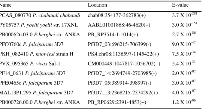
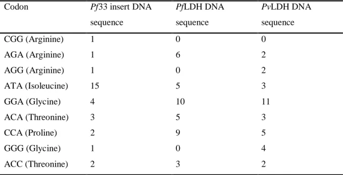
All different R/KxLxQ/E motif combinations were searched in the amino acid sequences of PFC0760c and P. A score is based on relative amino acid weights calculated from the amino acid composition of the protein sequence.
Results
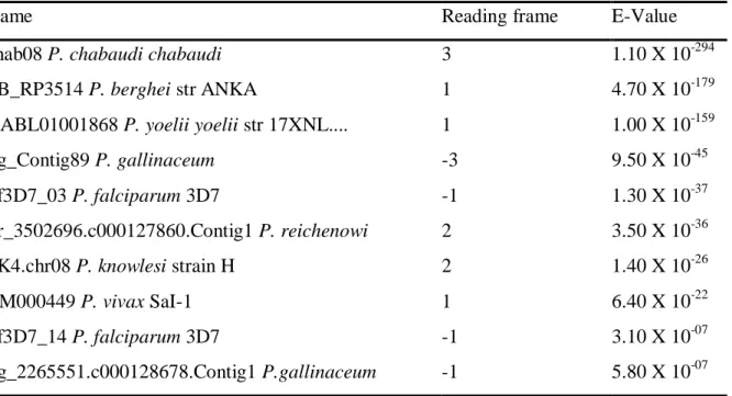
- BLAST searches and sequence alignments
- Expression levels
- Transmembrane domain prediction
- Subcellular location prediction
- Conserved domains
- Helical wheel plot of the conserved peptide sequence FKLGSCYLYIINRNLKEI
- Structure predictions
- T-cell epitope predictions
- Protein interaction
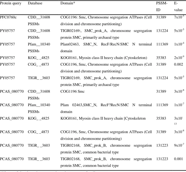
From the TMHMM prediction it appears that the C terminus of Pca 96 is located within a membrane and the N terminus is located outside the membrane. The predominant domain observed in each of the proteins was an SMC (structural maintenance of chromosomes) protein domain (SMC_prok_A, SMC_prok_B, SMC_N and SMC).
Discussion
Prediction of the structural folds present in PFC0760c and Pca 96 revealed several folds predicted to be part of the protein structure. Unfortunately, many of the proteins predicted to affect PFC0760c have yet to be assigned a function.
Recombinant expression of the Plasmodium falciparum protein PFC0760C
Introduction
Results
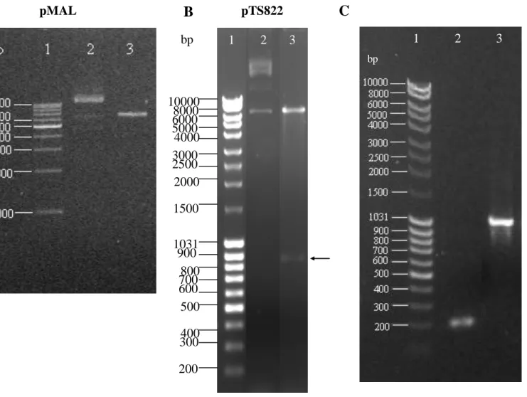
- Confirmation of the identity of the pMAL-c2X and pTS822 plasmids
- Optimisation of expression of the recombinant P. falciparum MBP fusion protein
- Theoretical molecular weight and pI of proteins
- Base pair composition of Plasmodium proteins
- Protein disorder prediction
- Purification of Pf33-MBP
- Factor Xa cleavage of Pf33-MBP
- Diethylaminoethyl (DEAE)-Sepharose TM ion exchange chromatography to
An attempt was made to optimize the expression of the Pf33-MBP fusion protein from the pTS822 vector. Large parts of the Pf33-MBP fusion protein (Figure 4.7 A) were predicted to be disordered, as was MBP (Figure 4.7 B).
- Sub-cloning of the P. falciparum insert into pGEX4T1
- Recombinant expression of GST and the GST-fusion protein (Pf33-GST)
- Purification of GST and Pf33-GST
PCR products corresponding to the size of the nonrecombinant pGEX4T1 (200 bp) did not possess the DNA insert (arrow). Analysis of the expression of the GST fusion protein, Pf33-GST, by SDS-PAGE and Western blotting.
Discussion
Mehlin et al., (2006) found that codon usage and base pair composition do not appear to be contributing factors towards poor expression of recombinant proteins. It is possible that MBP and Pf33 form a tight association even after Factor Xa cleavage as experienced by Ko et al.
Immunological studies
Introduction
Production of antibodies in chickens against synthetic peptides
- Selection of peptides
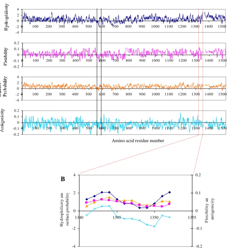
The position of the cysteine residue resulted in the C-terminus being predicted to be more immunogenic than the N-terminus by Predict 7 (Figure 5.2 B). The position of the conserved sequence FKLGSCYLYIINNRLKEI is located between the solid black lines, and the position of the peptide SDDDNRQIQDFE is between the dotted red lines.
Results
- Production of chicken anti-peptide antibodies against P. falciparum protein
- Measuring anti-peptide antibody titres in chickens
- Affinity purification of anti-peptide antibodies
- Evaluation of purified anti-peptide antibodies with ELISA
- Anti-Plasmodium lactate dehydrogenase antibodies
- Localisation of native P. yoelii yoelii protein, PY05757 and LDH with
- Western blotting of parasite material
- Purification of human anti-malaria antibodies
The anti-peptide SDDDNRQIQDFEC antibodies were pooled and affinity purified on a SulfoLink® column coupled to peptide SDDDNRQIQDFEC (Figure 5.5 C) (Section 2.33.2 and 2.34). The anti-peptide FKLGSCYLYIIINNRLKEI and CFKLGSCYLYIINRNLKEI antibodies typically eluted after 5-6 ml of 100 mM glycine, pH 2.8 (Figure 5.5 A and B).

Discussion
It is possible that the epitope FKLGSCYLYIIINNRLKEI was not available to interact with the antibodies. A Plasmodium lysate could be passed over an affinity matrix coupled with the anti-peptide antibodies to obtain the native protein.
General Discussion
In the present study, a number of proteins with known function were compared with PFC0760c, with respect to their mRNA expression levels (Le Roch et al., 2003) with the hope that these data provide further insight into the role that PFC0760c plays within the parasite life cycle. A computer algorithm to detect emerging patterns would be useful to assist in this analysis, but was beyond the scope of the current study.