AN ANALYSIS OF DOSE EFFECTIVENESS AND INCIDENCE OF LATE RECTAL
COMPLICATIONS OF HIGH DOSE-RATE BRACHYTHERAPY IN THE RADICAL TREATMENT OF CERVICAL CANCER
B.Rad (Diagnostic)
B.Rad Honours (Therapy)
Dissertation submitted in fulfillment of the requirement for the Degree MAGISTER TECHNOLOGIAE
RADIOGRAPHY (ONCOTHERAPY) in the
SCHOOL OF HEALTH TECHNOLOGY
FACULTY OF HEALTH AND ENVIRONMENTAL SCIENCES at the
CENTRAL UNIVERSITY OF TECHNOLOGY, FREE STATE BLOEMFONTEIN
Supervisor: DR. H. FRIEDRICH-NEL Co-supervisor: PROF. L. GOEDHALS
Bloemfontein Mei 2007
! " # $ " % &
I, DEIRDRÉ LONG, do hereby declare that this project submitted for the degree MASTER TECHNOLOGIAE;
RADIOGRAPHY (ONCOTHERAPY) in the SCHOOL OF HEALTH TECHNOLOGY, FREE STATE, is my own independent work that has not been submitted before, to any institution by me or anyone else as part of any qualification.
--- ---
Signature of student Date
Dedicated to my father:
Dedicated to my father:
Dedicated to my father:
Dedicated to my father:
Reginald Margin Ellerbeck
Blessed is the man who trusts Blessed is the man who trusts Blessed is the man who trusts Blessed is the man who trusts
In the Lord, In the Lord, In the Lord, In the Lord,
And whose hope is the Lord.
And whose hope is the Lord. And whose hope is the Lord.
And whose hope is the Lord.
For he shall be like a tree For he shall be like a tree For he shall be like a tree For he shall be like a tree
Planted by the waters, Planted by the waters, Planted by the waters, Planted by the waters, Which spreads out its roots Which spreads out its roots Which spreads out its roots Which spreads out its roots
bbbby the river, y the river, y the river, y the river, And will not fear when he And will not fear when he And will not fear when he
And will not fear when heat comes; at comes; at comes; at comes;
But its leaf will be green, But its leaf will be green, But its leaf will be green, But its leaf will be green, And will not be anxious in the year And will not be anxious in the year And will not be anxious in the year And will not be anxious in the year
of drought, of drought, of drought, of drought,
Nor will cease from yielding fruit.
Nor will cease from yielding fruit.
Nor will cease from yielding fruit.
Nor will cease from yielding fruit.
Jeremiah 17: 7,8
Jeremiah 17: 7,8 Jeremiah 17: 7,8
Jeremiah 17: 7,8
To my husband, Michael To my husband, Michael To my husband, Michael To my husband, Michael
And our son Jason And our son Jason And our son Jason And our son Jason ––––
Thank Thank Thank
Thank----you. you. you. you.
!& % ' "(
!& % ' "(
!& % ' "(
!& % ' "(
Soli deo Gloria
My grateful thanks are extended to the following people who assisted and stood by me these past two years:
Firstly all honor and glory to my Heavenly Father God, who gave me the wisdom and strength to complete my thesis;
My husband Michael, for all your love and support in so` many ways. Thank you for making time in your busy schedule for proof- reading my thesis;
Our son Jason, for understanding why “mommy is busy” and could not always give my full attention to you all the time;
Dr Hesta Friedrich-Nel, my study leader, for her invaluable guidance, time, continuous support and encouragement, as well for proof-reading this thesis;
Prof Louis Goedhals, my co-study leader and head of the Department of Oncotherapy for his quidance, continuous support and encouragement, supply of patient data and equipment, as well as proof reading this thesis;
Prof Gina Joubert, head of the Department of Biostatistics, UFS, and Maryn Brussouw for their invaluable assistance in analyzing the data;
Thys Doman, statistician of the Department of Oncotherapyl for his invaluable assistance;
My colleagues at the Department of Oncotherapy, especially my head, Mrs S.
Rossouw for her support, encouragement and patience;
My parents, Una and Reg Ellerbeck, I would like to thank you for the steadfast encouragement and unwavering support you have given me all through my studies.
CONTENTS
Ai LIST OF APPENDICES D
ii LIST OF TABLES E
iii LIST OF FIGURES H
CHAPTER ONE
1. AN OVERVIEW OF THE STUDY 1
1.1 Introduction. 1
1.2 Worldwide incidence of cervical cancer. 2
1.3 Cervical cancer in South Africa. 3
1.4 Cervical cancer at the Department of Oncotherapy, 4 Universitas, Bloemfontein.
1.5 Development of treatment protocols at the Department 6 of Oncotherapy, Bloemfontein.
1.6 High dose-rate brachytherapy. 8
1.7 Fractionation and dose effectiveness. 10
1.8 Problem statement. 12
1.9 The objectives of the current study. 13
1.10 The motivation and significance of the study. 13
1.11 Methodology. 14
1.11.1 Patient selection. 14
1.11.2 Calculations. 14
1.11.3 Research tool. 15
1.12 Data analysis. 15
1.13 Ethics. 15
1.14 Outcome of the study. 15
1.15 Arrangement of the dissertation. 16
1.16 Conclusion. 17
B CHAPTER TWO
2. LITERATURE REVIEW 18
2.1 Introduction. 18
2.2 Epidemiology. 19
2.3 Radiation therapy. 21
2.4 Recommendations of the American Brachytherapy 22 Society (ABS).
2.5 Dose effectiveness of fractionation schedules. 26
2.6 Biology of HDR brachytherapy. 29
2.7 Time-dose models. 32
2.8 Treatment techniques and dose specifications. 35
2.9 Late complications of organs at risk. 38
2.10 Future developments. 41
2.11 Conclusion. 42
CHAPTER THREE
3. METHODOLOGY 44
3.1 Introduction. 44
3.2 Research design. 44
3.3 Patient selection. 45
3.4 External beam radiotherapy. 46
3.5 High dose-rate – Intracavitary brachytherapy. 48
3.5.1 Insertion technique. 48
3.5.2 Treatment planning. 51
3.6 Calculations. 54
3.7 Research tool. 55
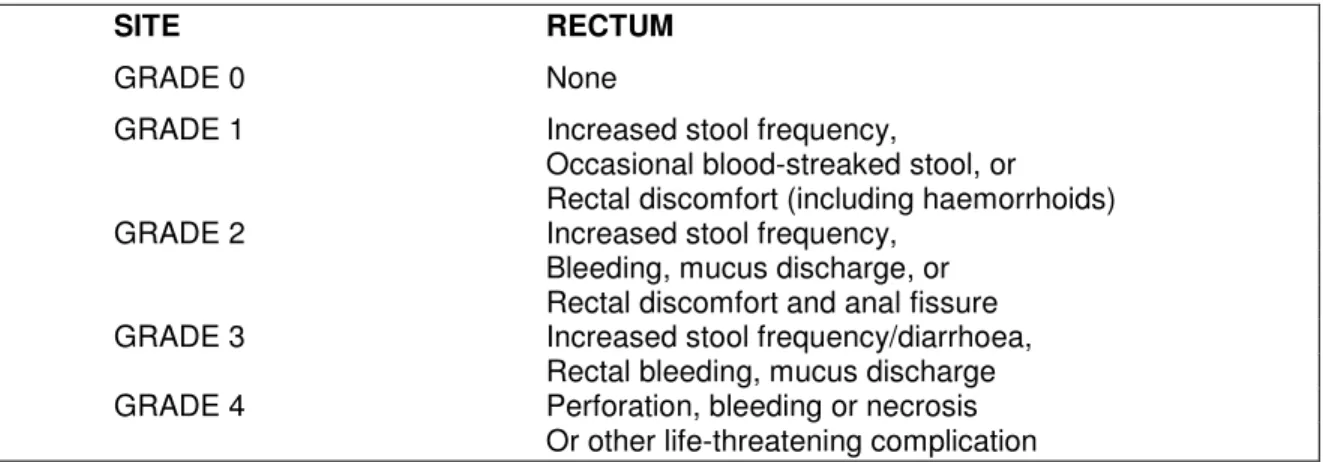
3.8 Scoring criteria for late radiation complications. 56
3.9 Statistical analysis and follow-up. 56
3.10 Conclusion. 57
C CHAPTER FOUR
4. RESULTS 58
4.1 Introduction. 58
4.2 Patient selection. 58
4.3 Patient demographics. 59
4.4 Tumour characteristics. 60
4.5 Radiation therapy treatment. 63
4.6 Biologically effective doses (BED). 65
4.6.1 BED values of EBRT. 65
4.6.2 BED values of HDR-ICBT. 65
4.6.3 The cumulative BED values of EBRT & HDR-ICBT. 66
4.7 Follow-up results. 66
4.7.1 Tumour recurrence. 66
4.7.2 Late radiation complications. 69
4.8 Metastases. 73
4.9 Survival. 75
4.10 Conclusion. 75
CHAPTER FIVE
5. DISCUSSION & CONCLUSION 76
5.1 Introduction. 76
5.2 High dose-rate brachytherapy. 76
5.3 Dose effectiveness. 77
5.4 Late radiation complications. 81
5.5 Recommendations. 83
5.6 Shortcomings of the study. 84
5.7 Conclusion. 85
6. SUMMARY. 87
7. OPSOMMING. 89
8. REFERENCES. 91
D
(" # $$ ! (
CHAPTER 1
Appendix I A Research tool: Patient data source form 100 Appendix I B Ethics: Letter of consent 106
CHAPTER 2
Appendix II A FIGO staging of carcinoma of the uterine cervix 108
Appendix C Abbreviations 110
Appendix D Certificate of language editing 112
E
) (" # " * (
CHAPTER 1
Table 1.1 Female patients treated for cancer at the Department of 4 Oncotherapy, Bloemfontein.
Table 1.2 Treatment protocols at Universitas Annex, stage II 6 cervical carcinoma: 1986-1990.
Table 1.3 Treatment protocol stage III cervical carcinoma: 7 1986-1990.
Table 1.4 Treatment protocol for FIGO stages I-III cervical cancer 8 patients treated at the Department of Oncotherapy,
Universitas Annex, Bloemfontein, from 1994-currently.
CHAPTER 2
Table 2.1 ABS suggested doses of EBRT and HDR-ICBT. 25 Table 2.2 Crude incidence of late complications according to 40 RTOG/EORTC-late radiation morbidity scoring scheme.
CHAPTER 3
Table 3.1 Criteria for patient inclusion. 45
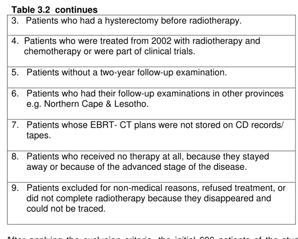
Table 3.2 Patient exclusion details. 45
Table 3.3 Treatment protocol for EBRT at the Department 47 of Oncotherapy, Bloemfontein.
Table 3.4 Treatment protocol for HDR-ICBT at the Department 53 of Oncotherapy, Bloemfontein.
F CHAPTER 4
Table 4.1 Patient treatment files suitable for study. 59
Table 4.2 Reasons for patient exclusion. 59
Table 4.3 Summary of race groups treated. 60
Table 4.4 FIGO staging vs.cumulative BED values to the tumour 63 and the rectum.
Table 4.5 Number of fractions received for HDR-ICBT. 64 Table 4.6 EBRT-BED values to the tumour Gy10 and the rectum Gy3. 65 Table 4.7 HDR-ICBT-BED values to the tumour Gy10 and the 66 rectum Gy3.
Table 4.8 Cumulative BED values for EBRT & HDR-ICBT. 66
Table 4.9 Probability of tumour recurrence. 67
Table 4.10 FIGO staging of patients with tumour recurrence. 68
Table 4.11 Summary of local infiltration. 68
Table 4.12 Tumour recurrence vs. BED10. 69
Table 4.13 Incidence of late radiation complications. 70 Table 4.14 FIGO staging of patients with late radiation complications. 70 Table 4.15 Site of late radiation complications. 71 Table 4.16 Crude incidence of first late radiation complications 71 according to the RTOG/EORTC scoring.
Table 4.17 Crude incidence of second late radiation complications 72 according to RTOG/EORTC scoring.
Table 4.18 Probability of late radiation complications. 73 Table 4.19 Late radiation complications vs. BED3. 73 Table 4.20 Patients with metastases at last follow-up. 74
Table 4.21 Probability of metastases. 74
G CHAPTER 5
Table 5.1 BED10 values of the Department of Oncotherapy, 78 Bloemfontein (1998-2003) and HDR fractionation
schedules for recently published studies.
H
) (" # # + (
CHAPTER 2
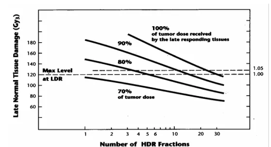
Fig. 2.1 Relationship between number of HDR fractions and late 31 normal tissue effects.
Fig. 2.2 Manchester system definition of points “A” and “B”. 35
CHAPTER 3
Fig. 3.1 The “four field box” technique. 47
Fig. 3.2 “Patslide” – Patient transfer apparatus. 49
Fig. 3.3 AP – radiograph. 51
Fig. 3.4 LAT – radiograph. 51
Fig. 3.5 Point “A”. 52
Fig. 3.6 Different rectum positions. 52
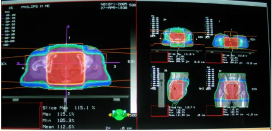
Fig. 3.7 Isodose curves generated with fixed relative dwell 54 weighting based on the Manchester System.
CHAPTER 4
Fig. 4.1 HIV/AIDS status of patients. 60
Fig. 4.2 Histology of uterine cervical cancer. 61 Fig. 4.3 Differentiation of uterine cervical cancer cells. 61 Fig. 4.4 Endophytic/exophytic type of tumours. 62 Fig. 4.5 FIGO staging of uterine cervical cancer patients. 62
Fig. 4.6 Energy chosen for EBRT treatment. 64
Fig. 4.7 Tumour recurrence of uterine cervical cancer patients. 67
Fig. 4.8 Organs infiltrated. 68
Fig. 4.9 Treatment of patients with tumour recurrence. 69 Fig. 4.10 Treatment for patients with late radiation complications. 72 Fig. 4.11 Sites of metastases for patients with uterine cervical cancer. 74
CHAPTER 1
AN OVERVIEW OF THE STUDY
1.1 Introduction
The first reference to the practice of gynaecology as a field of medicine is found in the Kahun papyrus from 2000 B.C. In a description of women`s diseases, Hippocrates, who is considered to be the father of both medicine and oncology, referred to cancer of the uterus as a disease with a poor prognosis. He recommended the following treatments for cancer:
“Regular evacuation of the bowels; the patient is to be given a bath each day with lukewarm water; narcotics such as opium given internally or injected into the uterus for pain relief; the application of leeches or red-hot irons or other caustic agents to the cervix” (Werner 1990). Since the discovery of X-rays by Wilhelm Conrad Roentgen in 1895, the treatment of uterine cervical cancer by means of radiotherapy has developed immensely.
Radiotherapy is a clinical modality that uses ionising radiation in the treatment of patients with malignant neoplasias. The aim of radiotherapy is to deliver a precisely measured dose of radiation to a defined tumour volume with as little damage as possible to surrounding healthy tissue, resulting in eradication of the tumour, a high quality of life and prolongation of survival at a competitive cost (Halperin, Schmidt-Ulrich, Perez & Brady 2004: 3). The current study will look at radiotherapy as treatment modality for patients diagnosed with uterine cervical cancer.
Uterine cervical cancer occurs in women around the world, irrespective of their age and race. The South African cervical cancer screening program implemented since 2001 are an attempt to facilitate early detection of cervical cancer through the Papanicolaou (PAP) smear (Mqoqi, Kellet, Sitas & Jula 2004: 22). Through the screening policy it has become evident that cervical cancer is the leading cancer among South African women. It is thus crucial to treat patients who have been diagnosed with uterine cervical cancer with the best modality currently available.
This current study will look at radiotherapy as the treatment modality administered to patients at the Department of Oncotherapy, Bloemfontein, diagnosed with uterine cervical cancer, since a new modality of high dose- rate (HDR)-intracavitary brachytherapy (ICBT) was implemented in 1994.
Treatment results are quantified and analysed in terms of tumour control and late rectal complications. This chapter provides an overview of the incidence of uterine cervical cancer, the development of treatment protocols at the Department of Oncotherapy, Bloemfontein, and the dose effectiveness of HDR-ICBT as an introduction to the problem statement and objectives for this study.
1.2 Worldwide incidence of cervical cancer
Worldwide, uterine cervical cancer is one of the most frequently occurring cancers in women, with more than 80% occurring in developing countries (Stewart & Kleihues 2003: 215). Carcinoma of the uterine cervix is the sixth most common malignant neoplasm amongst women in the world, after carcinoma of the breast, lung, colorectum, endometrium and ovary (Perez & Kavanagh 2004: 1800). From a worldwide perspective, uterine cervical cancer remains one of the biggest causes of cancer death in women. Globocan 2000 estimated that in the year 2000, the number of patients diagnosed with this disease was 470 606, of whom 233 372 died
as a result (Parkin, Bray, Ferlay & Pisani 2001: 153). The American Cancer Society estimated that in 2001 there were 12 900 new cases of invasive uterine cervical cancer in the United States and 4 400 deaths from the disease, in addition to over 50 000 cases of carcinoma in situ (Perez & Kavanagh 2004: 1800).
The epidemiology of uterine cervical cancer is strongly related to the standard of living of populations from the different world regions (Parkin et al. 2001: 153). It follows then that for developed countries, in addition to a lower incidence of the disease, most cases of uterine cervical cancer are detected at a pre-invasive stage where the rates of cure approach 100%
with local therapies. On the other hand, developing countries present a different picture, with most cases being diagnosed with locally advanced disease. This requires a much more aggressive treatment approach and, in general, the survival prospects are not very encouraging (Duenas- Gonzalez, Cetina, Mariscal & De la Garza 2003: 390). Nag et al. (2002) reported that cervical cancer is the commonest type of cancer in many developing countries (Nag, Dally, de la Torre, Tatsuzaki, Kizilash, Kurusun, Pinillos, Pokrajac, Sur & Levin 2002: 298). Developing countries with a high incidence of cervical cancer are Zimbabwe, Uganda, South Africa, Gambia and Algeria. Black Zimbabwean women have the highest incidence rates of 55 per 100 000 (Mqoqi et al. 2004: 22).
1.3 Cervical cancer in South Africa
In South Africa, a total of 6 061 and 4 944 uterine cervical cancer cases were reported to the National Cancer Registry (NCR) in 1998 and 1999 respectively. The NCR (established in 1986) is the main cancer data source, which publishes pathology-based cancer incidence data in South Africa. These are the most recent statistics available and comprise 20%
and 17% respectively of all cancer cases reported in females over two
years. Consequently, uterine cervical cancer was the leading cancer in women in 1998 and the second leading cancer in 1999 (Mqoqi et al. 2004:
22). The incidence of uterine cervical cancer also differs among the different population groups in South Africa. Statistics indicate that in 1999 a total of 4 944 cases were observed, of whom 4 127 were black, 390 coloured, 371 white and 56 Asian. The statistics for 2000 – 2004 at the Department of Oncotherapy, Bloemfontein, has indicated that Black South African women were the most affected by this disease (Doman 2006).
1.4 Cervical cancer at the Department of Oncotherapy, Universitas Annex, Bloemfontein
The incidence rate of cervical cancer at the Department of Oncotherapy, Universitas Annex, Bloemfontein (Free State, South Africa) is high according to the statistics (Doman 2006). The statistics from 2000 to 2004 in Table 1.1 indicate that a total of 6 285 female patients received treatment for cancer over a five year period from January 2000 to December 2004. Of these, 2 117 patients (33.7%) were diagnosed with cervical cancer (FIGO stages I-IV) and were treated accordingly.
Table 1.1 Female patients treated for cancer at the Department of Oncotherapy, Bloemfontein (Doman:2006)
White % of
whites % of
females Black % of blacks % of
females Total % of
Total % of females
2004 cervix 29 4.25 8.92 389 24.13 43.37 418 18.21 34.21
Females 325 897 1222
2003 cervix 26 4.70 7.78 402 23.52 46.26 428 18.92 35.58
Females 334 869 1203
2002 cervix 36 5.57 11.39 351 22.43 37.70 387 17.50 31.03
Females 316 931 1247
2001 cervix 38 9.69 10.83 412 27.23 42.13 450 20.41 33.86
Females 351 978 1329
2000 cervix 43 6.44 12.50 391 24.15 41.60 434 18.89 33.80
Females 344 940 1284
Total cervix 172 1945 2117
Females 1670 4615 6285
Average 34 6.13 10.29 389 24.29 42.21 423 18.79 33.70
Treatment modalities for uterine cervical cancer differ according to the disease. Surgery, chemotherapy and radiotherapy, or a combination thereof, are currently being used to treat patients diagnosed with uterine cervical cancer. Radiotherapy plays an important role in the treatment of uterine cervical cancer. A combination of megavoltage external beam radiotherapy (EBRT) and intracavitary brachytherapy (ICBT) is the accepted definitive mode of treatment of International Federation of Gynecologists and Obstetricians (FIGO) stages I-III cervical cancer. The curative potential of radiotherapy in the management of carcinoma of the cervix is enhanced by the use of intracavitary brachytherapy, which delivers a high radiation dose directly to the tumour while sparing the surrounding normal tissues (Patel, Rai, Mallick & Sharma 2005: 125). The choice of radiation modality depends on the efficacy, disease site, equipment availability, treatment duration, expertise and radiation safety considerations (Nag et al. 2002: 298).
How often HDR brachytherapy is used depends on how common a particular cancer is in that country and whether that site can be effectively treated by HDR brachytherapy. Currently, more than 1 000 units exist in the world, including almost 400 in developing countries. The use of HDR- ICBT for uterine cervical cancer patients is the most common indication for brachytherapy, and gynaecological brachytherapy can account for up to 100% of the brachytherapy practice in some developing countries (Nag et al. 2002: 298-299).
The statistics shown in Table 1.1 therefore clearly indicate the need to introduce HDR-ICBT into the treatment schedule for FIGO stages I-III cervical cancer patients with curative intent. In April 1994 the standard prescribed protocol for FIGO stages I-III cervical cancer patients at the Department of Oncotherapy, Bloemfontein, South Africa, changed with the
installation of a high dose-rate (HDR) Ir192 brachytherapy afterloading unit, described in the next section.
1.5 Development of treatment protocols at the Department of Oncotherapy, Bloemfontein
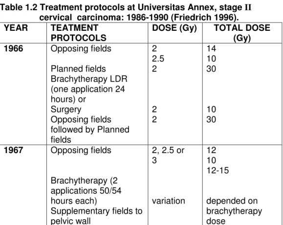
Radiotherapy treatment was started in Bloemfontein during 1960 by a diagnostic radiologist, Dr R Tahan. A kilovoltage apparatus for teletherapy (RT 250, Philips) and radium sources (Radium-226) for brachytherapy were the main treatment units during those years. The duration of the brachytherapy (Radium-226) was 72 hours and the patient received three fractions (Friedrich 1996: 16). The treatment protocols from 1966 to 1990 for stages IIb and III cervical carcinoma are summarised in Tables 1.2 &
1.3 respectively (Friedrich 1996: 17).
Table 1.2 Treatment protocols at Universitas Annex, stage II cervical carcinoma: 1986-1990 (Friedrich 1996).
YEAR TEATMENT
PROTOCOLS DOSE (Gy) TOTAL DOSE
(Gy) 1966 Opposing fields
Planned fields Brachytherapy LDR (one application 24 hours) or
Surgery
Opposing fields followed by Planned fields
2 2.5 2
2 2
14 10 30
10 30
1967 Opposing fields
Brachytherapy (2 applications 50/54 hours each)
Supplementary fields to pelvic wall
2, 2.5 or 3
variation
12 10 12-15
depended on brachytherapy dose
1969 Fractionation 3 x per week
1970 Double dose 3.5 to 7 45 - 54
1972 to
1990 Opposing fields
Brachytherapy (2 x 50 h) Surgery or
supplementary fields or Opposing fields,
treatment plan and brachytherapy LDR booster
3
3
12 -15
54 - 60
Table 1.3 Treatment protocol stage III cervical carcimoma: 1996-1990
YEAR TREATMENT
PROTOCOLS DOSE (Gy) TOTAL DOSE
(Gy) 1966 Opposing fields
Planned field length = 15cm
Brachytherapy LDR booster or booster plan
2
2
10
30 1972 Field length (planned
fields)=12cm 3 or
3.5 57 -60
42 -45 1973 –
1974 Field length (planned fields)=10cm
1975 –
1990 Field length (planned
fields)=8cm 3 57 - 60
In April 1994, the Department of Oncotherapy, Bloemfontein, implemented a high dose-rate (HDR) brachytherapy treatment system, an Ir-192 Nucletron Microselectron afterloading source, using the ring applicator.
After careful analysis of the available data on fractionation schedules, the standard prescribed protocol for treating patients for uterine cervical cancer (FIGO stages I-III) was established. The advent of HDR brachytherapy in the department, which has the advantages of individualised dosimetry, outpatient treatment and elimination of radiation exposure of medical personnel, brought a convenient treatment option for
patients with uterine cervical cancer, permitting treatment of a large number of patients (10-15 patients weekly).
The fractionation schedule applicable to the study is shown in Table 1.4.
Patients with FIGO stages I-III uterine cervical cancer are treated with a combination of EBRT- 2 Gy/fraction x 25 fractions (50Gy to whole pelvis) and HDR-ICBT given in 4-6 fractions, once weekly (2 Gy/fraction, normalized to the highest rectum dose point), to achieve a minimum total dose of 15 Gy to point A, as opposed to the previous protocols mentioned.
Point A is defined as a geometric point in relationship to the cervical os and uterine axis.
Table 1.4 Treatment protocol for FIGO stages I-III cervical cancer patients treated at the Department of Oncotherapy, Universitas Annex, Bloemfontein, from 1994 up to the time of writing
EBRT- Dose per fraction (Gy)
EBRT- No.
of fractions
EBRT-
WholePelvic Dose (Gy)
HDR-ICBT- Dose per fraction (Gy)
HDR-ICBT- No.
of fractions
HDR-ICBT- Total dose to tumour –min.15Gy
2 25 50 Gy 2
(2Gy to highest rectum dose point)
4-6 Cumulative recorded dose to point A
1.6 High dose-rate brachytherapy
Since Margaret Cleaves first performed intracavitary brachytherapy for cancer of the cervix in 1903, the radiotherapy of cervical cancer has traditionally been based on low dose-rate (LDR) intracavitary brachytherapy. HDR brachytherapy was developed to overcome some potential disadvantages of LDR brachytherapy, especially in the treatment of cervical cancer (Ferrigno, Nishimoto, Dos Santos Novaes, Pellizzon, Maia, Fogarolli & Salvajoli 2005: 1108). High dose-rate brachytherapy
allows for shorter treatment times, resulting in reduced hospitalisation costs owing to outpatient therapy, a reduced risk of applicator movement during therapy, and a larger throughput of patients in a busy department (Nag, Erickson, Thomadsen, Orton, Demanes & Petereit 2000: 202).
Studies reported by Falkenberg, Kim, Meleth, De Los Santos & Spencer (2006: 50) and Nag et al. (2000: 202) have compared LDR brachytherapy to HDR brachytherapy and have demonstrated comparable local control, survival and morbidity. To achieve disease control equivalent to that with LDR brachytherapy, changes in the dose/fraction schedule and strict attention to normal tissue doses when using HDR brachytherapy are mandatory. The biological disadvantage of HDR-ICBT in the treatment of cervical cancer is that short treatment times of HDR-ICBT do not allow for the repair of non-lethal damage in normal tissue, or the redistribution of cells in the cell cycle, or reoxygenation of the tumour cells. Multiple treatments are thus required (Nag 2004: 620). These radiobiological disadvantages of HDR-ICBT can, however, be overcome through adequate fractionation (Nag et al. 2000: 201).
The primary concern of using HDR brachytherapy is a potential late toxicity due to large doses per fraction (5 - 9 Gy/fraction). Late responding tissues such as the rectum and bladder have a greater capability of repair than tumour or early responding tissues, but this repair does not occur as fully as that of the same tissues treated with low doses. Fractionation and dose adjustments of the total dose are crucial factors in lowering the frequency of late radiation complications to the rectum, such as rectal bleeding, without compromising the treatment (Patel et al. 2005:125).
1.7 Fractionation and dose effectiveness
Although a large number of fractionation schedules are used worldwide for HDR brachytherapy, the optimal schedule has yet to be decided. Petereit and Pearcey (1999: 364) analysed the fractionation schedules of 24 published articles and came to the conclusion that there is no optimal fractionation schedule available, but suggested that reasonable fractionation schedules should be based on single institutional experience with accurate reporting. Members of the American Brachytherapy Society (ABS) with expertise in HDR cervical brachytherapy conducted a literature review, and, supplemented with their clinical experience and biomathematic modeling, formulated guidelines for HDR brachytherapy for cervical carcinoma. These recommendations are based on the Patterns of Care studies which showed that recurrences and complications decreased when brachytherapy was used in addition to EBRT (see Literature Review, Nag et al. 2000: 202). The International Atomic Energy Agency (IAEA) has also published recommendations specifically for implementing HDR, Ir192 brachytherapy in developing countries (Nag et al.
2002: 298).These recommendations correlate with those of the ABS and were thus relevant to the implementation of some of the recommendations into the EBRT and HDR-ICBT treatment at the Department of Oncotherapy, Bloemfontein.
Although there is a marked variation among institutions in the dose and fractionation used for cervical HDR-ICBT, most centers use a schedule of approximately 1.8 to 2 Gy per fraction for 25 fractions EBRT and 6-8 Gy per fraction for HDR in four to six fractions (Orton, Seyedsadr & Somnay 1991: 1425). Analysing different treatment schedules emphasizes the need to deliver biologically effective doses of radiation to ensure the highest probability of tumour control in the pelvis, because the salvage rate in patients who fail with isolated pelvic recurrences are less than optimal even after pelvic exenteration (Perez & Kavanagh 2004: 1858).
Consequently, because of a lack of personal or documented experience, radiation oncologists frequently resort to the use of bio-effect dose models to convert from LDR to HDR (Nag 2004: 610). This is the best available model for the quantitative assessment of clinical problems, primarily because it allows a distinction to be made between the fractionation and dose-rate sensitivities of early and late responding tissues (Clark, Souhami, Roman, Chappell, Evans & Fowler 1997: 989). Calculating the biologically effective dose to point A using the linear quadratic formula with an / of 10 is critical to compare fractionation schedules (Petereit &
Fowler 2003: 1159). In general, the / values for tumour and early- responding tissues are approximately 10 (Gy10), and for late-responding tissues 3 to 5 (Gy3-5) (Petereit & Pearcey 1999: 360). The values derived are not actual doses, but biologically effective ones that take into consideration dose-rate and impact of fraction size.
In uterine cervical cancer, the response to radiotherapy is clearly dose- dependent. As the dose increases, so too does the probability of tumour control. However, the risk of damage and late complications in normal tissues also increases with the dose (Stewart & Viswanathan 2006: 908).
The lower the dose-rate of radiation a cell is exposed to, the greater the likelihood of repair. Late-reacting normal tissues seem more capable of repair than tumours; at a given therapeutic dose, the tumour is preferentially killed over normal tissue. The duration of LDR treatment (several days) allows for non-lethal damage repair. The short treatment time of HDR brachytherapy prohibits this repair during the actual irradiation. However, if an interval of more than 24 hours is maintained, normal tissues can undergo full repair. Therefore, LDR may allow recovery of more normal tissues during treatment, but HDR may offer the advantage of increased cytotoxicity to the tumour (Stewart & Viswanathan 2006: 909).
1.8 Problem statement
The radiation treatment protocol for carcinoma of the uterine cervix stages I-III had to be adjusted at the Department of Oncotherapy, Universitas Annex, Bloemfontein, in April 1994 to incorporate HDR-ICBT with EBRT.
The standard prescribed treatment protocol implemented at Universitas Annex since 1994 has thus not been evaluated or analysed for dose effectiveness and late radiation complications considering the biologically effective dose (BED) administered to cervical cancer patients. As a wide range of fractionation schedules exists worldwide with as yet no optimum schedule, there is a need to deliver biologically effective doses of irradiation to ensure the highest probability of tumour control in the pelvis with minimum late rectal complications.
The aim of the current study was to calculate the biologically effective dose for the fractionation schedule implemented at the Department of Oncotherapy, Universitas Annex, Bloemfontein, since 1994, to determine whether the fractionation schedule delivered BED`s that lead to local tumour control without severe late rectal complications. These cumulative BED`s, calculated for the combination of EBRT and HDR-ICBT, to the tumour Gy10 and the rectum Gy3 for cervical cancer patients will enable the Department of Oncotherapy, Universitas Annex, Bloemfontein, for the first time to compare the fractionation schedule results with those of other institutions worldwide.
Analysis of the current treatment schedule, regarding the calculated BED, will indicate whether the total dose given to the planned tumour volume (PTV) by means of EBRT and the gross tumour volume (GTV) by means of HDR-ICBT has led to local tumour control with negligible late toxicity to the rectum.
1.9 The objectives of the current study
(i) To analyse the dose effectiveness of the fractionation schedule retrospectively by calculating the cumulative biologically effective dose (Total BED) of the combination of EBRT and HDR-ICBT for FIGO stages I-III uterine cervical cancer patients treated at the Department of Oncotherapy, Bloemfontein from January 1998 to December 2003.
(ii) To analyse retrospectively the incidence of late radiation complications as a result of the particular fractionation schedule used, by utilising the Radiation Therapy Oncology Group (RTOG)/European Organization for Research and Treatment of Cancer (EORTC) grading system, grade 0-5 (0 means an absence of radiation effects and 5 means the effects led to death). See Table 2.2.
1.10 The motivation and significance of the study
As the incidence of cervical cancer is high in developing countries such as South Africa it is essential to treat those affected with the best modality currently available (Mqoqi et al. 2004: 22). Radiotherapy plays an important role in the treatment of carcinoma of the uterine cervix and a combination of EBRT and HDR-ICBT is the accepted definitive mode of treatment (Patel et al. 2005).
Establishing BED values for the first time to the tumour (Gy10) and the rectum (Gy3) by using the LQ model in the fractionation schedule applicable to the study will be significant in enabling the Department of Oncotherapy, Universitas Annex, to compare the results of this study with those reported in the literature worldwide. Whether the BED values obtained could be used as a predictor of local tumour control and of rectal toxicity in the current treatment protocol for cervical cancer will be indicated after statistical analyses have been done. The outcome of the
study will thus clearly indicate whether the total dose given to the planned tumour volume (PTV) and gross tumour volume (GTV) has been a biologically effective dose for local tumour control with negligible late toxicity.
Other studies were done on patients with cervical carcinoma at the Department of Oncotherapy, Universitas Annex (Friedrich 1996). The focus of Friedrich`s study was to calculate LQ (Linear Quadratic) values for early and late responding tissues, using larger doses per fraction (3 Gy). This study will be unique in calculating the cumulative biologically effective dose of the fractionation schedule applicable to the study, and analysing the tumour control and late radiation complication rate according to the cumulative dose and calculated BED for FIGO stages I-III cervical carcinoma.
1.11 Methodology 1.11.1 Patient selection
A list of patients treated with EBRT and HDR-ICBT for cervical cancer FIGO stages I-III was obtained from the department’s HDR-ICBT patient treatment book. All patient treatment files from 1998-2003 were analysed so as to include in this retrospective study only patients with complete dosimetric data and a minimum follow-up period of two years.
1.11.2 Calculations
The linear quadratic model was used to calculate the biologically effective doses for all the patients who met the inclusion criteria for this retrospective study. It is a mathematically simple way to quantify biological responses to different fractionation schedules (Clark et al. 1997: 989).
1.11.3 Research tool
A data source form was designed as a research tool to capture the relevant data from the patient treatment files (Appendix ). Patient demographics, fractionation schedule details, tumour control, late radiation complications, metastatic disease and patient survival details were obtained from the patients treatment files and follow-up notes.
1.12 Data analysis
The data collected for this retrospective study was processed by the Department of Biostatistics, University of the Free State, Bloemfontein.
Results were summarised by frequencies and percentages (categorial variables) and means, standard deviations, minima and maxima (numerical variables). The software used was SAS 9.1.3 Service Pack 3.
1.13 Ethics
The proposal for this retrospective study was presented to the Ethics Committee at the University of the Free State at the meeting held on 14 March 2006 and attained the necessary approval. ETOVS NR 31/06. A letter of consent (Appendix ) permitting the use of patient records for the retrospective study was obtained from the Head of Clinical Services, Universitas Annex.
1.14 Outcome of the study
The results obtained from this study of patients treated for FIGO stages I- III cervical cancer by using a combination of EBRT and HDR-ICBT are applied as follows:
• Documentation of the biologically effective dose (BED) values for future use in treating cervical cancer patients and comparison of the
BED values to the tumour Gy10 and rectum Gy3 with those of other international institutions.
• Implementation of a RTOG/EORTC scoring sheet in the patient follow- up files for future research on the crude incidence of late radiation complications
• Use of the data to compile a report for a thesis for M.Tech.
qualification.
• Presentation of the results of the thesis at congresses and/or seminars.
• Publication of data in relevant medical journals.
1.15 Arrangement of the dissertation
CHAPTER 1: An overview of the study
This chapter contains an introduction to the relevance of the study, incidence of cervical cancer, development of treatment protocols, HDR- ICBT, fractionation and dose effectiveness, the problem statement, objectives, motivation and the outcome of the study.
CHAPTER 2: Literature review
The literature review covers the epidemiology of uterine cervical cancer, staging, radiation therapy, ABS recommendations, biology of HDR brachytherapy, time-dose models, treatment techniques, late radiation complications and future developments of HDR brachytherapy.
CHAPTER 3: Methodology
The methodology of the study discusses the methods and materials used for patient selection, EBRT, HDR-ICBT insertion technique and treatment planning, calculations of the biologically effective dose, the research tool, scoring of late radiation complications, statistical analysis and follow-up examinations of uterine cervical cancer patients.
CHAPTER 4: Results
The results of the current study are shown in this chapter. The results as well as other information gathered by the data source form regarding patient selection, patient demographics, tumour characteristics, tumour control, metastases and survival are reflected in this chapter.
CHAPTER 5: Discussion and conclusion
The dose effectiveness and the incidence of late radiation complications of the fractionation schedule of EBRT and HDR-ICBT used in treating uterine cervical cancer patients at the Department of Oncotherapy, Bloemfontein are discussed.
1.16 Conclusion
HDR-ICBT for carcinoma of the cervix is widely used because of its advantages over LDR brachytherapy. Although a large number of fractionation schedules are in use for HDR-ICBT, the optimal schedule has yet to be decided (Patel et al. 2005: 125). Because there are few established guidelines for its clinical use the American Brachytherapy Society (ABS) formed a committee to issue guidelines specifically for the use of HDR-ICBT for cervical carcinoma (Nag et al. 2000: 202). The literature review in chapter 2 focuses on these recommendations and other published studies regarding the use of EBRT and HDR-ICBT for cervical carcinoma.
CHAPTER 2
LITERATURE REVIEW
2.1 Introduction
Fletcher and his co-workers stated over 55 years ago: “We are still in an empirical era in the use of radium therapy in cancer of the cervix”
(Fletcher, Shalek, Wall & Bloedorn 1952: 935). In the same way, Orton (1986) stated 21 years ago that apart from a change from radium to cesium, they were still in that empirical era regarding the many innovations that have occurred in the use of external beam radiotherapy in comparison to the treatment philosophies concerning intracavitary cervix treatments. The latter were dictated primarily by tradition and well- established convention. Unfortunately, there was evidence indicating that many of these established conventions were by no means optimal in terms of late complications (Orton 1986: 37). Orton also stated that there is a temptation to judge treatment “success” by avoiding complications rather than by long-term survival. He proclaimed that because fractionation and dose rate patterns to the vulnerable pelvic organs vary considerably from patient to patient, doses need to be expressed in terms of some type of “biologically effective dose” units and not simply Gy (Orton 1986: 37).
Treating patients who have been diagnosed with uterine cervical cancer in the most effective manner requires extensive research of the most recent literature on the subject. This literature review scrutinises the appropriate evidence supporting the use of HDR-ICBT in combination with EBRT as the accepted definite mode of treatment for uterine cervical cancer and
correlates it with the dose effectiveness and late radiation complications found in the study. This literature study entailed reviewing the relevant literature in textbooks and published articles to ascertain the work being done in this field and the relevant methods being used. Key words such as
“HDR brachytherapy” and “uterine cervical cancer” were used to retrieve the relevant data from the Internet. The epidemiology of patients diagnosed with uterine cervical cancer provides a background to the study.
2.2 Epidemiology
The incidence of uterine cervical cancer differs among different population groups in South Africa. Since the beginning of the National Cancer Registry (NCR) in 1986, uterine cervical cancer has been the leading cancer in black women. A total of 5 069 cancers were reported on black females in 1998, with 4 342 reported in 1999, comprising, on average, about 84% of all uterine cervical cancer cases reported in the two years.
The second highest rates of 29 per 100 000 and 26.4 per 100 000 in the two consecutive years were recorded among coloured women. Asian women had the lowest cervical cancer incidence rates of 11 per 100 000 in 1999. Uterine cervical cancer in white women peaked at a significantly lower Age-Specific Incidence Rate (ASIR) of 34.5 per 100 000 and at a younger age (60-64) than in black and coloured women where ASIR peaked between the ages of 65 and 69 (Mqoqi et al. 2004: 22).
According to Perez (1998) the incidence of uterine cervical cancer is substantially higher among women in low socio-economic classes. Uterine cervical cancer is more frequent in women who had first sexual intercourse at an early age, have a history of sexual promiscuity, or have had a large number of pregnancies (Perez 1998: 1734). Considering the epidemiology of uterine cervical cancer in South Africa, it is thus of utmost
importance to treat women who have been diagnosed with uterine cervical cancer with the best curative modality available.
The initial staging system of uterine cervical cancer proposed in 1929 by a subcommittee of the League of Nations was revised in 1937 and 1950.
These functions were taken over by International Federation of Gynecologists and Obstetricians (FIGO) in collaboration with the World Health Organization (WHO) and the International Union Against Cancer.
The staging recommendations were last revised in 1995 (Shepherd 1995:
319). See Appendix for the FIGO staging classification: Cervical Carsinoma. This FIGO staging (being refined e.g. IIIb1R) is used at the Department of Oncotherapy, Universitas Annex, Bloemfontein. The FIGO staging system is based on clinical evaluation (inspection, palpation, colposcopy), roentgenographic examination of the chest, kidneys, and skeleton, and endocervical curettage and biopsies.
Different stages have different local control outcome as indicated by a study done by Patel et al. (Patel et al. 2005: 125). They reported on 121 patients with stages I-III uterine cervical cancer who were treated with EBRT and HDR-ICBT. The actuarial local control rate at 5 years was 100% for stage I, 80% for stage II and 67,2% for stage III uterine cervical cancer patients. Toita et al. (2003) also stated that in their study on uterine cervical cancer patients treated with EBRT and HDR-ICBT, the 5-year local control rate for stage II was 87% and stage III was 50% (Ferrigno et al. 2005: 1113). Thus it seems as if the local tumour control of a patient diagnosed with uterine cervical cancer is influenced by the FIGO staging.
The probability of rectal complications shows a correlation for dose- response, while clinical stage is not associated with increased risk of late rectal sequalae (Chen, Liang, Yeh, Yang, Shiau & Lin 2004: 667).
2.3 Radiation therapy
Since the introduction of the first remote afterloading machine in 1962, designed by Rune Walstam at the Radiumhemmet in Stockholm, with a three 50 mg and one 9 mg radium tubes, modern brachytherapy techniques of afterloading and dosimetry optimisation have made revolutionary strides in treating cancer patients (Mould, Batterman, Martinez & Speiser 1994: 8).
Radiation therapy of uterine cervical cancer patients has traditionally been based on low dose-rate (LDR) intracavitary brachytherapy for almost 100 years. However, concerns about its disadvantages, such as the exposure of medical staff to radiation, prolonged treatment time, risk of applicator movement and mandatory hospitalisation, led to the development of HDR brachytherapy (Shakespeare, Lim, Lee, Back, Mukherjee & Lu 2006: 277).
Because of the high incidence of uterine cervical cancer in developing countries, it is the most common indication for gynecological brachytherapy and can account for up to 100% of the brachytherapy practice (Nag et al. 2002: 298). Due to the short treatment times needed, HDR machines are capable of treating larger numbers of patients than LDR techniques. The shorter treatment times of HDR brachytherapy also reduce the risk of applicator movement and reduce hospitalisation costs because of using outpatient therapy instead. Additionally, in HDR brachytherapy, late tissue complications might be minimised more effectively than in LDR brachytherapy, because greater normal tissue displacement (e.g. bladder anteriorly and rectum posteriorly) is possible because of the short treatment times and available retraction devices (Nag et al. 2000: 202).
Other advantages of HDR-ICBT treatment include eliminating radiation to care givers and visitors. HDR brachytherapy also eliminates the need for source preparation and transportation and because there is only one
source, there is minimal risk of losing a radioactive source (Nag 2004:
620). HDR sources are of smaller diameter than the cesium sources that are used for intracavitary LDR brachytherapy and therefore reduce the need for dilatation of the cervix, thus reducing the need for heavy sedation or general anesthesia. HDR brachyhterapy also improves treatment dose distribution optimisation. Technical advances in HDR brachytherapy and its advantages over LDR brachytherapy made this treatment modality, in combination with EBRT, an essential component of the curative treatment of uterine cervical cancer (Nag et al. 2002: 299).
2.4 Recommendations of the American Brachytherapy Society (ABS) A literature analysis was done by Petereit et al. (1999) of the University of Wisconsin, USA, on the fractionation schedules of 24 articles published using the linear quadratic model to determine if doses can be correlated with local control and complications. Their conclusion was that a dose response relationship could not be identified for tumour control or late tissue complications, mainly because most of the HDR publications reported inadequate details of the dose fractionation schedules. It was also reported that the optimal fractionation schedule for treating uterine cervical cancer using HDR brachytherapy is still unknown, and presently can be based on single institutions with significant experience (Petereit, Sarkaria, Potter & Schink 1999: 359).
In order to address this problem, in 2000, the American Brachytherapy Society (ABS) published recommendations regarding the treatment of patients diagnosed with uterine cervical cancer with fractionation schemes which utilises the combination of EBRT and HDR-ICBT (Nag et al. 2000:
202). A retrospective study was done by Shakespeare et al. (2006) to assess the suggested ABS guideline tolerability in an Asian population.
The study indicated good local control and survival outcomes. The
conclusion of the study was that it is reasonable and safe to follow the guidelines laid out by the ABS (Shakespeare et al. 2006: 281). The following are some recommendations and guidelines given by the ABS:
General recommendations
The ABS recommends that brachytherapy be included as a component of the definitive radiation therapy for uterine cervical carcinoma, based on the Patterns of Care studies that show that recurrences and complications are decreased when brachytherapy is used in addition to EBRT (Nag et al.
2000: 202). Nag et al. reported that the relative doses given by EBRT compared to brachytherapy depend upon the initial volume of disease, the ability to displace the bladder and rectum, the degree of tumour regression during pelvic irradiation and institutional preference. Patients with larger tumour volumes receive 20-30 Gy of EBRT before HDR-ICBT treatment is initiated to allow for tumour shrinkage of tumor and a more favourable anatomic geometry for applicator placement. Because the overall treatment duration would be unduly prolonged if HDR treatments were begun after completion of EBRT, the HDR-ICBT is interdigitated during the course of EBRT. However, EBRT is not given on the day of a HDR-ICBT treatment. Typically, if the vaginal geometry is optimal, HDR brachytherapy begins after 2 weeks of EBRT. HDR-ICBT is given once a week, with the EBRT continued on the other four days of the week. These recommendations have been implemented at the Department of Oncotherapy, Universitas Annex, Bloemfontein.
External beam radiotherapy
The ABS recognises that the whole pelvic EBRT dose varies from institution to institution. The HDR fraction size and number depends on the EBRT dose (Nag et al. 2000: 203). Some institutions prefer to limit the whole pelvis dose for patients with early disease and to perform the first intracavitary insertion after 20 Gy, with further EBRT delivered with a
central block in place. If a lower EBRT dose is chosen for patients with early disease, the ABS recommends increasing the HDR fraction size and number of fractions. However, the individual fraction size should be kept to less than 7.5 Gy due to reports of higher toxicity with larger fractions. Most institutions, which include the Department of Oncotherapy, Bloemfontein, prefer to deliver 40-50 Gy of EBRT to the entire pelvis. In these cases, the brachytherapy dose is decreased. If EBRT doses greater than 45-50 Gy are to be given, the treatment fields should be coned down after the initial 45-50 Gy in an effort to exclude small bowel (Nag et al. 2000: 203).
Dose specification
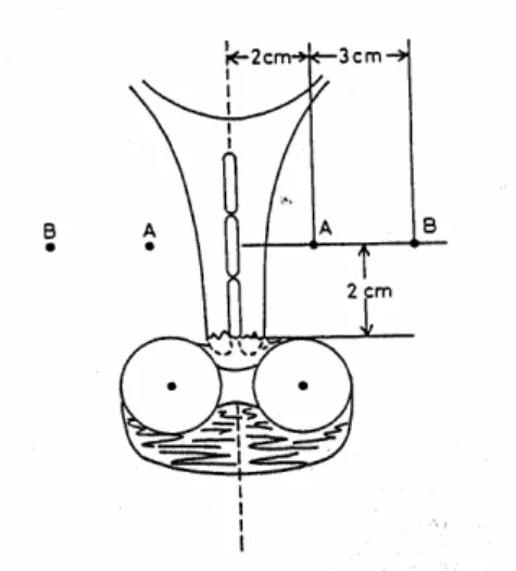
Ideally, the dose should be prescribed to the individual patient`s target volume. Unfortunately, many facilities lack the capability to determine the volume at risk, nor is there sufficient information in the literature to establish a better delineated target volume than the customary, Manchester System defined Point A. For HDR brachytherapy, the ABS has provided an applicator-based definition of point A that has been named point H (Nag et al. 2000: 204). Point H was created by the ABS to standardise the dosimetry process in the computerised treatment planning era by defining point A in relation to the applicator as seen on radiographs, as apposed to the cervical os.
Dose recommendations
The ABS recommends that the goals are to treat uterine cervical cancer with a combined EBRT and HDR dose to point A, to at least a total LDR equivalent of 80-85 Gy for early stage disease and 85-90 Gy for advanced stage disease (Nag et al. 2000: 201-211). The pelvic sidewall dose recommendations are 50-55 Gy for early lesions and 55 -65 Gy for advanced ones. Every attempt should be made to keep the bladder and rectal doses below 80 Gy and 75 Gy LDR equivalent doses, respectively.
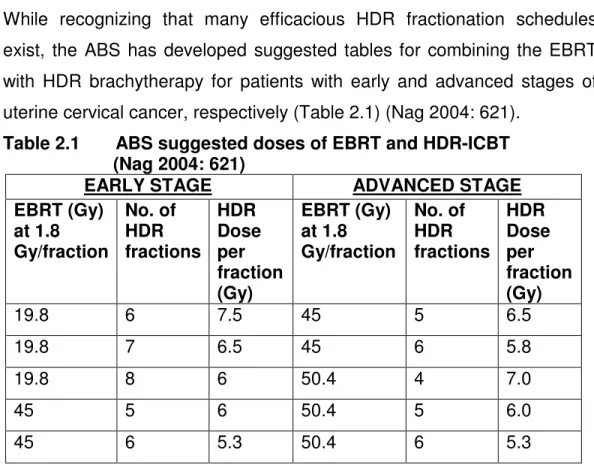
While recognizing that many efficacious HDR fractionation schedules exist, the ABS has developed suggested tables for combining the EBRT with HDR brachytherapy for patients with early and advanced stages of uterine cervical cancer, respectively (Table 2.1) (Nag 2004: 621).
Table 2.1 ABS suggested doses of EBRT and HDR-ICBT (Nag 2004: 621)
EARLY STAGE ADVANCED STAGE
EBRT (Gy) at 1.8 Gy/fraction
No. of HDR fractions
HDR Dose per fraction (Gy)
EBRT (Gy) at 1.8 Gy/fraction
No. of HDR fractions
HDR Dose per fraction (Gy)
19.8 6 7.5 45 5 6.5
19.8 7 6.5 45 6 5.8
19.8 8 6 50.4 4 7.0
45 5 6 50.4 5 6.0
45 6 5.3 50.4 6 5.3
Treatment schedules integrating external beam radiation therapy and brachytherapy were initially designed with regard to the disease stage and volume (Perez & Kavanagh 2004: 1840). In most centers, as the tumour volume or stage increases, the external beam dose plays a more prominent role in the total dose, whereas the number of HDR fractions and the dose per HDR fraction are decreased.
Although there is a marked variation in the dose and fractionation used for cervical HDR-ICBT, most centers use a schedule of approximately 1.8/2 Gy per fraction for 25 fractions EBRT and 6-8 Gy per fraction in four to six fractions (Orton et al. 1991: 1425). The fractionation schedule applicable to this retrospective study, used at the Department of Oncotherapy, Bloemfontein, is 2Gy/fraction for 25 fractions of EBRT and a dose of 2 Gy prescribed to the highest rectum dose point, administered in 4-6 fractions of HDR-ICBT. The dose to point A varies for each HDR-ICBT treatment given as a result of the changes in the rectum position for each treatment.
The standard prescribed protocol for the HDR-ICBT treatment requires a minimum total dose of 15 Gy to point A, achieved in 4-6 fractions.
2.5 Dose effectiveness of fractionation schedules
The use of HDR brachytherapy initially faced criticism and concerns with regard to its effectiveness and late toxicity. However, over the past few years, numerous published studies have demonstrated comparable local control, survival and morbidity with HDR when compared with LDR (Shakespeare et al. 2006: 278). A study on HDR vs. LDR by Falkenberg et al. (2006) confirmed equivalence in pelvic control, cause-specific survival and overall survival, as well as late morbidity between LDR and HDR brachytherapy (Falkenberg et al. 2006: 54). The following are some results reported in the literature on the clinical outcome of different institutions when using HDR-ICBT in combination with EBRT in their treatment schedule for uterine cervical cancer patients.
In 1991, Lanciano et al. (1991) reported their findings on pre-treatment and treatment factors associated with improved outcome in squamous cell carcinoma of the uterine cervix (Lanciano, Won & Coia 1991: 667-676).
Lanciano et al. (1991) compared the outcome of the 1973 and 1978 Patterns of Care surveys. It indicated that on 1558 patients treated using EBRT with or without low dose-rate brachytherapy at different institutions throughout the United States, no dose-response relationship was seen to a dose at point A of < 75, 75-85, or > 85Gy in stage I and II disease.
Although a dose-response relationship was seen with a dose > 85 Gy in stage III disease, a dose > 85 Gy was also associated with higher rate of complications.
A retrospective study was done by Chen et al. (2000) to see whether patient, treatment and dosimetric factors could be correlated to the risk of
developing late rectal complications in patients with uterine cervical cancer. The fractionation schedule consisted of EBRT of 40-44 Gy/20-22 fractions given within 4-5 weeks to the whole pelvis, after which the dose was boosted up to 54-58 Gy with central shielding. HDR-ICBT consisted of 3-4 insertions at doses of 5-7.2 Gy and the cumulative rectal biologic equivalent dose was calculated. The study indicates that patients who have stages IIb-IVa disease, a cumulative rectum dose greater than 65 Gy, or who are of 70 years of age and older, are at risk for late rectal sequelae and should be adequately assessed so that the treatment dose can be adjusted to prevent late rectal complications (Chen, Liang, Yang, Liu & Lin 2000: 960).
The Brazilian Experience of Ferrigno et al. (2001) is based on the analysis of dose effectiveness and late radiation complications. One hundred and thirty-eight patients with FIGO stages II and III were treated with 45 Gy of EBRT in 25 fractions and HDR brachytherapy was performed during EBRT with a dose of 24 Gy in 4 weekly fractions of 6 Gy to point A. The overall survival, disease-free survival and local control at 5 years was 53.7%, 52.7%, and 62%, respectively. The five-year actuarial incidence of rectal, bladder and small bowel late complications was 16%, 11% and 14%, respectively. Patients treated with a cumulative BED at rectum points above 110 Gy3 had a higher but not statistically significant five-year actuarial rate of complications at these organs (Ferrigno, dos Santos, Pellizon, Maia, Fogarolli, Gentil & Salvajoli 2001: 1123).
Sood et al. (2002) reported on the predictive value of the linear quadratic model in the treatment of cervical cancer using HDR brachytherapy. In this retrospective study 49 patients were treated with EBRT of 45 Gy (1.8 Gy/fraction) to the whole pelvis and HDR-ICBT consisting of a total of 18-19 Gy given in 2 fractions of 9-9.5 Gy.
Twenty-three patients received concomitant cisplatin-based
chemotherapy. The report indicated that in patients treated with radiotherapy alone, a BED10>89 Gy indicated a trend toward a better local control rate. This difference was not observed in patients receiving chemotherapy. A BED3<100 Gy3 was associated with negligible late toxicity. The 4-year local control rate was 80% and 83%
and disease-free survival rate of 75% and 70% with and without chemotherapy, respectively (Sood, Garg, Advadhani, Gorla, Malhotra, Guha, Deore & Vikram 2002: 1377)
Another analysis was performed by Toita et al. (2003) on dose effectiveness and fractionation schedule. Eighty-eight patients with cervical cancer were treated with EBRT of 2 Gy/fraction to a total dose of 40 Gy, and HDR-ICBT was performed once a week with a total point A dose of 18 Gy. The three-year actuarial pelvic control rate was 82% for all 88 patients: 96% for early stage and 76% for advanced disease. No significant dose-response relationship was observed by the treatment schedule and cumulative BED at point A for both early and late disease.
All patients treated with 86.4 Gy10 at point A suffered both proctitis and enterocolitis. Patients with cumulative BED at rectal point 100 Gy3 had a significantly higher incidence of proctitis (Toita, Kakinohana, Ogawa, Adachi, Moromizato, Nagai, Maehama, Sakumoto, Kanazawa &
Murayama 2003: 1344).
The report published by Wang et al. (2004) compared two linear quadratic model-based iso-effect fractionation schemes of HDR brachytherapy for cervical cancer. Five hundred and forty-one women were categorised into two groups according to the two iso-effect schemes used. Group 1 consisted of 254 patients treated with EBRT plus 7.2 Gy HDR-ICBT to point A for three fractions. Group 2 consisted of 284 patients treated with EBRT plus 4.8 Gy HDR-ICBT for five fractions. Overall, 66 patients developed pelvic recurrence. Of these, 53 patients had central recurrence:
24 (9.4%) were in group 1 and 29 (10.1%) in group 2. The actuarial survival rate for Groups 1 and 2 was 63.5% and 56.1% at five years and at ten years 47.8% and 49.3%, respectively. The incidence of high-grade complications remained unchanged, 8% vs. 7%. Multivariate analysis revealed that the fractionation scheme (three fractions vs. five fractions) was a significant factor influencing the proctitis rate, but not the local pelvic control rate, overall survival rate, or cystitis rate. The report concluded that the treatment results of the two groups showed similar outcomes while the complications decreased, and that the linear quadratic model correctly predicted the outcome. Biologically, the manipulation of the fraction size in their study suggested that the sensitivity of the late responding tissue to the fractional change from 7.2 Gy to 4.8 Gy in HDR- ICBT is high and detectable clinically (Wang, Huang, Sun, Chen, Fang, Hsu, Changchien & Leung 2004: 179). The cumulative BED for these published studies correlates well for local tumour control Gy10 (range: 80- 100 Gy10) and for negligible late toxicity Gy3 (range: 100-120 Gy3).
Although HDR brachytherapy has been used for more than 30 years in the treatment of uterine cervical cancer, the optimal time, dose and fractionation have yet to be established through systemic clinical trials (Sood et al. 2002: 1377). Toita et al. also stated that an optimum treatment schedule has not yet been clearly determined (Toita et al. 2003 : 1344). Regarding the results reported by all the above-mentioned authors on dose effectiveness of the different fractionation schedules for EBRT and HDR-ICBT, it is still unclear as to which schedule is the optimum fractionation schedule.
2.6 Biology of HDR brachytherapy
The primary disadvantage of HDR brachytherapy is of radiobiological concern. The high dose-rate leading to short treatment times does not
allow for the repair of non-lethal damage in normal tissue, or the redistribution of cells in the cell cycle, or re-oxygenation of the tumour cells; hence, multiple treatments are required (Nag 2004: 620). The radiobiological disadvantage can, however, be overcome through adequate fractionation, e.g. 4/5 fractions of 2 Gy/fraction prescribed to the highest rectum dose point, which is the treatment protocol used in this retrospective study.
Radiobiologically, the change from low dose rates to HDR requires a reduction in overall dose and the introduction of fractionation.
Fractionation introduces logistic problems for brachytherapy because multiple treatment exposures of HDR radiation, 24 hours apart, require either repeat implant procedures or a very stable, carefully verified implant remaining in situ for the duration of the fractionated treatment. Appropriate dose reductions have also been required, and the considerable range of schedules used for HDR brachytherapy reflects the uncertainty in this area (Hoskin & Bownes 2006: 209).
To achieve tumour control with HDR equivalent to that with LDR brachytherapy, attention to the dose/fraction schedule and to normal tissue doses is mandatory. In general, the / values for tumour and early- responding tissues are approximately 10 (Gy10) and for late-responding tissues 3-5 (Gy3-5). The values derived are not actual doses but biologically effective ones that take into consideration dose-rate and impact of fraction size (Perez & Kavanagh 2004: 1839).
Figure 2.1 illustra