1
Complications of Laparoscopic Cholecystectomy:
Addington Experience
Dr SZ Mbatha
Student number: 206526595
Submitted in partial fulfilment of the requirements for the degree of Master of Medicine in the Department of Surgery.
UNIVERSITY OF KWAZULU-NATAL 2014
Supervisor
Mr F. Anderson
2
3
Dedication
Dedicated to my wife and family for their unwavering support.
4
Acknowledgements
1. My Supervisor Mr F. Anderson for all the guidance during this process.
2. Ms Fikile Nkwanyana – Biostatistitian
3. Staff at Medical Records at Addington Hospital
4. Colleagues in the unit for their encouragement and support
5
TABLE OF CONTENTS
Page
List of figures……… 6
List of tables………... 7
Abbreviations………... 8
Abstract………. 9
Chapter 1 – Introduction………... 10
Chapter 2 – Literature Review………. ……… 12
2.1 Gallstone disease………. ……… 12
2.2 Laparoscopic Cholecystectomy……….. ……… 17
2.2.1 Operative technique……….. ……… 20
2.2.2 Technical considerations……….. ……… 24
2.2.3 Complications………. ……… 32
Chapter 3 - Patients and methods………... 37
Chapter 4 – Results……… 40
Chapter 5 - Discussion and conclusion………... 51
References……….. 56
Appendix……….. 61
Psychomotor decision making model……… 61
Table of outcomes and demographic………. 62
Approach to BD injuries flow diagram...………. 63
List of possible complication of lost gall stones………64
Other references……… 65
6
FIGURES
Page
Figure 1 – Cholesterol stones……….. 14
Figure 2 – Pigment stones………. 14
Figure 3 – Port placement ……… 22
Figure 4 – Normal biliary anatomy………... 25
Figure 5 – Variations of cystic duct anatomy………. 26
Figure 6 – Variations in cystic artery……… 27
Figure 7 – Critical view of safety drawing……….. 28
Figure 8 – Critical view of safety image……….. 29
Figure 9 – Pie chart of presentations……….. 42
7
TABLES
Page
Table 1 – Risk factors for cholesterol gallstone formation……… 13
Table 2 – Indications and contraindications of LC……….... 18
Table 3 - Advantages and disadvantages of LC………..…… 19
Table 4 – Bismuth classification of bile duct strictures………. 33
Table 5 – Strasberg classification of biliary injuries………. 34
Table 6 – Patient demographics……….. 40
Table 7 – HIV and presentation……… 41
Table 8 – Demographics, Presentation and Outcomes……… 43
Table 9 – Timing and Presentations……… 45
Table 10 – Timing and Complications………. 46
Table 11 – Comparison of complications with other studies……… 50
Table 12 – Patient numbers in similar studies……… 50
Table 13 – Potential predisposing factors in bile duct injuries………. 53
8
Abbreviations:
LC – Laparoscopic cholecystectomy
ERCP – Endoscopic Retrograde CholangioPancreatogram CBD - Common Bile Duct
CBDS – Common bile duct stones OJ – Obstructive Jaundice
US - Ultrasound
CVS – Critical view of safety
IOC - Intra-operative Cholangiogram AC – Acute Cholecystitis
GSP – Gallstone Pancreatitis BC – Biliary Colic
HIV – Human Immunodeficiency Virus
MRCP – Magnetic Resonance Cholangiopancreatogram GB – Gallbladder
GA – General anaesthetic
9
Abstract
Background
Laparoscopic cholecystectomy is a common surgical procedure performed for complicated gallstones. The timing of cholecystectomy is controversial with a trend toward early
cholecystectomy in patients with acute cholecystitis. This study examined the presentation, timing of cholecystectomy and outcomes in a resource constrained environment.
Methods
A retrospective analysis of laparoscopic cholecystectomies performed from January 2010 to June 2011. The mode of presentation, ERCP (endoscopic retrograde
cholangiopancreotogram) rate, and timing of cholecystectomy, complications and morbidity were analysed.
Results
One hundred and sixty seven patients were evaluated. The mean age was 44(17-78) years and 93% were female and 7% male. There were 44%, 24%, 21% and 14% who presented with biliary colic, pancreatitis, acute cholecystitis and jaundice respectively. They had laparoscopic cholecystectomies after a mean 34(4-90) days and 9(5.4%) patients required conversion to an open cholecystectomy. Complications occurred in 16.2% and bile duct injuries and bile leaks in 0.6% and 1.6% respectively. One patient died.
Conclusions
Most patients had delayed laparoscopic cholecystectomy. There was no difference in outcomes for the different presentations and the complications are similar to other reports in the literature.
10
CHAPTER 1
INTRODUCTION
Laparoscopic cholecystectomy (LC) has over past two decades become the operation of choice for the treatment of gallstone disease and has become one of the most commonly performed surgical operations. Our unit performs over a hundred of laparoscopic
cholecystectomies in a year. Current literature indicates that the complication rate for LC is low, but still higher than that found in open cholecystectomy. Our institution is one of the training hospitals in the Durban Functional Region and with surgeons of differing levels of experience and expertise from registrars to senior consultants.
In a South African environment laparoscopic cholecystectomy is performed in regional hospitals with a referral pattern from primary and district services. There are delays in referral and surgery because of a booking system which may extend into months. These delays also impact negatively on timeous pre-operative evaluation.
Complications of LC arise as a result of the operation and can either be local or systemic.
Local complications are the most common, well researched in the literature and are specific to LC in particular (i.e. biliary injuries) or to laparoscopic surgery in general (i.e. access related injuries); whereas systemic complications are general complications associated with undergoing a surgical procedure. Our study will focus on the local complications.
11 Aim of study
The aim of the study is assess our unit’s complication rate in comparison to international reports on the most common complications and whether our patient demographics and timing of operation influence the outcomes of our laparoscopic cholecystectomies’.
Specific objectives
• Laparoscopic cholecystectomy is one of the most commonly performed
operations in our unit. We want to assess whether the complication rate is at an acceptable level. If it is not, are there any factors that can be identified that require refinement and improvement.
• Patients in our community tend to present to hospital late in the course of their illness. Does that than worsen our perioperative morbidity and complications.
• Addington hospital is a training institution for undergraduate and postgraduate medical students and junior consultants; it is also a training facility for nurses. Do these factors increase our complication rate?
We performed an audit to analyse our patients and outcomes. We hypothesised that though we operate in a resource constrained environment our outcomes should be similar to the international norm. Our audit looked at outcomes of LC in the whole spectrum of gallstone disease.
12
CHAPTER 2
LITERATURE REVIEW
2.1 Gallstone disease 1, 2, 3, 4
Gallstones were found in 9.7 %( 2.5-13.9%) of the population in South Africa 1.
Gallstones arise as a result of crystallization of bile contents like cholesterol, calcium bilirubinate, calcium salts: phosphate, carbonate, palminate. 2, 3 .There are two groups of gallstones: cholesterol stones and pigment stones. Pigment stones are further subdivided into black and brown stones.
Cholesterol stones
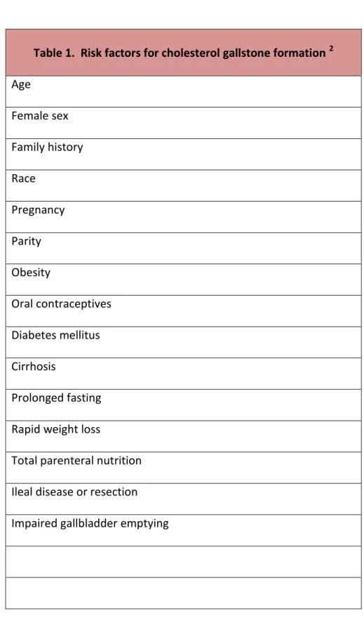
Cholesterol stones are the most common in Western society. They are formed by super saturation of cholesterol in bile, they contain ≥ 70% cholesterol in variable amounts , have different sizes and different shapes variable colour depending on amount of pigment.. 2, 3. . Table 1. Below illustrates the risk factors for cholesterol stone formation. 2
Pigment stones
Black pigment stones are composed of calcium bilirubinate, carbonate or phosphate. They are small, multifaceted, brittle and sometimes speculated, associated with haemolytic disorders. They develop exclusively in gallbladder; have less than 20% cholesterol.
Brown stones are commonly found in Asia. They are soft, mushy, brownish-yellow stones formed in gallbladder or bile ducts due to bile stasis and bacterial infection. They are formed
13
by precipitation of calcium bilirubinate with dead bacterial cell bodies. The biliary infection is caused by b-glucuronidase-producing bacteria.
Table 1. Risk factors for cholesterol gallstone formation 2
Age
Female sex Family history Race
Pregnancy Parity Obesity
Oral contraceptives Diabetes mellitus Cirrhosis
Prolonged fasting Rapid weight loss
Total parenteral nutrition
Ileal disease or resection
Impaired gallbladder emptying
14
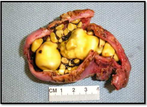
Figure 1: Cholesterol stones, numerous, yellow, irregular, and variable sized
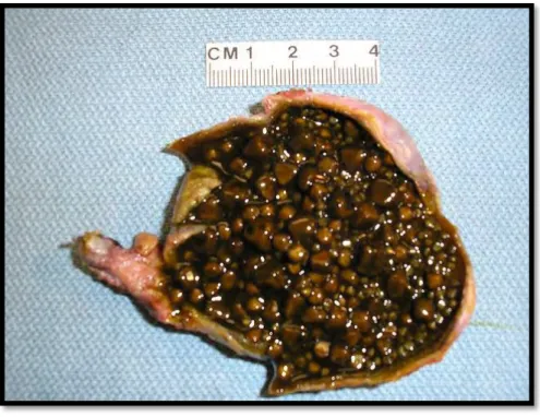
Figure 2: Pigment stones, numerous, brown, irregular, and variable sized.
Source: Qiao's Pathology (Art and Science in Medicine).
Jian-Hua Qiao, MD, FCAP, Los Angeles, CA, USA
15 Manifestation of gallstone disease
The vast majority of gallstones are asymptomatic and remain asymptomatic, with only 10%
and 20% developing symptoms in 5 and 20 years respectively. 2, 3
Gallstone disease is one of the most common diseases affecting the digestive tract. 2 Symptomatic gallstones may be in the gallbladder, bile ducts or both. The most common manifestations of gallstones are biliary colic, acute cholecystitis, pancreatitis and jaundice.
Biliary colic occurs as a result of stone obstructing the outlet at Hartmann’s pouch with contraction of gallbladder against a closed orifice. 2 It presents with recurrent right upper quadrant or epigastric pain. The pain is commonly post-prandial, increasing in the first 30 minutes and lasting a few hours. 2, 3, 4
Acute cholecystitis is inflammation of the gallbladder as a result of a stone impacted and remaining impacted in the cystic duct. It presents with severe unremitting right upper quadrant pain lasting more than 12-24hrs. The patient will also have signs of systemic inflammation like fever and leucocytosis, sometimes a positive Murphy’s sign. If the obstruction is prolonged and without treatment then bacterial infection supervenes with development of gallbladder empyema, gangrenous gallbladder or perforation.
Gallstone pancreatitis occurs as a result of small stone passing through common bile duct and occluding pancreatic duct. This obstruction causes increased ductal pressure with activation of pro-enzymes and auto-digestion leading to acute pancreatitis.
Obstructive jaundice (OJ) results from passage of stone into and obstructing the common bile duct (CBD). It may be transient when stone dis-impacts and floats or passes into the duodenum. OJ may also be due to Mirrizi’s syndrome where gallbladder stone in Hartmann’s pouch compresses or erodes into CBD. A serious complication of obstructive jaundice is
16
ascending cholangitis, as a result of infection of static bile. It’s characterized by Charcot’s triad of jaundice, fever, right upper quadrant pain or more severe form known as Reynold’s pentad when there is associated confusion and shock.
Investigations
Blood investigations include liver function test to rule out cholestasis, amylase and lipase in suspected gallstone pancreatitis.
Ultrasound (US) is the gold standard imaging modality for diagnosis of gallstones and gallstone complications.
US features of acute cholecystitis include gallbladder stones with thickened gallbladder wall and pericholecystic fluid.
OJ will also have dilated intrahepatic and extra-hepatic ducts in the presence of gallstone if the obstruction is prolonged. Sometimes bile duct stones can be detected with US but may be obscured by duodenal gas.
Endoscopic Retrograde Cholangiopancreatogram (ERCP) is useful for biliary decompression and bile duct clearance in patients with ductal stones, acute cholangitis, and non-resolving gallstone pancreatitis.
Another evaluation will include upper endoscopy to assess and exclude gastro-oesophageal reflux and peptic ulcer disease.
17 2.2 Laparoscopic cholecystectomy (LC)
Laparoscopic cholecystectomy is one of the most commonly performed operations in general surgery 5 and is now considered the standard of care for symptomatic gallstone disease
The first documented open cholecystectomy was performed in 1882 by Carl Langebuch of Germany; it remained the treatment of choice for gallstone disease for about 103 years. 6, 7. Then on the 12 September 1985 the first LC was performed by a German surgeon Dr Erich Mϋhe. In France Phillipe Mouret of Lyon started using the procedure in 1987 and in North America J Barry Mc Kernan and William B Saye performed their first LC in 1988. The French were for some time credited with performing the first LC. Dr Mϋhe was eventually acknowledged in his home country only in 1992 and was officially recognized as the
originator of LC in the Society of American Gastrointestinal Endoscopic Surgeons (SAGES) conference in 1999 in San Antonio, Texas after delivering a presentation about his
experiences with the first cases. 7 The procedure gained popularity amongst surgeons and was rapidly adopted based on anecdotal evidence presented by those who were performing the operation before there were any formal studies assessing its effectiveness and safety. It was pushed by physician competition, media, industry, high praise by patients and patient demand. 5, 8, 9 Studies were conducted and released from the early 1990’s looking at the complications, advantages and efficacy compared to open cholecystectomy which till the late 80’s was the mainstay of treatment for symptomatic or complicated gallstones. Other studies have subsequently been conducted looking at the timing of the procedure, looking at
complication rate and outcomes in early versus late cholecystectomy.
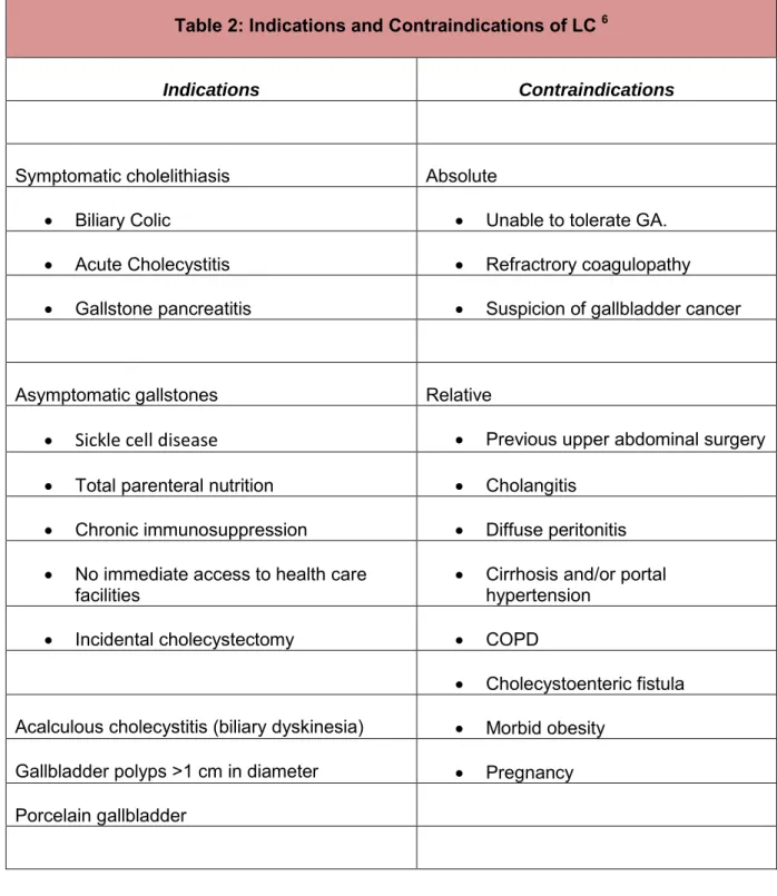
Cholecystectomy for asymptomatic gallstones is not recommended because of low rate of developing symptoms of about 1- 2% per year. Prophylactic cholecystectomy in only
18
indicated for certain specific conditions like patients with Sickle cell anaemia and calcified porcelain GB 6 . A summary of indications and contraindications are listed in Table 2 below 6
Table 2 – Indications and Contraindications of Laparoscopic Cholecystectomy 6
Table 2: Indications and Contraindications of LC 6
Indications Contraindications
Symptomatic cholelithiasis Absolute
Biliary Colic Unable to tolerate GA.
Acute Cholecystitis Refractrory coagulopathy
Gallstone pancreatitis Suspicion of gallbladder cancer
Asymptomatic gallstones Relative
Sickle cell disease
Previous upper abdominal surgery Total parenteral nutrition Cholangitis Chronic immunosuppression Diffuse peritonitis
No immediate access to health care
facilities Cirrhosis and/or portal
hypertension Incidental cholecystectomy COPD
Cholecystoenteric fistula Acalculous cholecystitis (biliary dyskinesia) Morbid obesity
Gallbladder polyps >1 cm in diameter Pregnancy Porcelain gallbladder
19 Advantages of laparoscopic cholecystectomy
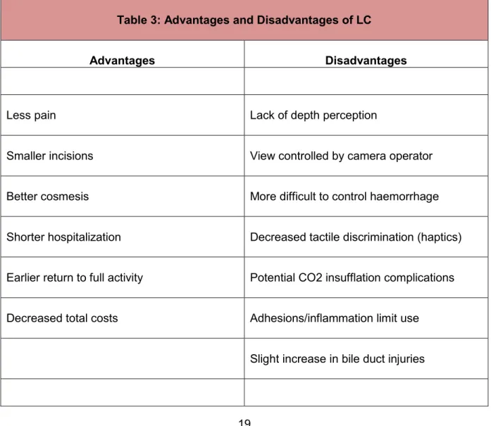
The laparoscopic approach has certain advantages over the open approach which has led to its universal adoption as gold standard for gallstone disease, and of course it also has its own disadvantages. A multitude of studies have identified these advantages to include better pain control , shorter hospital stay , early return to normal activity and work, less post- operative pain ,better cosmesis with smaller scars 8. The significant disadvantage is the purported increase incidence of bile duct injuries compared to the open procedure.
Table 3: Advantages and disadvantages of LC 6
Table 3: Advantages and Disadvantages of LC
Advantages Disadvantages
Less pain Lack of depth perception
Smaller incisions View controlled by camera operator
Better cosmesis More difficult to control haemorrhage
Shorter hospitalization Decreased tactile discrimination (haptics)
Earlier return to full activity Potential CO2 insufflation complications
Decreased total costs Adhesions/inflammation limit use
Slight increase in bile duct injuries
20 2.2.1 Operative technique
Pre-operative evaluation
Clinical evaluation – patients presents with symptomatic gallstones or with complications of gallstones. Physical examination assess for complications of gallstones, comorbidities, previous upper abdominal operations.
Laboratory evaluation as mentioned above include-liver function test, clotting profile, urea and electrolytes, full blood count.
Anaesthesia
Operation is conducted under general anaesthesia
Position
With patient under anaesthesia the table is then positioned in reverse Trendelenburg (30 degree) with left lateral tilt (15 degree)
Surgeon position
In the American technique (which is normally used in our unit) the surgeon stands on the left side of patient with the camera operator on same side and second assistant on the right side. The video monitor is positioned on the opposite side of the surgeon; if a second monitor is available it’s placed on the same side as surgeon. In the French technique the surgeons stands between the patients legs which are abducted.
21 Abdominal access and port placement
Abdominal access and establishing pneumoperitoneum can be achieved either by closed or open techniques. Closed technique using Veress needle which is then replaced by a port inserted blindly. Hasson open technique – small incision at umbilicus to open into abdomen and port inserted under vision. Or entry via a small incision using optical port with
transparent tip that allows direct visualization with laparoscope during entry. The three (3) following ports are inserted under vision with laparoscope.
Port placement
4 ports are used: 2 x 10mm – 11mm ports and 2 x 5mm ports - see Figure 3 below for port position. Proper placement of ports greatly facilitates the ease with which the operation can be conducted and progress.
22 Figure 3 - Port placement in LC 10
23 Dissection
Dissection should be started at known structures i.e. gallbladder. Dissection starts with opening up the investing peritoneum of the lowest part of the Hartmann’s pouch. The dissection should be conducted towards and at the Calot’s triangle aiming to obtaining the critical view of safety to avoid any injury. Once the surgeon is satisfied with proper
identification of the structures the cystic duct and artery are than doubly ligated proximally (i.e. CBD side) with clips and single ligated distally (gallbladder side). Care should be taken before clipping that the common bile duct is not tented up and that the proximal clip is not at the cysto-choledocus junction. Cystic artery should ideally be clipped prior to duct as this allows more length gain on cystic duct before applying clips. Once ligated and gallbladder freed it is dissected ante-grade to remove it from the rest of the liver bed. During this part of dissection aberrant Duct of Luschka are sometimes encountered. Dissection should be conducted in the areolar avascular plane and avoid going into liver bed as this leads to excessive bleeding and sub-hepatic collections leading to abscess formation post- operatively.
Retrieval and closure
The gallbladder is then retrieved via the 10-11mm port. If there was perforation or spillage or severe inflammation it can be removed using extractor endo-bag. During retrieval the sheath and skin incision might need to be extended to facilitate ease of removal and avoid rupture of gallbladder with stone spillage.
Irrigation above and below the liver followed with suctioning is recommended especially when there has been spillage of infected bile and/or stones, but care must be taken not to lose any spilled stones.
Port incisions are closed with monofilament non-absorbable sutures, if the sheath had to be extended it is closed with Vicryl 1.0 suture before suturing skin.
24 2.2.2 Technical considerations
Following a proper operative technique is essential in improving outcomes and decreasing the rate of complications. Salient technical consideration include proper port placement and patient position, dissection to obtain critical view of safety and proper identification of anatomy, careful and judicious use of diathermy and early conversion to open procedure when difficulties are encountered. Conversion to open procedure should not be seen as failure or a complication but a necessary step to avoid complications. “Conversion to an open procedure should not be considered a complication when marked inflammatory changes or difficulties in outlining the confluence of the cystic and common bile duct lead to this decision” 11
Biliary system anatomy and its variations:
Proper knowledge of normal anatomy and anatomical variations of the extra-hepatic biliary tree and the Calot’s and hepatocystic triangles is of significant surgical importance and crucial to safely performing the LC and minimizing complications. The normal anatomy is only present in a third of patients. It is imperative for any surgeon performing LC to be aware of these variations and be able to identify them when they occur so as to prevent injuries.
“Besides the experience of the surgeon, anatomical variations play an important role in the development of bile duct injuries”. 5 Figures 4-6 below demonstrate the normal anatomy and its variants.
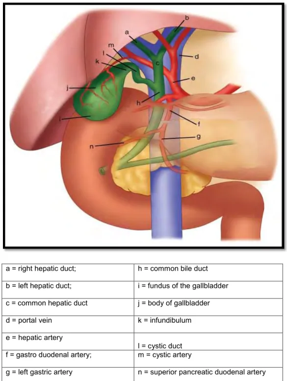
25
Figure 4: Normal biliary anatomy: - Anterior aspect of the biliary anatomy 3
a = right hepatic duct; h = common bile duct b = left hepatic duct; i = fundus of the gallbladder c = common hepatic duct j = body of gallbladder
d = portal vein k = infundibulum
e = hepatic artery
l = cystic duct f = gastro duodenal artery; m = cystic artery
g = left gastric artery n = superior pancreatic duodenal artery
Note: the situation of the hepatic bile duct confluence anterior to the right branch of the portal vein, the posterior course of the right hepatic artery behind the common hepatic duct.
The same peritoneal lining that covers the liver covers the fundus and the inferior
Source of Images : Brunicardi FC, Andersen DK, Billiar TR, Dunn DL, Hunter JG, Matthews JB, Pollock RE: Schwartz’s Principles of Surgery, 9th Edition : http// accessmedicine.com - Copyright @ The McGraw-Hill Companies, INC.
26 Figure 5: Variations of the cystic duct anatomy.
Variations of the cystic duct anatomy.
A. Low junction between the cystic duct and common hepatic duct B. Cystic duct adherent to common hepatic duct
C. High junction between the cystic and the common hepatic duct D. Cystic duct drains into hepatic duct
E. Long cystic duct that joins common hepatic duct behind the duodenum F. Absence of cystic duct
G. Cystic duct crosses posterior to common hepatic duct and joins it anteriorly.
H. Cystic duct courses anterior to common hepatic duct and joins it posteriorly.
Source of Images : Brunicardi FC, Andersen DK, Billiar TR, Dunn DL, Hunter JG, Matthews JB, Pollock RE: Schwartz’s Principles of Surgery, 9th Edition : http// accessmedicine.com - Copyright @ The McGraw-Hill Companies, INC.
27
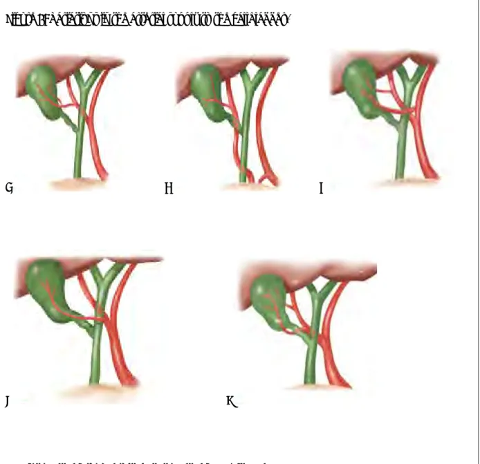
Figure 6: Variations in the arterial supply to the gallbladder.
A B C
D E
A. Cystic artery from right hepatic artery, about 80–90%.
B. Cystic artery from right hepatic artery (accessory or replaced) from superior mesenteric
artery, about 10%.
C. Two cystic arteries, one from the right hepatic, the other from the left hepatic artery, rare D. The cystic artery branching from the right hepatic artery and running anterior to the
common hepatic duct, rare
E. Two cystic arteries arising from the right hepatic artery, rare
Source of Images : Brunicardi FC, Andersen DK, Billiar TR, Dunn DL, Hunter JG, Matthews JB, Pollock RE: Schwartz’s Principles of Surgery, 9th Edition : http// accessmedicine.com - Copyright @ The McGraw-Hill Companies, INC.
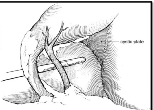
28 Critical view of safety
A common cause of bile duct injuries is misidentification of structures. Critical view of safety (CVS) is a method of identification of only two and only two structures (cystic duct and artery) entering the gallbladder together with at least the base of cystic plate/gallbladder liver bed , before clipping and cutting anything. 12 It is important to understand that the critical view of safety is not a method of dissection but a temporally static identification of critical structures in their entire circumferences before clipping and ligating. Although CVS will allow proper identification of structures it does not protect against injury when dissection is
persisted with despite adverse local conditions as in case of severe inflammation. Therefore in cases of severe inflammation alternative methods like conversion to open procedure, Intra operative cholangiogram should be considered. 12
Figures 7 and 8 below demonstrate the critical view of safety
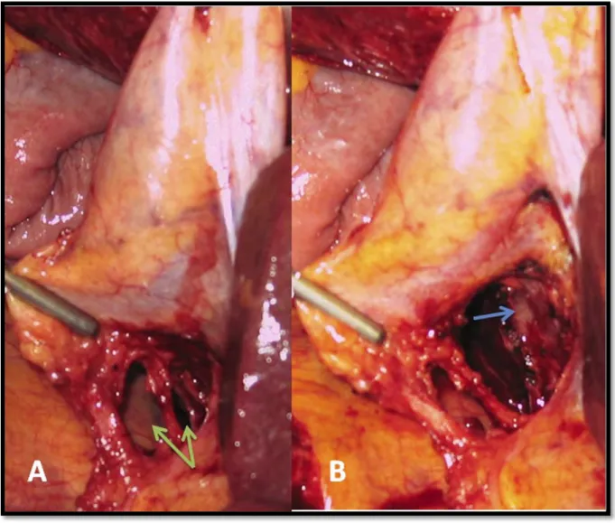
Figure 7: - Critical view of safety12
2 structures entering GB, and can get around their circumference
29 Figure 8 12
Figure 8: Difference between 2 “windows” and critical view of safety (CVS). 12
(A)Dissection has led to the creation of 2 windows, 1 between the cystic duct and artery and 1 between the artery and the liver (arrows). This dissection does not fulfil the criteria of CVS because the cystic plate cannot be clearly identified.
(B) CVS: - Arrow Points to whitish clearly identified cystic plate.
30 Severe inflammation
Another factor that raises technical difficulty during surgery is the disease process itself and distortion of anatomy directly related to the disease. Previous studies have suggested that acute cholecystititis , shrunken fibrotic gallbladder, liver cirrhosis are associated with increased difficulty of dissection with higher conversion rates and , increased common bile duct injuries. 9, 11 .
The timing of laparoscopic cholecystectomy in patients with acute cholecystitis remains controversial. Early operations are conducted within 7 days of onset of symptoms (optimal timing is thought to be within 72 hrs. from onset of symptoms) during the initial presentation whereas delayed/ interval LC is performed at 6 – 12 weeks after initial presentation following an initial course of conservative management with antibiotics and intravenous fluids. In the acute phase there’s inflammation and oedema which may facilitate adhesion’s dissection; in the delayed phase, after initial treatment, inflammation and oedema is replaced by fibrotic adhesions which occasionally makes dissection difficult.13 A meta-analysis conducted by Siddiqui et al found that in the early group operative time was longer with shorter overall hospital stay, but there was no difference in the overall morbidity and conversion rates between early and late groups.14 In western environments early cholecystectomy is commonly practised.
Surgeon’s laparoscopic experience
Surgeon’s laparoscopic experience has been shown to be inversely correlated to incidence of common bile duct injuries which tend to be higher in surgeons with little experience in the procedure, but these lesions decreased as surgeons gained more experience.5, 9, 11. “The risk for common bile duct injuries was reduced if the surgeon had an experience of >100 laparoscopic cholecystectomies”. 11
31
Laparoscopic surgery requires highly developed psychomotor skills that are crucial not only in performing the technical aspects of the operation but also in the decision making process during the dynamic process of the operation. Psychomotor skills involve cognitive and motor functions which are learned behaviour, but are also influenced by daily life’s events and challenges. Several studies have demonstrated that acquisition of laparoscopic
psychomotor skills can be expedited by training in virtual reality simulation and/or video trainers. This training improves proficiency and decreases risk of causing iatrogenic injuries in real operations more especially in the young inexperienced surgeons. Appendix A below shows an example of processes in psychomotor surgical decision making37.
Intraoperative cholangiogram (IOC)
The use of Intra-operative cholangiography has been advocated in patients suspected of having choledocholithiasis and to decrease the rate of common bile duct injuries by defining the biliary anatomy. However its use is controversial. Z-graggen et al 11 in their study found that the incidence of common bile duct injuries , overall morbidity, re-operation rates and mortality were all comparable between patients who underwent and those who did not undergo routine intra-operative cholangiography; and their results indicated that there were no advantages for patients undergoing intraoperative cholangiography; however they did find that intraoperative cholangiography allowed diagnosis of common bile duct injuries in 75% of patients . 11 Whilst MacFayden et al stated 5 “In this series, 41.5% of the patients had intra-operative cholangiography performed with an 82.7% success rate. However, it cannot be concluded from this data whether or not the performance of IOC offers early detection of bile duct injuries, although it is very useful in identifying anatomical variations and
choledocholithiasis.” 5
32 2.2.3 Complications
Common complications cited in the literature include:
Bile duct injury and bile leaks
Haemorrhage
Gallbladder perforation with or without spilled stones
Bowel injury
Port site surgical site infections
Port site hernia
Abscesses
Retained gallstones
Bile duct injury and leaks
Bile duct injuries and bile leaks are the most serious complications of laparoscopic cholecystectomy and have been found in 0,5% and 0.38% respectively. 5 Factors that predispose to these injuries include anatomical variations, thermal injuries from diathermy damaging bile duct nutrient arteries which may lead to late stenosis, acute cholecystitis, short cystic duct, misapplication of metal clips with partial or complete transections. 5 The prevention of bile duct injury is improved with the use of an intraoperative cholangiogram 15 and ensuring a critical view of safety prior to ligation of the cystic artery and duct. 16.
Magnetic Resonance Cholangiopancreatogram (MRCP) is considered best standard non- invasive imaging modality for suspected bile duct injuries, however CT scan is superior in detecting intra-abdominal postcholecystectomy collections. 17 MRCP delineates both the proximal and distal system allowing for accurate identification of the exact site and length of injury and helps in planning corrective surgery. 17
33 Classification of bile duct injuries
Table 4 and Table 5 below show the classifications of bile duct injuries.
Bismuth classification of bile duct injuries, developed before the advent of laparoscopic cholecystectomy, classifies injuries according to their location in relation to hepatic duct bifurcation. 5, 16
Strasberg classification was designed based on spectrum of injuries occurring during laparoscopic era and is the most detailed classification where all types of injuries are identified including minor leaks.
Table 4: Bismuth classification of biliary stricture 1. Low common hepatic duct stricture—hepatic duct stump > 2 cm 2. Mid common hepatic duct stricture—hepatic duct stump < 2 cm
3. Hilar stricture with no residual common hepatic duct—hilar confluence intact 4. Destruction of hilar confluence—right and left ducts separated
5. Involvement of aberrant right sectoral duct alone or including common duct
34
Table 5 :The Strasberg Classification of Biliary Injuries
A. Injury to small duct in continuity with biliary system, either from the liver bed or cystic duct B Injury to sectoral duct* with consequent obstruction
C Injury to sectoral duct* with consequent bile leak from a duct not in continuity with the biliary system
D Injury to the lateral aspect of an extra-hepatic duct E1 Stricture located ≥2 cm from the bile duct confluence E2 Stricture located ≤2 cm from the bile duct confluence E3 Stricture located at the bile duct confluence
E4 Stricture involving the right and left bile ducts E5 Complete occlusion of all bile ducts
*Usually an aberrant right hepatic duct
Haemorrhage
Bleeding can occur as a result of vascular injury or bleeding from liver bed following dissection. Vascular injuries can be access related injuries with Veress needle or ports or may occur during dissection. Access related injuries involve epigastric vessels, mesenteric vessels, omental vessels. 17 Major central blood vessels are also at risk. During laparoscopic dissection the most commonly injured vessel is right hepatic artery because of its course anterior to common bile duct and is sometimes mistaken for a cystic artery. Right hepatic artery injury may be some form of transection which might require conversion to open procedure to control bleeding. Occlusion of the artery may not be immediately noticed but
35
present later with deranged liver function test, right lobe atrophy or even liver failure.
Delayed bleeding may occur as a result of slipped cystic artery clip. 17.
Bleeding from liver bed results from deep a dissection stripping the liver capsule either as a result misidentification of the correct plane or due to severe inflammation or cirrhosis.
Gallbladder perforation and spilled stones
Gallbladder perforation is one of the most common complications. Perforation with only bile leakage extends the operative time but with proper irrigation and suction does not cause major problems. But spilled gallstones have to be removed as they might lead to problems in future. It is estimated that gallstones are dropped in approximately 10% of laparoscopic cholecystectomies, although reporting is poor. 18. Stones should always be retrieved but invariable some stones do get lost in the abdomen. 18 The most commonly reported complications of lost stone were abscesses ( abdominal wall, intra-abdominal, sub-hepatic and sub-phrenic).19 The true frequency of late abscesses as a result of dropped or lost gallstones is unknown but estimated to be around 0.3% – 0.6%. 18 Pneumoperitoneum has ability to carry spilled stone to any nook or crevice in the abdominal cavity, so these late abscesses/ inflammatory mass can be found anywhere. Pigment stones seem to be main causes of abscesses; typical bacteria include E. coli, klebsiella pneumoniae and
enterococcus. The most effective and simplest treatment is open drainage with removal of the stone or stone fragments. Minimally invasive techniques are usually not sufficient as they do not remove the cause of the abscess which is the stone. 18
Principles of intra-operative management of perforated gallbladder are containment of spillage by closing the hole with grasper, clips or endoloop; copious irrigation and use of suction devices; removal of as many stones as possible while avoiding spreading them to
36
deeper areas, retrieval of perforated bladder and stones with endo-bag, proper documentation for future ease of diagnosis should late complications arise. 19
Bowel injury
Bowel injuries constitute some of the most serious complication, and may go unrecognized for some time after the procedure. Bowel injury occurs during access with port and Veress needle. Or it may occur during dissection commonly with diathermy. Access related injuries tend to increase in patients with previous abdominal operations because of adhesions.
Thermal injuries with diathermy tend to involve either duodenum or transverse colon because of their proximity to gallbladder. These bowel injuries can be managed
laparoscopically if recognized immediately, or may require conversion to open procedure.
Thermal injuries may declare only later on, days after operation because of progression and perforation of a previously unrecognised area of ischaemia.
37
CHAPTER 3
PATIENTS AND METHODS
We performed a retrospective Chart review of patients who underwent a laparoscopic cholecystectomy from January 2010 to June 2011. A list of all patients who underwent laparoscopic cholecystectomies during this period was compiled from theatre register and database. The compiled list was submitted to the medical records department for them to retrieve the charts, which were then collected and analysed. All patients with complete charts were included in the study. Because of the poor filing system not all the charts could be located. Patients who were scheduled for and underwent open cholecystectomies were excluded. We analysed the patient’s demographics, mode of presentations, comorbidities, preoperative endoscopic retrograde cholangiopancreatogram (ERCP), and timing of
cholecystectomy, conversion rate, perioperative complications and mortality.
Patients presented with four major presentations namely biliary colic, acute cholecystitis, gallstone pancreatitis or obstructive jaundice. The presence of gallstones was confirmed with preoperative abdominal ultrasound. Laboratory evaluation included liver function tests, full blood count, international normalised ratio (INR), amylase, and urea and electrolytes. Those patients with biliary colic without cholestasis were scheduled for the next available slot for laparoscopic cholecystectomy.
Patients with symptomatic gallstones were assessed for cholestasis at presentation and those with cholangitis had an urgent ERCP and biliary decompression. In the others with
38
cholestasis serial monitoring of the liver function tests was performed and in those who resolved at 5-7 days, laparoscopic cholecystectomy was scheduled as soon as possible.
Failure to resolve is an indication for preoperative ERCP.
Patients with acute cholecystitis that present to hospital more than 72 hrs from onset of symptoms are treated with therapeutic antibiotics and those who resolve are scheduled for operation in 6 weeks.
In this series, four ports were used with emphasis on trying as much as possible to obtain the critical view of safety, lateral retraction of gallbladder neck, before clipping and ligation of cystic duct and artery. This is the technique taught and practiced in the unit, and all our surgeons are encouraged to employ this as a standard technique. The necessary equipment to conduct intraoperative cholangiography was not available and therefore not performed.
Statistics
All the results were compiled into a Microsoft Excel spread sheet. Statistical analysis was conducted using the IBM SPSS package Version 2.0. Descriptive analysis was conducted and chi-square tests (Pearson and Fischer exact) applied to assess for statistical
significance between various associations. Results were considered statistical significant if the p-value < 0.05.
39 Ethical considerations
Our study is a retrospective review. Patients were not informed of the study, however their identity was protected and not be publicised. The study was approved by Biomedical Research Ethic Committee of the University of KwaZulu-Natal and ethics committees of Addington Hospital and Provincial department of health.
Limitations of the study 1. Small study.
2. Short period assessed
3. Only one unit being assessed
4. Access to charts – some charts were not found due to our filling system and loss.
Some of the charts may be incomplete.
40
CHAPTER 4
RESULTS
Clinical records of only 167 from total of 210 (80%) consecutive patients that had
laparoscopic cholecystectomy during this period could be located and found to be complete.
This was considered adequate for our analysis. The age, ethnic and gender distribution is illustrated in Table 6. The mean age was 44.3 (17-78) years with standard deviation of 14.3.
Table 6: Demographics
Ethnicity n (%)
African 79(47.3%)
Indian 66(39.5%)
Mixed 11(6.6%)
Caucasian 11(6.6%)
Gender
Females 156(93.4%)
Males 11(6.6%)
Age distribution
17 -29 26 (15.6%)
31-39 48(28.7%)
40-59 65(38.9%)
≥ 60 28(16.8%)
41
Ninety five (57%) had comorbidities. The commonest morbidity was hypertension and/or diabetes with fifty four (32.3%) patients.
There were 20(12%) patients who had HIV infection. Seven (35%) of the 20 HIV infected patients had jaundice at presentation with p = 0.009 which is statistically significant.
Six had acute cholecystitis, two gallstone pancreatitis and five biliary cholic. Only 9 were on anti-retroviral therapy and their CD4 count ranged from 118 to 502. Three of the HIV positive patients sustained complications p value = 0.978
Table 7: HIV and Presentation
Presentation n Percent p-value
AC 6 30 % P =0.377
GSP 2 10% P=0.164
BC 5 25 P=0.064
OJ 7 35% P=0.009
Two patients had hypercholesterolemia.
Eleven had gastro-oesophageal reflux disease. The rest had miscellaneous comorbidities which were non-contributory to the disease process.
42 Presentations
The breakdown of presentations is represented in the pie chart below - Figure 9 and Table 8 below demonstrates the presentation, demographics and outcomes. The majority of our patients (n=74 / 44.3%) presented with biliary colic. About five of the patients were recorded as having dual presentations 3 had obstructive jaundice with pancreatitis and two had acute cholecystitis with pancreatitis. There was no difference in the mean age of patients with the different presentations of gallstone disease. Four(11%) of the 35 patients with acute
cholecystitis presented within 72hrs and had cholecystectomies within a week of admission as a result of a failed strategy of initial conservative management. The rest presented later than 72 hours and had symptom resolution with antibiotics. The other patients had
laparoscopic cholecystectomies after a mean 34(4-90) days without a difference in the 4 modes of presentation.
Figure 9: Pie chart of presentations
35
40 74
23
Presentations
AC GSP BC OJ
43
Table 8: Patient Demographics, Presentations, outcomes
n (%) Acute
Cholecystiti Gallstone
pancreatitis Obstructive
jaundice Biliary cholic Age
17-29yrs 26(15.6) 6 10 3 9
30 -39yrs 48(28.7) 12 7 9 21
40-59yrs 65(38.9) 13 15 10 28
≥60yrs 28(16.8) 4 8 1 16
Ethnic group
African 79(47.3)% 18 22 16 25
Mixed ethnicity 11(6.6%) 5 0 2 4
Indian 67(40.1%) 11 12 5 41
Caucasian 10(5.6%) 1 6 0 4
Gender
Female 156(93%) 33 38 22 68
Male 11(7%) 2 2 1 6
Co-morbidity
DM +/- HPT 54(32%) 7 17 6 25
HIV 20(12%) 6 2 7 5
Miscellaneous 21(12.6%)
Outcomes
ERCP 40(24%) 3 24 15 0
CBD stones 15(9%) 3 4 8 0
Conversions 9(5.4%) 3 4 1 1
Complications 27(16.2%) 5 7 7 9
Mortality 1(0.6%) 1 0 0 0
Total 167 35 40 23 74
44
A total of 40 patients had preoperative ERCP. Eleven (27.5%) of the forty had complications with a p-value = 0.026 .Twenty four (24) were patients with gallstone pancreatitis who showed persistent cholestasis and no resolution of symptoms after 5 -7 days of
conservative treatment, however only 4 were found to have CBDS. Sphincterotomy and ductal clearance was performed in all the patients at ERCP, as per standard protocol of the ERCP unit. Fifteen (65%) of 23 patients with obstructive jaundice had ERCP prior to surgery, the others showed resolving cholestasis on serial liver function test. Eight of the fifteen (15) had CBD stones. No other causes of obstructive jaundice were identified in those patients who did not have choledocolithiasis, they were considered to have passed stones after transient impaction in CBD.
CBD stones were found in total of 15 patients (see table 8). Two of those patients had retained stones, the first had presented with gallstone pancreatitis and pre-operative ERCP was normal however she had persistent bile leak post operatively and at ERCP found to have stone and stricture, the second had presented with acute cholecystitis with Mirrizi’s syndrome managed by open cholecystectomy, she had persistently elevated enzymes one month after the operation, the post-operative ERCP showed a retained stone which was extracted.
Timing of the operation
The timing of the operation from time of presentation varied widely, biliary colic patients were booked on next available slate date which typically fell several weeks away. These patients presented with symptom duration usually longer than a month, therefore all the 74 patients with biliary colic were operated on more than 6 weeks from onset of symptoms.
45 Table 9: Timing and Presentation
Out of the 35 Acute cholecystitis 23(66%) were done within the 2- 6 weeks from initial presentation after successful resolution of symptoms with a course of intravenous antibiotic.
The antibiotic regimen used was flagyl and zinacef with analgesia and fluid replacement. Six had their operation within a week, one was the 30 year old woman who did not respond to conservative management despite normal liver function test, she was found to have a gangrenous gallbladder. Another was a 50yr old diabetic female who also did not respond well to treatment and was taken to theatre on day 7, she was found to have Type 1 Mirrizi syndrome. She was converted to open cholecystectomy and had a long post-operative course because of retained CBD stone which was extracted endoscopically. Those done at
˃ 6weeks did not have any adverse outcomes as a result of the delay, one patient had significant hypothyroidism which needed to be medically treated and stabilized enough for her to withstand the operation at 3 months. The timing in gallstone pancreatitis was similar to acute cholecystitis.
Table 9:- Timing and presentations
n(%) Acute
cholecystitis
Gallstone pancreatitis
Obstructive jaundice Timing
≤1 week 10(11%) 6 4 0
2-6 weeks 56(60%) 23 25 12
>6 weeks 27(29%) 6 10 9
46
Thirty five (35) patients with gallstone pancreatitis had delayed operations, with the majority having their operations in the 2-6week period. Patients were initially managed conservatively with serial LFT monitoring. Those who resolved were booked electively and 24 required ERCP and sphincterotomy prior to surgery. Only one of the 24 had their operation within one week of admission. There was no reported recurrence of pancreatitis in the delayed group.
ERCP and sphincterotomy allowed for the delayed cholecystectomy.
Timing and complications:
A total of 27 patients had complications and their breakdown in terms of timing is
demonstrated in table 9 below. Timing did not seem to influence the complication rate; it was similar between all three groups.
Table 10: Timing and Complications
Timing Complications
Weeks Total(n) N Percentage
˂ 1 week 10 2 20%
2 – 6 weeks 56 10 18%
˃ 6 weeks 27 6 22%
47 Intra-op findings
At surgery 34% (n=57) had inflamed gallbladders and 16 of those had complications. Four females had fibrotic shrunken gallbladders. Eight patients had gallbladder empyema. There were two gangrenous gallbladders, a young female who had an uneventful peri-operative course and an elderly lady who died.
11% had dense adhesions around the gallbladder and 7.8% aberrant anatomy – 3 aberrant arteries, 7 cystic duct anomalies, and two intrahepatic gallbladders.
Complications
The complication rate was 16.2%. The spectrum and prevalence of complications and comparisons to other published data is illustrated in Table 11. Sixteen (59%) of the patients with complications had an inflamed gallbladder at surgery (p = 0.003) and 7(38.9 %) had adhesions (p= 0.012). Six patients (3.6%) had gallbladder perforations with stone spillage.
One of these had multiple stones with gallbladder empyema, she developed postoperative pneumonia and hypo-adrenalism which were successfully managed, and her stones had been successfully retrieved at time of operation. None of the other patients had
complications associated with spilled stones at follow –up.
One patient (0.6%) sustained common bile duct injury. She was 38 years old and presented with gallstone pancreatitis. She had a cholecystectomy after 6 weeks and was found to have an inflamed gallbladder adherent to the bile ducts. A bile duct injury was noted intra-
operatively and converted to open operation where successful Roux-en- Y hepatico-
jejunostomy was performed. She had a bile leak from the anastomosis and was taken back for a relaparatomy at day 3. The anastomosis was reinforced and she had an uneventful
48
recovery thereafter. Her last review date was 7 months after the operation and she was doing well and discharged.
Three patients (1.8%) had bile leaks postoperatively. They presented with obstructive jaundice with cholangitis, acute cholecystitis and pancreatitis. The first developed bile peritonitis post-operatively, she was taken for diagnostic laparoscopy where a slipped cystic duct ligaclip was identified as source of the leak, and was successfully managed
laparoscopically. The second had minor bile duct injury managed by ERCP with papillotomy.
She had a long (2 weeks) hospital stay due to persistent low volume leak. However at 2 month review her bile fistula had completely sealed and she was well. And the third patient had a Mirrizi type stricture, dense adhesions, and fibrotic gallbladder with empyema and had to be converted to an open cholecystectomy. She had a retained stone at ERCP and
required a hepatico-jejunostomy because of an associated distal bile duct stricture. She recovered after a prolonged hospital stay.
Bowel injury occurred in 2 patients. Both were converted to open procedures after injury to the colon and small bowel was noted respectively. The first one had an umbilical hernia, sustained a small colonic injury with entry port, the injury was identified immediately and successfully repaired, the hernia was also repaired. She developed umbilical site surgical site sepsis which was managed conservatively. The other patient had hostile abdomen from previous abdominal surgery for colonic cancer. Both had uneventful postoperative causes.
Three patients had significant bleeding. Two were from the liver bed and were managed laparoscopically and the third from a port sight which required exploration to ligate the offending vessel.
49
Nine of 167(5%) patients required conversion to an open cholecystectomy for various reasons: bile duct injury ( n=1) ,inflamed GB with empyema and colonic injury( n=1) , lateral tear of dilated cystic duct (n=1) , mirrizi syndrome ( n=3) , adhesions from previous
laparotomy (n=2), sealed GB perforation( n=1) with dense adhesions.
At review after discharge 1 patient had a pelvic fluid collection which was successfully managed expectantly. Another patient had persistent cholestasis without retained stones at ERCP. This had resolved at 6 months. Another patient with HIV infection had tuberculosis with a mass lesion in the right iliac fossa confirmed by CT scan and colonoscopy. In another port sepsis was successfully managed with antibiotics and another a retained stones was managed by ERCP. One patient developed an umbilical site hernia at 4 months.
One patient died. This was a 76 year old female with hypertension who presented with acute cholecystitis and non-resolving sepsis with antibiotics. She was taken to theatre and a gallbladder empyema with a sealed perforation, dense adhesions to colon and duodenum were found, initially only cholecystotomy performed because of septic shock and intra-
operative instability. She was resuscitated in ICU and taken back to theatre in 48hrs where a completion open cholecystectomy was successfully performed. She died three days later as a result of severe sepsis.
Complications and presentation
Table 8 above demonstrates complication rate for different presentation modes. There was no statistically significant difference between the different presentation and complication rate although obstructive jaundice had a higher complication rate as a group.
50
Table 11: Comparison of Complications with other studies
Table 12 – Patient numbers in similar studies.
Authors Patient numbers No of centres Time period studied
Jatzko et al 700 OC/ 704 LC Single 1991 -1993 for LC
Asoglu et al 1158 Single 1991 – 2001
Deziel et al 77,604 4292
Z-graggen et al 10,174 82 1992 -1995
Duca et al 9542 Single 1992-2001
Kanakala et al 2117 Single 1998 – 2007
Giger et al 22,953 114 units 1995 – 2003
Complication Present study Deziel. et al (%) 20 Duca et al (%) 21 Z’graggen et al (%) 11
n %
Total (%) 27 16.2% -
Bile duct injury 1 0.6% 0.6 0.1 0.31
Bile leaks 3 1.8% 0.29 0.57 0.9
Bowel injury 2 1.2% 0.14 - -
Spilled stones 6 3.6% - - 5.71
Haemorrhage 3 1.8 0.25 2.3 -
Wound sepsis 1 0.6 - 1.4 1.1
Port site hernia 1 0.6 - 0.13 -
Conversion rate 9 5.4 - 1.9 8.2
Mortality 1 0.6 0.04 0.1 0.2
51
CHAPTER 5 DISCUSSION
Laparoscopic cholecystectomy is the standard of care for symptomatic gallstones. In training institutions laparoscopic cholecystectomies are performed by trainees and surgeons with varying levels of experience. Because of retrospective nature of our study we could not accurately determine the level of expertise of each operating surgeon. However junior surgeons most often operate under the supervision of senior surgeons. The overall complication rate and rate of conversions did not seem to be increased as a result of our institution being a training facility.
Murphy et al in their multivariate analysis of predictors of complications in LC found that advanced age, multiple co-morbidities, and male sex were independent predictors of increased risk of complications; presence of inflammation and urgent LC were associated with increased complication rate, but neither the hospital nor surgeon volume (number of cases done per year) were associated with increased risk of complications. 22 It is important to note that Murphy et al looked at volume and not experience of the surgeon. Other studies have also found that male gender and high American Society of Anaesthesiologist (ASA) score, severity of local inflammation were independent risk factors associated with increased systemic complications and mortality. 23, 24
Gallstones complicate by causing biliary colic, acute cholecystitis, gallstone pancreatitis and jaundice by biliary obstruction related to the stone or stricturing as is the case in Mirizzi’s syndrome. Acute cholecystitis varies from a mild inflammation to severe disease associated with systemic sepsis and/or empyema, gangrene and perforation of the gallbladder. In patients presenting with jaundice, cholangitis and systemic sepsis urgent biliary
52
decompression may be required prior to removal of the gallbladder. In severe acute pancreatitis biliary obstruction and the systemic effects require urgent initial attention.
The timing of cholecystectomy in these settings may be influenced by the mode of
presentation. Most studies on the timing of cholecystitis have focused on patients with acute cholecystitis. The timing of cholecystectomy in acute cholecystitis has evolved with good results demonstrated in patients operated on within 72 hours 25 and 24hours 26. When LC is performed by an experienced surgeon in acute cholecystitis, it is a safe and effective procedure whether done early or delayed. Although operating in the earlier group requires modification of technique and extends operative time. 13 In this study the timing was not influenced by the mode of presentation and there was no difference in the complication rates.
Early cholecystectomy may result in more conversions to open surgery 27, 28 whereas delays in cholecystectomy are associated with potential recurrent symptoms and hospital
admissions 29. In this study one patient who had known biliary colic presented with acute cholecystitis whilst awaiting cholecystectomy. In a meta-analysis that analysed early (within 7 days) versus late (≥6 weeks) cholecystectomy for acute cholecystitis, early
cholecystectomy was associated with a reduced total hospital stay but longer operation time and no difference in overall postoperative morbidity.14 The circumstances within our health care system dictate that the majority of our patients will fall into the category of delayed laparoscopic cholecystectomies.
The means available to prevent bile duct injury are routine intraoperative cholangiography and ensuring a critical view of safety. The routine use of intraoperative cholangiography is controversial 30 and the critical view of safety may be more effective with less cost. 27
53
Cholangiography may then be limited to assessing for suspected bile duct stones. 28. In this unit ERCP is performed prior to surgery in suspected bile duct stones.
Table 12 below shows the potential risk factors for bile duct injury. We had one injury identified intra-operatively and 3 bile leaks as discussed above. Common hepatic duct/common bile ducts are the most common injuries. 5 The two most common
predisposingfactors are acute cholecystitis with severe local inflammation; where common bile duct is confused with cystic duct; and surgeon’s lack of experience. 21
Table 13 : Potential predisposing factors in bile duct injuries 5
Anatomical variations
Absent or short cystic duct
Cystic duct arising from Right hepatic duct
Aberrant or accessory right hepatic duct
Acute or chronic inflammation Attempt to control haemorrhage Improper technique
Tenting of the common bile duct
Improper placement of mental clips on the cystic duct
Hole made in cystic or common ducts during dissection
54
About 25 -36 % of iatrogenic injuries are detected during LC, the others are recognized during the early post-operative course or may present later as strictures. 31 These injuries should always be referred to centres with experience in hepatobiliary surgery or if
recognised intra-operatively help of an experienced hepatobliary surgeon should be sought so as to prevent further injury. For some leaks and strictures, successful management can be achieved endoscopically with ERCP sphincterotomy and stenting, more complex injuries will require surgical repair. Surgical repair involves repair over T-tube for small injuries without tissue loss recognized at the time of LC to hepaticojejunostomy for more complex injuries and strictures. 31.
Adhesions and severe inflammation, whether chronic or acute, resulted in anatomical distortions and difficulty obtaining the critical view of safety. A multitude of studies have demonstrated that the most common reason for conversion was failure to identify anatomy at Calot’s triangle and failure to make significant progress during dissection. Five of the nine conversions had severe inflammation with three having Mirizzi’s syndrome. The conversion rate of 5% compares to a previous study with 3.6%. 32 It is accepted that conversion is not failure or a complication but is used to avoid further complications. It is however associated with increased analgesic requirements, longer hospital stays and increased overall
morbidity. 33
Gallbladder perforation with stone spillage occurs in 5-40% of laparoscopic procedures and results in complications in <1% of cases. Routine conversion to an open procedure is not recommended.34, 35. Zehetner et al in their review of studies with more than 500 LCs found that the incidence of gallbladder perforations was 18.3% with spilled stones at 7.3%.19 In this series stone pillage occurred in 6(3.6%) patients without complications at 1 month review.
55
A significant number of HIV infected patients presented with obstructive jaundice in this study, 35% with p value = 0.009 which was statistically significant but due to small numbers it is difficult to draw significant conclusions. However there was no correlation between HIV infection and complications. .AIDS cholangiopathy which is associated with advanced HIV infections with very low CD4 counts (<50-100) 36 was suspected in one female at ERCP and EUS. She was on antiretroviral therapy with CD4 count of 334. She had an uneventful cholecystectomy 5 days after ERCP and discharged 2 days later. Fifty per cent of the HIV infected patients had inflamed gallbladders at surgery (p=0.111) and 15% developed complications (p=0.978).
Despite the delays to surgery the recovery is rapid with 148 (87%) patients discharged within 72 hours of the postoperative period
Conclusion
Our complications rate is comparable to other published studies.
Few patients present early with complicated gallstones in this study, late presentations however did not seem to influence our outcomes.
Complication rates were similar between the different operative timing periods. The only mortality was in a patient with severe sepsis who failed the initial strategy of conservative management with antibiotics. There is no difference in the complication and conversion rates between the different complications of gallstones and