Design, synthesis and evaluation of novel levodopa pro-drugs for the treatment of Parkinson's disease
M Strydom
orcid.org/0000-0002-7622-6706
Thesis submitted in fulfilment of the requirements for the degree Doctor of Philosophy in Pharmaceutical Chemistry
at the
North-West University
Promoter: Prof A Petzer Co-promoter: Prof JP Petzer
Graduation May 2018
Student number: 21087113
National Research Foundation acknowledgement
This work is based on the research supported in part by the National Research Foundation of South Africa (Grant specific unique reference numbers (UID) 85642, 96180). The Grant holders acknowledge that opinions, findings and conclusions or recommendations expressed in any publication generated by the NRF supported research are that of the authors, and that the NRF accepts no liability whatsoever in this regard.
TABLE OF CONTENTS
List of abbreviations and acronyms i
List of figures vi
List of tables ix
Abstract 1
Uittreksel 3
Preface 5
Declaration 6
Letter of agreement 7
Chapter 1: Introduction 8
1.1 Background 8
1.2 Lazabemide 10
1.3
L-Dopa prodrugs 11
1.4 Propargylamine MAO inhibitors 12
1.5 Rationale of this study 13
1.6 Study objectives 14
Chapter 2: Literature review 15
2.1 Introduction 15
2.2 Parkinson’s disease 15 2.3 Background, pathology and symptoms of PD 16
2.4 Mechanisms of pathogenesis in PD 17
2.5 Treatment of PD 20
2.5.1
L-Dopa therapy 20
2.5.2 DA agonists 20
2.5.2.1 Apomorphine-type DA receptor agonists 20
2.5.2.2 Ergot-type DA receptor agonists 21
2.5.2.3 Other small-molecule DA receptor agonists 22
2.5.3 Aromatic amino acid decarboxylase (AADC) inhibitors 22 2.5.4 Catechol-O-methyltransferase (COMT) inhibitors 23
2.5.5 Monoamine oxidase (MAO) inhibitors 24
2.5.6 Other agents for PD therapy 25
2.5.6.1 Anticholinergic agents 25
2.5.6.2 Adenosine receptor antagonists 26
2.5.6.3 Serotonin 5-HT1A agonists 27
2.5.6.4 Glutamate antagonists 27
2.6
L-Dopa 28
2.7 Monoamine oxidase inhibitors 31
2.7.1 Background 31
2.7.2 MAO-B inhibitors 32
2.7.2.1 Selegiline 32
2.7.2.2 Rasagiline 33
2.7.2.3 Lazabemide 34
2.7.2.4 Safinamide 35
2.7.3 MAO-A inhibitors 36
2.7.3.1 Moclobemide 36
2.7.3.2 Brofaromine 36
2.7.3.3 Befloxatone 37
2.7.4 The structure of MAO-A and MAO-B 37
2.7.4.1 The structure of MAO-B 38
2.7.4.2 The structure of MAO-A 40
2.8 Prodrugs 42
2.8.1 Background of prodrugs 42
2.8.2
L-Dopa and DA prodrugs 44
2.9 Conclusion 48
References 49
Chapter 3: The design and evaluation of an
L-dopa-lazabemide prodrug
for the treatment of Parkinson’s disease 62
Abstract 63
3.1 Introduction 64
3.2 Materials and methods 67
3.2.1 The synthesis of lazabemide 67
3.2.2 The synthesis of the
L-dopa-lazabemide prodrug 67
3.2.2.1 Chemicals and instrumentation 67
3.2.2.2 The synthesis of
L-dopa(TBDMS)
267
3.2.2.3 The synthesis of Boc-
L-dopa(TBDMS)
268
3.2.2.4 The conjugation of protected
L-dopa with lazabemide 68 3.2.2.5 Removal of the TBDMS and Boc protective groups from Boc-
L-
dopa(TBDMS)
2-lazabemide 68
3.3 Determination of physicochemical and biological properties 69
3.3.1 Materials and instrumentation 69
3.3.2 Ethics consideration 69
3.3.3 Shake-flask method for logD determination 69
3.3.4 Determination of solubility 70
3.3.5 Determination of ionization constant, pKa 70
3.3.6 Determination of toxicity towards cultured cells 71 3.3.7 High performance liquid chromatography (HPLC) 71 3.3.8 Determination of passive diffusion permeability 72
3.3.9 Determination of chemical stability 72
3.3.10 Determination of plasma and tissue stability 72
3.4 Animal studies 73
3.5 Results 73
3.5.1 Synthesis of the
L-dopa-lazabemide prodrug 73
3.5.2 Physicochemical properties 77
3.5.2.1 LogD 77
3.5.2.2 Solubility 78
3.5.2.3 Ionisation constant 78
3.5.2.4 Cell viability 78
3.5.2.5 Passive diffusion permeability 79
3.5.2.6 Chemical stability 80
3.5.2.7 Plasma and tissue stability 81
3.5.3 In vivo effect of the prodrug on brain monoamines 83
3.6 Discussion and conclusion 85
Acknowledgements 85
Conflict of interest 85
References 86
Chapter 4: An investigation of the in vitro reversibility of MAO inhibition by
lazabemide 93
Abstract 94
4.1 Introduction 95
4.2 Experimental section 98
4.2.1 The synthesis of lazabemide 98
4.2.2 Measurement of IC
50values 99
4.2.3 Dialysis 99
4.3 Results 100
4.3.1 The synthesis of lazabemide 100
4.3.2 IC
50values for the inhibition of human MAO 100 4.3.3 Reversibility of MAO-B inhibition by dialysis 101
4.4 Discussion and conclusion 102
Acknowledgements 103
Conflict of interest 103
References 103
Chapter 5: The synthesis and property evaluation of novel
L-dopa prodrugs for the treatment of Parkinson’s disease 108
Abstract 109
5.1 Introduction 110
5.2 Materials and methods 114
5.2.1 The synthesis of the
L-dopa prodrugs 114
5.2.1.1 Chemicals and instrumentation 114
5.2.2 The synthesis of
L-dopa prodrugs 8-11 114
5.2.2.1
L-Dopa-4-pyridylmethylamine hydrochloride (8) 114 5.2.2.2
L-Dopa-2-(4-pyridyl)ethylamine hydrochloride (9) 115 5.2.2.3
L-Dopa-2-(2-pyridyl)ethylamine hydrochloride (10) 115 5.2.2.4
L-Dopa-3-phenyl-1-propylamine hydrochloride (11) 115
5.3 Determination of physicochemical and biochemical properties
115
5.3.1 Materials and instrumentation 115 5.3.2 Shake-flask method for logD determination 116 5.3.3 Determination of ionisation constant, pKa 116 5.3.4 Determination of toxicity towards cultured cells 116 5.3.5 High performance liquid chromatography (HPLC) 117 5.3.6 Determination of passive diffusion permeability 117
5.3.7 Determination of chemical stability 117
5.3.8 Determination of plasma and tissue stability 117
5.3.9 Ethics consideration 118
5.4 Results 118
5.4.1 Synthesis of the
L-dopa prodrugs 118
5.4.2 Physicochemical properties 120
5.4.2.1 LogD 120
5.4.2.2 Ionisation constant 121
5.4.2.3 Cell viability 122
5.4.2.4 Passive diffusion permeability 122
5.4.2.5 Chemical stability 123
5.4.2.6 Plasma and tissue stability 125
5.5 Discussion and conclusion 127
Acknowledgements 128
Conflict of interest 128
References 129
Chapter 6: The synthesis and evaluation of novel propargylamine MAO inhibitors incorporating the pyridyl moiety 149
Abstract 150
6.1 Introduction 151
6.2 Experimental section 156
6.2.1 Materials and methods 156
6.2.2 The synthesis of propargylamine compounds, 1a-c and 2a-
d 157
6.2.2.1 N-(4-Pyridylmethyl)propargylamine (1a) 157
6.2.2.2 N-(2-Pyridylmethyl)propargylamine (1b) 157
6.2.2.3 N-(3-Pyridylmethyl)propargylamine (1c) 158
6.2.2.4 Bis(propargyl)(pyridine-4-ylmethyl)amine (2a) 158 6.2.2.5 Bis(propargyl)(pyridine-2-ylmethyl)amine (2b) 158 6.2.2.6 Bis(propargyl)(pyridine-3-ylmethyl)amine (2c) 158 6.2.2.7 Bis(propargyl)[2-(pyridine-2-yl)ethyl]amine (2d) 158
6.2.3 Measurement of IC
50values 158
6.2.4 Molecular modelling 159
6.3 Results 160
6.3.1 The synthesis of the propargylamine compounds 160 6.3.2 IC
50values for the inhibition of human MAO 160
6.3.3 Molecular modelling 162
6.4 Conclusion 165
Acknowledgements 165
Conflict of interest 166
References 166
Chapter 7: Conclusion 175
7.1 Introduction 175
7.2 Specific findings and conclusions 176
7.3 Future recommendations 178
Addendum 1: Permission to reproduce figures 179
LIST OF ABBREVIATIONS AND ACRONYMS
A
AADC Aromatic L-amino acid decarboxylase ALDH Aldehyde dehydrogenase
ANOVA Analysis of variance
APCI Atmospheric-pressure chemical ionisation A
2AAdenosine A
2Areceptor
Å ångström
B
Boc
2O Di-t-butyl dicarbonate
BOP Benzotriazol-1-yloxytris(dimethylamino)-phosphonium hexafluorophosphate
C
Caco-2 Heterogeneous human epithelial colorectal adenocarcinoma cells Ca
2+Calcium
CNS Central nervous system
COMT Catechol-O-methyltransferase CO
2Carbon dioxide
CoQ10 Coenzyme Q
10/ubiquinone
CTZ Chemoreceptor trigger zone CYP2B6 Cytochrome P450 2B6 CYP2C19 Cytochrome P450 2C19Cys Cysteine
D
D1 Dopamine receptor 1
D2 Dopamine receptor 2
D3 Dopamine receptor 3
D4 Dopamine receptor 4
Da Dalton
DA Dopamine
DAT Dopamine transporter
DATATOP (R)-deprenyl and tocopherol antioxidative therapy of Parkinsonism DBU 1,8-Diazabicyclo[5.4.0]undec-7-ene
DH Dopamine -hydroxylase
DMEM Dulbecco's modified Eagle medium DNA Deoxyribonucleic acid
DOPAC Dihydroxyphenyl acetic acid
E
EDTA Ethylenediaminetetraacetic acid EI Electron ionisation
ER Extended-release
F
FAD Flavin adenine dinucleotide FDA Food and drug administration
G
GABA gamma-Aminobutyric acid GPCR G-protein-coupled receptors
g Gram
H
H Hour
H
2O
2Hydrogen peroxide HCl Hydrochloric acid
HeLa Henrietta Lacks (uterine cell variety) 5-HIAA 5-Hydroxyindole acetic acid
HPLC High-performance liquid chromatography
5-HT Serotonin
HRMS High resolution mass spectrometry HVA Homovanillic acid
I
IC
50Inhibitor concentration at 50% inhibition Ile Isoleucine
i.p. Intraperitoneal
K
K
iInhibitor constant
L
LAT Large amino acid transporter LBs Lewy bodies
Leu Leucine
L-Dopa Levodopa
LogD Partition coefficient of ionisable solutes LogP Partition coefficient of permeability LNAA Large neutral amino acid
Lys Lysine
M
MAO Monoamine oxidase MHz Megahertz
MilliQ Millipore corporation ultrapure water Min Minutes
MPTP 1-Methyl-4-phenyl-1,2,3,6-tetrahydropyridine MS Mass spectrometry
MTT 3-(4,5-Dimethylthiazol-2-yl)-2,5-diphenyltetrazolium bromide
N
NA Noradrenaline NE Norepinephrine NMM N-Methylmorpholine NMDA N-Methyl-D-aspartate
NMR Nuclear magnetic resonance
O
O
2Oxygen
3-O-MD 3-Methoxy-4-hydroxy-
L-phenylalanine
P
PAMPA Parallel artificial membrane permeability assay PBS Phosphate buffered saline
P
ePermeability coefficient pKa Acid dissociation constant pH Power of hydrogen
Phe Phenyl
PNMT Phenylethanolamine-N-methyltransferase PD Parkinson’s disease
Ppm (δ) Parts per million PS Parkinson se siekte Pro Proline
PTAPT Peptide transporter associated prodrug therapy
Q
QSAR Quantitative structure-activity relationship
S
SD Standard deviation
SNpc Substantia nigra pars compacta
T
TBDMS Tert-butyldimethylsilyl TFA Trifluoroacetic acid TH Tyrosine hydroxylase THF Tetrahydrofuran
TLC Thin layer chromatography Trp Tryptophane
Tyr Tyrosine
U
UCH-L1 Ubiquitin C-terminal hydrolase L1
UPDRS Unified Parkinson's disease rating scale UPS Ubiquitin-proteosome system
UV/Vis Ultraviolet-visible
α
α-syn α-synuclein
µ
µM Micro molar
% Percentage
LIST OF FIGURES
Figure 1.1 Dopamine biosynthesis from
L-Dopa and the MAO-B catalysed
catabolism thereof. p. 8
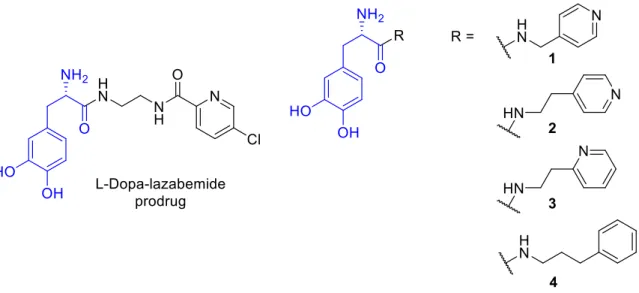
Figure 1.2 The structures of the
L-dopa-lazabemide prodrug as well as the other 4 carrier-linked
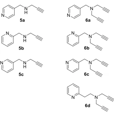
L-dopa prodrugs of this study. p. 9 Figure 1.3 The structures of the propargylamine compounds that were synthesised
and investigated in the current study. p.10
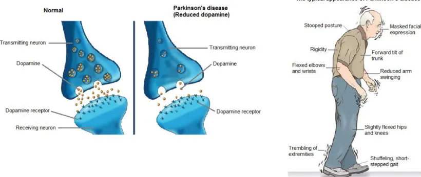
Figure 2.1 A normal vs. a PD dopaminergic neuron and the typical appearance of
PD. p. 16
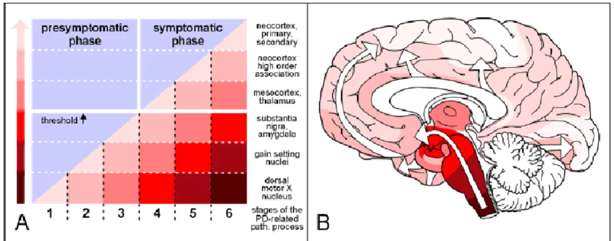
Figure 2.2 Parkinson’s disease presymptomatic and symptomatic phases. p. 17 Figure 2.3 Neuropathology of Parkinson’s disease. p. 18 Figure 2.4 Mechanism of neurodegeneration in PD. p. 19 Figure 2.5 An illustration of the breakdown of
L-dopa in the body. p. 20
Figure 2.6 The structure of apomorphine. p. 21
Figure 2.7 The structure of cabergoline. p. 21
Figure 2.8 The structure of ropinirole. p. 22
Figure 2.9 An illustration of the breakdown of
L-dopa in the presence of an AADC
inhibitor in the body. p. 23
Figure 2.10 An illustration of the breakdown of
L-dopa in the presence of a COMT
inhibitor in the body. p. 24
Figure 2.11 A schematic diagram of dopaminergic neurotransmission and the role of
MAO-inhibitors. p. 25
Figure 2.12 The structure of istradefylline. p. 26
Figure 2.13 The structure of memantine. p. 27
Figure 2.14 The structure of
L-dopa. p. 28
Figure 2.15 The metabolism of
L-dopa and its major decarboxylated product DA.
p. 30
Figure 2.16 The structure of selegiline. p. 33
Figure 2.17 The structure of rasagiline. p. 33
Figure 2.18 The structure of lazabemide. p. 35
Figure 2.19 The structure of safinamide. p. 35
Figure 2.20 The structure of moclobemide. p. 36
Figure 2.21 The structure of brofaromine. p. 37
Figure 2.22 The structure of befloxatone. p. 37
Figure 2.23 The molecular structure of MAO-B and the amine binding site. p. 40 Figure 2.24 The molecular structure of MAO-A and the amine binding site. p. 41 Figure 2.25 A schematic diagram of the rational for prodrug design. p. 44
Figure 2.26 The structure of L-α-methyldopa. p. 46
Figure 2.27 The structures of A) α-methyldopa-Phe and B) α-methyldopa-Pro. p. 46 Figure 2.28 The structure of an imidazoline-4-one ring as potential
L-dopa prodrug.
p. 47
Figure 2.29 Chemical structures of di- and tripeptide prodrugs of
L-dopa. p. 47 Figure 2.30 The structures of selected dopamine prodrugs. p. 48 Figure 3.1 The structures of
L-dopa, lazabemide and the
L-dopa-lazabemide
prodrug. p. 64
Figure 3.2 The structures of selegiline, rasagiline and safinamide. p. 66 Figure 3.3 The synthetic route to lazabemide. p. 73 Figure 3.4 The protection of
L-dopa and the synthesis of the
L-dopa-lazabemide
prodrug. p. 75
Figure 3.5 Atom numbering scheme for the
L-dopa-lazabemide prodrug. p. 76 Figure 3.6 The chemical stability of the
L-dopa-lazabemide prodrug at different pH
values. p. 81
Figure 3.7 The metabolic stability of the
L-dopa-lazabemide prodrug in human and
rat plasma. p. 82
Figure 3.8 The metabolic stability of the
L-dopa-lazabemide prodrug in rat brain
and liver homogenates. p. 83
Figure 3.9 The concentrations of selected monoamines and metabolites in the striatum of mice following oral and intraperitoneal (i.p.) treatment with saline (S),
L-dopa (LD),
L-dopa and carbidopa (LD/C), or the
L-dopa-
lazabemide prodrug (P). p. 84
Figure 4.1 The structures of selegiline, rasagiline and safinamide. p. 94 Figure 4.2 The structures of lazabemide, Ro 41-1049 and Ro 16-6491. p. 97 Figure 4.3 The adduct that forms with the inhibition of human MAO-B by N-(2-
aminoethyl)-p-chlorobenzamide (Ro 16-6491). p. 97
Figure 4.4 The synthesis of lazabemide. p. 100
Figure 4.5 Sigmoidal curves for the inhibition of human MAO-A and MAO-B by
lazabemide. p. 101
Figure 4.6 The reversibility of the inhibition of human MAO-B by lazabemide.
p. 101 Figure 5.1 The structures of
L-dopa and other compounds discussed in the text.
p. 111 Figure 5.2 The structures of selected experimental prodrugs of
L-dopa. p. 112 Figure 5.3 The structures of the
L-dopa prodrugs (8–11) examined in this study.
p. 113 Figure 5.4
L-Dopa dipeptides with the amino acids, leucine, valine and
phenylalanine. p. 119
Figure 5.5 The protection of
L-dopa and the synthesis of the
L-dopa prodrugs, 8–
11.
p. 120
Figure 5.6 The chemical stability of the
L-dopa prodrugs (8–11) at different pH
values. p. 124
Figure 5.7 The metabolic stability of the
L-dopa prodrugs in rat plasma. p. 126 Figure 5.8 The metabolic stability of the
L-dopa prodrug in rat brain and liver
homogenates. p. 126
Figure 6.1 The structures of known propagylamine inhibitors. p. 151 Figure 6.2 The possible structure of the covalent N(5) flavocyanine adduct
following inactivation of MAO-B by pargyline. p. 152 Figure 6.3 Potential mechanisms for the reaction of propargylamine inhibitors with
the FAD of MAO to form covalent N(5) flavocyanine adducts. p. 153 Figure 6.4 The X-ray crystal structure (1GOS) of MAO-B inactivated by pargyline.
p. 155 Figure 6.5 The structures of the propargylamine compounds that were
synthesised and investigated in the current study. p. 156 Figure 6.6 Synthetic route to the propargylamine compounds 1a–c and 2a–d.
p. 160 Figure 6.7 Sigmoidal curves for the inhibition of human MAO-A and MAO-B by
pargyline, selegiline and 1a. p. 161
Figure 6.8 The proposed reversible interactions between pargyline and 1a, and
MAO-A. p. 164
Figure 6.9 The proposed reversible interactions between pargyline and 1a, and
MAO-B. p. 165
LIST OF TABLES
Table 3.1 The logD values of the
L-dopa-lazabemide prodrug and lazabemide
at different pH values. p. 77
Table 3.2 The solubility of the
L-dopa-lazabemide prodrug in water and
aqueous buffer at pH 7.4. p. 78
Table 3.3 The percentage viable cells remaining after treatment with the
L- dopa-lazabemide prodrug and
L-dopa. p. 79 Table 3.4 The permeability (Pe) of the
L-dopa-lazabemide prodrug, lazabemide
and
L-dopa at selected pH values. p. 80
Table 5.1 The logD values of the
L-dopa prodrugs (8–11) at different pH
values. p. 121
Table 5.2 The ionisation constants (pKa) of the
L-dopa prodrugs (8–11). p. 121 Table 5.3 The percentage viable cells remaining after treatment with
L-dopa
and the
L-dopa prodrugs (10 and 11). p. 122 Table 5.4 The permeability (Pe) of
L-dopa, the
L-dopa prodrugs (8–11) and the
carrier molecules at selected pH values. p. 123 Table 6.1 The human MAO inhibition potencies of 1a–c, 2a–d and reference
inhibitors pargyline and selegiline. p. 162
Abstract
Parkinson’s disease is a slowly progressive neurodegenerative disorder of unknown cause that selectively affects the dopaminergic, extrapyramidal nigrostriatal pathway. Parkinson’s disease is a mid- or late life disease, presenting most often at ages 55-65, affecting 1-2% of the population over the age of 65. Current therapy is essentially symptomatic, and L-dopa, the direct precursor of dopamine, is the treatment of choice in more advanced stages of the disease. The oral bioavailability of L-dopa is estimated to be about 10% and less than 1% of the administered oral dose reaches the brain unchanged. In an attempt to overcome the problems with peripheral L-dopa metabolism, delivery difficulties and insufficient conversion of L-dopa to dopamine in the brain tissue, L-dopa prodrugs are proposed in this study.
An L-dopa-lazabemide prodrug was thus proposed to overcome the problems associated with L-dopa absorption and delivery to the brain. Lazabemide, a monoamine oxidase (MAO) B inhibitor, slows depletion of dopamine stores and elevates dopamine levels produced by exogenously administered L-dopa. L-Dopa was linked at the carboxylate with the primary aminyl functional group of lazabemide via an amide, a strategy which is anticipated to protect
L-dopa against peripheral decarboxylation and possibly also enhance the membrane permeability of the prodrug. Selected physicochemical and biochemical properties of the prodrug were determined. Although oral and i.p. treatment of mice with the prodrug did not result in an enhancement of striatal dopamine levels, DOPAC (Dihydroxyphenyl acetic acid) levels were significantly depressed compared to saline, L-dopa and carbidopa/L-dopa treatment.
Secondly, in another attempt to overcome L-dopa’s limited bioavailability and brain penetration, the present study synthesises four carrier-linked prodrugs of L-dopa in which 4- pyridylmethylamine, 2-(4-pyridyl)ethylamine, 2-(2-pyridyl)ethylamine and 3-phenyl-1- propylamine are linked to the carboxylate of L-dopa. Key physicochemical and biochemical parameters of the prodrugs were evaluated in an attempt to assess the potential of these prodrugs as vehicles to enhance the absorption and central delivery of L-dopa.
Although the development of lazabemide has been discontinued, this compound is still used as reference MAO-B inhibitor in the in vitro screening of experimental MAO inhibitors. The third section of the present study aimed to characterise the in vitro MAO inhibition properties of lazabemide with respect to potency, isoform selectivity and reversibility. The results show that lazabemide is a selective inhibitor of human MAO-B with an IC50 value of 0.091 µM. For
human MAO-A, lazabemide exhibits an IC50 of >100 µM. Interestingly, dialysis restores MAO- B activity only to a very small extent following inhibition by lazabemide, which shows that, in vitro inhibition persists and lazabemide may be viewed as an irreversible MAO-B inhibitor.
Irreversible MAO inhibitors of the well-known propargylamine class include drugs that have been used clinically such as pargyline, selegiline and rasagiline, specifically for the treatment of depression and as adjuvants to L-dopa in Parkinson’s disease. Due to their importance as MAO inhibitors, the fourth part of the present study synthesises a small series of novel propargylamine compounds that incorporate the pyridyl moiety. Pyridyl-derived propargylamines have not yet been investigated as potential MAO inhibitors. This study finds that the pyridyl-derived propargylamines do not inhibit either of the human MAO isoforms.
Key words: Parkinson’s disease, L-dopa, lazabemide, prodrugs, propargylamine
Uittreksel
Parkinson se siekte (PS) is ‘n stadig vorderende neurodegeneratiewe siekte van onbekende oorsaak wat die dopaminergiese, ekstrapirimidale nigrostriatale baan selektief affekteer. PS kom voor in die middel tot laat fase van lewe, om en by die ouderdom van 55-65 en affekteer 1-2% van die populasie oor die ouderdom van 65. Huidige behandeling is hoofsaaklik simptomaties en L-dopa, die direkte voorganger van dopamien, is die behandeling van keuse in meer gevorderde stadiums van die siektestoestand. Die orale biobeskikbaarheid van L- dopa is beraam om 10% te wees, waarvan slegs 1% van die toegediende orale dosis die brein onveranderd bereik. Om te probeer om die probleme met die perifere L-dopa metabolisme, aflewerings probleme en onvoldoende omskakeling van L-dopa na dopamien in die brein te oorkom, is L-dopa progeneesmiddels voorgestel vir hierdie studie.
‘n L-dopa-lazabemied progeneesmiddel is daarom voorgestel om die probleme wat geassosieer is met L-dopa absorpsie en aflewering in die brein te oorkom. Lazabemied is ‘n monoamienoksidase (MAO) B inhibeerder wat die uitputting van dopamienstore vertraag en verhoog dopamienvlakke wat geproduseer word deur ekstern toegediende L-dopa. L-Dopa was by die karboksilaat verbind met die primêre aminiel funksionele groep van lazabemied via ‘n amied. Hierdie strategie verwag dat die L-dopa beskerm sal word teen perifere dekarboksilasie en moontlik ook die membraandeurlaatbaarheid van die progeneesmiddel verbeter. Sekere fisieschemiese en biochemiese eienskappe van die progeneesmiddel is vasgestel. Ten spyte daarvan dat oraal en intraperitoneaal behandelde muise met die progeneesmiddel, nie die striatale dopamienvlakke verhoog het nie, was DOPAC (Dihidroksiefeniel asynsuur) vlakke aansienlik laer in vergelyking met die sout, L-dopa en karbidopa/L-dopa behandelde muise.
Tweedens, in nog ‘n poging om die beperkde biobeskikbaarheid en breinpenetrasie van L- dopa te oorkom, het die huidige studie 4 draergekoppelde L-dopa progeneesmiddels gesintetiseer waaraan 4-piridielmetielamien, 2-(4-piridiel)etielamien, 2-(2-piridiel)etielamien en 3-feniel-1-propielamien verbind is aan die karboksielaat van L-dopa. Sleutel fisieschemies en biochemiese grense van die geneesmiddel is geëvalueer in ‘n poging om die potensiaal van hierdie progeneesmiddels as voertuie om die absorpsie en sentrale aflewering van L- dopa, vas te stel.
Ten spyte daarvan dat die ontwikkeling van lazabemied gestaak is, word hierdie verbinding steeds gebruik as ‘n verwysing MAO-B-inhibeerder in die in vitro keuring van eksperimentele
MAO-inhibeerders. Die derde gedeelte van die huidige studie het gemik om in vitro MAO- inhiberende eienskappe van lazabemied te karakteriseer met betrekking tot sterkte, isoform- selektiwiteit en omkeerbaarheid. Die resultate het aangedui dat lazabemied ‘n selektiewe inhibeerder van menslike MAO-B is, met ‘n IC50 waarde van 0.091 µM. Lazabemied toon ‘n IC50 waarde van >100 µM vir menslike MAO-A. ‘n Interessante observasie is dat met dialise, MAO-B aktiwiteit herstel tot ‘n baie klein mate na inhibisie met lazabemied, wat wys dat in vitro inhibisie voortgaan en lazabemied kan gesien word as ‘n onomkeerbare MAO-B-inhibeerder.
Onomkeerbare MAO-inhibeerders van die bekende propargielamiengroep sluit geneesmiddels in wat al klinies gebruik is soos pargilien, selegilien en rasagilien, spesifiek vir die behandeling van depressie en as ‘n toevoeging tot L-dopa in PS. Omdat hulle belangrike MAO-inhibeerders is, het die vierde gedeelte van die huidige studie ‘n klein reeks nuwe propargielamien verbindings gesintetiseer wat ‘n piridielgedeelte bevat. Piridiel-afgeleide propargielamiene is nog nooit voorheen ondersoek as potensiele MAO-inhibeerders nie.
Hierdie studie het bevind dat die piridiel-afgeleide propargielamiene nie een van die menslike MAO isoforms geinhibeer het nie.
Sleutelwoorde: Parkinson se siekte, L-dopa, lazabemied, progeneesmiddels, propargielamien.
Preface
This doctoral thesis is submitted in article format. All four articles are research articles and have been prepared for submission to academic journals. All the scientific research for this thesis was conducted by Mrs Monique Strydom. The relevant contribution of co-authors are also stated as well as permission granted by the respective journals and authors for the inclusion of these articles and figures in this thesis.
I would like to express my sincere gratitude towards the North-West University, especially towards the School of Pharmacy and Prof Jeanetta du Plessis for granting me the opportunity to pursue and complete my doctoral studies. During the period of this study, their assistance, guidance and support were of great worth, and I would like to thank all the people who aided to complete this thesis and fulfil my dreams. These people include members of the Sasol Centre for Chemistry for the numerous NMR and MS spectra, especially Dr Johan Jordaan en Mr Andre Joubert. Prof JL du Preez at the Analytical Technology Laboratory for his assistance in recording HPLC spectra and Mr Francois Viljoen from the division of Pharmacology for assistance with the monoamine determinations. Me Antoinette Fick and Mr Hylton Buntting and all the other NWU Vivarium staff for assistance with the animal studies. Thank you to the National Research Foundation for their highly appreciated financial support. This doctoral study and thesis would not have been a reality if it was not for my promoter, Prof A Petzer, and co-promoter, Prof JP Petzer, I am truly grateful for their treasured and respected assistance during the whole study period.
Lastly, I want to express my utmost thanks to my husband, Tiaan Strydom, parents Deon and Annette Hoon and all my friends and family for wiping away my tears and giving me words of encouragement when there were hardships, and celebrating with me when there were victories. This thesis can attest to 2 Cor 12:9, which states “My grace is sufficient for you, for (My) power is perfected in weakness.”
“All our dreams can come true if we have the courage to pursue them." Walt Disney
Declaration
This thesis is submitted in fulfilment of the requirements for the degree of the Philosophiae Doctor in Pharmaceutical Chemistry, at the School of Pharmacy, North-West University.
I, Monique Strydom hereby declare that the dissertation with the title: Design, synthesis and evaluation of novel levodopa pro-drugs for the treatment of Parkinson's disease is my own work and has not been submitted at any other university either whole or in part.
M. Strydom
Letter of agreement
Privaatsak X6001, Potchefstroom Suid-Afrika, 2520
Tel: (018) 299-1111/2222 Web: http://www.nwu.ac.za Oktober 2017
To whom it may concern,
Dear Sir/Madam
CO-AUTHORSHIP ON RESEARCH PAPERS
The undersigned as co-authors of the research articles listed below, hereby give permission to M. Strydom to submit these articles as part of the degree PhD in Pharmaceutical Chemistry at the North-West University, Potchefstroom campus.
The design and evaluation of an L-dopa-lazabemide prodrug for the treatment of Parkinson’s disease
An investigation of the in vitro reversibility of MAO inhibition by lazabemide
The synthesis and property evaluation of novel L-dopa prodrugs for the treatment of Parkinson’s disease
The synthesis and evaluation of novel propargylamine MAO inhibitors incorporating the pyridyl moiety
Yours sincerely,
Prof. A. Petzer Prof. J.P. Petzer Mr. F. Viljoen
Chapter 1 Introduction
1.1. Background
Parkinson’s disease (PD) is a progressive, neurodegenerative disorder which is caused by the loss of dopaminergic neurons from the substantia nigra pars compacta in the brain (Dauer
& Przedborski, 2003). The dopaminergic neurons which degenerate in PD are specifically those of the nigrostriatal pathway which deliver dopamine to the striatum. The resulting functional deficit of dopamine (DA) in the striatum is responsible for the motor symptoms observed in PD (Dauer & Przedborski, 2003). Ever since it’s early clinical use in the 1960s, L- dopa has remained the most effective treatment for PD (Freitas et al., 2016; Poewe & Antonini, 2015). L-Dopa (3,4-dihydroxy-L-phenylalanine) is a naturally occurring amino acid first isolated from the bean of Vicia faba in 1910–1911 (Hornykiewicz, 2010). In humans, dietary and clinically administered L-dopa is absorbed from the gastrointestinal tract via the amino acid transport machinery. L-Dopa also gains access to the central nervous system (CNS) via amino acid transporters at the blood-brain barrier and thus dietary amino acids may compete with L- dopa for transport in the intestine and at the blood-brain barrier (Camargo et al., 2014). L-Dopa is extensively metabolised with approximately 70% of the oral dose undergoing pre-systemic decarboxylation to DA by the enzyme, aromatic L-amino acid decarboxylase (AADC), present in the stomach, lumen of the intestine, kidney and liver (Khor & Hsu, 2007; Contin & Martinelli, 2010). Another prominent metabolic pathway for L-dopa is 3-O-methylation by hepatic catechol-O-methyltransferase (COMT) to yield 3-O-methyldopa (Nutt & Fellman, 1984). L- Dopa thus has a short half-life of approximately 0.7 to 1.4 h (Contin et al., 1990).
Figure 1.1: DA biosynthesis from L-dopa and the MAO-B catalysed catabolism thereof.
To overcome the poor bioavailability (~1%) of L-dopa, novel prodrugs will be designed, synthesised and evaluated. These prodrugs will be designed to improve the absorption of L- dopa from the gastrointestinal tract, protect L-dopa against peripheral metabolism and will allow L-dopa to permeate the blood-brain barrier more readily. In particular, L-dopa will be conjugated to a variety of carrier molecules including lazabemide, a selective and high affinity inhibitor of monoamine oxidase (MAO) B. Lazabemide will have the additional advantage that it may conserve the depleted supply of DA in the brain and enhance DA levels derived from
L-dopa. Such L-dopa prodrugs may significantly improve the treatment of PD. Based on the interest and therapeutic potential of L-dopa prodrugs, the present study synthesises four carrier-linked prodrugs of L-dopa in which 4-pyridylmethylamine, 2-(4-pyridyl)ethylamine, 2- (2-pyridyl)ethylamine and 3-phenyl-1-propylamine are linked to the carboxylate of L-dopa.
Figure 1.2: The structures of L-dopa-lazabemide prodrug and four additional L-dopa prodrugs investigated in this study.
Irreversible MAO inhibitors of the propargylamine class are well-known and include drugs that have been used clinically such as pargyline, selegiline and rasagiline, specifically for the treatment of depression and as add on therapy to L-dopa in Parkinson’s disease. Based on the academic and clinical interest in propargylamines as MAO inhibitors, the present study synthesises a small series of novel propargylamine compounds that integrate the pyridyl moiety. Pyridyl-derived propargylamines have never before been investigated as potential MAO inhibitors. As part of an initial exploratory approach, N-(4-pyridylmethyl)propargylamine (5a), N-(2-pyridylmethyl)propargylamine (5b) and N-(3-pyridylmethyl)propargylamine (5c) were considered for this study (Fig. 1.3). Also included in this study are the N,N-dipropargyl analogues, compounds 6a–d, isolated during the synthesis of 5.
Figure 1.3: The structures of the propargylamine compounds that were synthesised and investigated in this study.
1.2. Lazabemide
Lazabemide [Ro 19-6327; N-(2-aminoethyl)-5-chloro-2-pyridinecarboxamide], a MAO-B specific inhibitor, was discovered in the 1980s (Cesura et al., 1990; Cesura et al., 1999).
Lazabemide and related N-(2-aminoethyl)carboxamides (e.g. Ro 41-1049, Ro 16-6491) have the distinction of acting as mechanism-based inhibitors with a reversible mode of action. These inhibitors exhibit an initial competitive mode of binding, but are subsequently activated by MAO to form reversible adducts with the enzyme. The result is rapid and comprehensive MAO-B inhibition with enzyme activity only returning to baseline values 36 h after drug discontinuation (Dingemanse et al., 1997; Fowler et al., 1993). Following inhibition with irreversible MAO-B inhibitors, the recovery period can be 40 days (Fowler et al., 2005; Fowler et al., 2015).
Furthermore, for a pharmacological effect >90% MAO-B should be inhibited (Ramsay et al., 2016; Fowler et al., 2005). A dose of at least 0.4 mg/kg lazabemide given every 12 h provides
>90% inhibition of brain MAO B in patients with early PD (Fowler et al., 1993). Unfortunately, the development of lazabemide has been discontinued due to liver toxicity (Berlin et al., 2002).
The mechanism by which lazabemide inhibits MAO-B is not completely understood. The present study proposes to characterise the in vitro MAO inhibition properties of lazabemide with respect to potency, isoform selectivity and reversibility.
The present study also proposes a novel L-dopa-lazabemide prodrug to overcome the problems associated with L-dopa absorption and metabolism. As mentioned, lazabemide is a specific inhibitor of MAO-B, with a reversible mechanism-based mode of action (Cesura et al., 1990; Cesura et al., 1999; Binda et al., 2003; Edmondson et al., 2004). MAO-B inhibitors such as lazabemide are considered useful agents in the therapy of PD and are frequently combined with L-dopa (Youdim et al., 2006). By blocking the central MAO-B-catalysed metabolism of DA, these drugs are thought to slow the depletion of dopamine stores and to elevate DA levels produced by exogenously administered L-dopa (Finberg et al., 1998). In addition to enhancing the absorption and delivery of L-dopa, lazabemide released after activation of the prodrug will further bolster DA levels derived from L-dopa.
1.3. L-Dopa prodrugs
L-Dopa is extensively metabolised with approximately 70% of an oral dose undergoing pre- systemic decarboxylation to DA by the enzyme, AADC, present in the stomach, lumen of the intestine, kidney and liver (Khor & Hsu, 2007; Contin & Martinelli, 2010). Another prominent metabolic pathway for L-dopa is 3-O-methylation by hepatic COMT to yield 3-O-methyldopa (Nutt & Fellman, 1984). L-Dopa thus has a short half-life of approximately 0.7 to 1.4 h (Contin et al., 1990). Despite these and other shortcomings, L-dopa is used as DA replacement therapy in PD and since its first use in the 1960s, remains the most effective treatment (Freitas et al., 2016; Poewe & Antonini, 2015). To enhance bioavailability and limit peripheral metabolism, L-dopa is co-administered with inhibitors of AADC such as carbidopa or benserazide (Seeberger & Hauser, 2015). This greatly enhances the systemic bioavailability of an oral L-dopa dose. The metabolism of L-dopa may be further reduced and efficacy enhanced by administering COMT inhibitors such as entacapone (Nutt, 2000; Learmonth et al., 2004; Nissinen et al., 1992). Several experimental prodrugs of L-dopa have also been designed and evaluated (Di Stefano et al., 2011). For example, a prodrug in which L-dopa is linked via a biodegradable carbamate to entacapone has been reported (Savolainen et al., 2000; Leppänen et al., 2002). In this respect, prodrugs with benserazide linked to L-dopa have also been designed (Di Stefano et al., 2006).
Based on the interest and therapeutic potential of L-dopa prodrugs, the present study synthesises four carrier-linked prodrugs of L-dopa in which 4-pyridylmethylamine, 2-(4- pyridyl)ethylamine, 2-(2-pyridyl)ethylamine and 3-phenyl-1-propylamine are linked to the carboxylate of L-dopa (Fig. 1.2). The key physicochemical and biochemical parameters of the prodrugs will subsequently be evaluated to assess the potential of these prodrugs as vehicles to enhance the absorption and central delivery of L-dopa. These selected carriers will be linked
to L-dopa at the carboxylate with the primary aminyl functional group. This would protect the carboxylic acid of L-dopa against peripheral decarboxylation and possibly enhance passive diffusion permeability by elimination of the carboxylate charge. Additionally, unlike L-dopa, the prodrugs do not contain the carboxylate group, which is known to reduce membrane permeation of small organic compounds (Gleeson, 2008; Manallack et al., 2013).
1.4. Propargylamine MAO inhibitors
Propargylamine compounds are well known to act as inhibitors of the MAO enzymes and have been used in the clinic to treat neuropsychiatric and neurodegenerative disorders such as major depressive disorder and PD (Youdim et al., 2006). In this regard, the propargylamine compound, clorgyline, is a MAO-A specific inhibitor while selegiline and rasagiline, also propargylamines, exhibit specificity for MAO-B. Pargyline, in turn, is a non-specific propargylamine inhibitor (Youdim et al., 2006). In depressive illness and PD, MAO inhibitors act by reducing the MAO-catalysed metabolism of the relevant neurotransmitters and thereby elevating neurotransmitter levels in the brain (Ramsay et al., 2016). Thus, MAO-A inhibitors are used for the treatment of depression since they enhance central levels of serotonin and noradrenaline (Lum & Stahl, 2012), while MAO-B inhibitors block the metabolism of central DA and are applied in PD therapy (Youdim et al., 2006; Youdim & Bakhle, 2006). In this respect, MAO-B inhibitors are often used as adjuvants to L-dopa, the direct metabolic precursor of DA, in an effort to further enhance DA levels in the brain. Currently, selegiline and rasagiline are registered for the treatment of PD while pargyline, now discontinued, has been used as an antihypertensive drug.
Based on the academic and clinical interest in propargylamines as MAO inhibitors, the present study synthesises a small series of novel propargylamine compounds that incorporate the pyridyl moiety. Pyridyl-derived propargylamines have not thus far been investigated as potential MAO inhibitors. As part of a preliminary exploratory approach, N-(4- pyridylmethyl)propargylamine (5a), N-(2-pyridylmethyl)propargylamine (5b) and N-(3- pyridylmethyl)propargylamine (5c) will be considered for this study (Fig. 1.3). Also included in this study are the N,N-dipropargyl analogues, compounds 6a–d, isolated during the synthesis of 5a–c. This study thus investigated the effect of the pyridyl moiety on the MAO inhibition properties of pyridyl-derived propargylamines.
1.5. Rationale for this study
In an effort to overcome the problems with peripheral L-dopa metabolism, delivery difficulties and insufficient conversion of L-dopa to DA in the brain tissue, DA prodrugs and L-dopa prodrugs have been previously proposed (Di Stefano et al., 2008). From these studies, a promising approach that emerged was the design of a L-dopa prodrug in which L-dopa is linked to benserazide, a peripheral decarboxylase inhibitor (Di Stefano et al., 2006). The L-dopa- benserazide prodrug was designed to improve the absorption of L-dopa from the gastrointestinal tract and then release L-dopa and benserazide in the peripheral tissues. This approach has the advantage over L-dopa monotherapy because benserazide inhibits the peripheral metabolism of L-dopa and thereby increases the available L-dopa for uptake into the brain. The most important disadvantage of this approach is that the prodrug does not deliver L-dopa in the brain. Subsequently L-dopa does not diffuse freely across the blood-brain barrier, and is dependent upon uptake by the L-amino acid transport system, its bioavailability to the brain remains meager. To overcome these problems, the present study proposes new
L-dopa prodrugs. A potential approach is to design a L-dopa prodrug in which L-dopa is conjugated to a suitable carrier molecule. Ideally the carrier molecule confers good properties to the prodrug such as good solubility, high permeability and a LogD of 1-3. This may yield a prodrug with excellent physicochemical properties and good potential to deliver L-dopa to the brain (Hoon, 2013). The envisioned prodrugs may have the following advantages:
(1) Enhanced lipophilicity and therefore absorption from the gastrointestinal tract.
(2) Protection against peripheral decarboxylation in the liver since the carrier is linked to the carboxyl group of L-dopa. Peripheral inhibitors of amino acid decarboxylase (carbidopa and benserazide) may therefore not be necessary.
(3) Based on the enhanced lipophilicity of the prodrugs, the prodrugs may, in contrast to L- dopa, diffuse freely across the blood-brain barrier.
(4) Since L-dopa and the carrier drugs are linked via a more stable amide, more time is allowed for the prodrugs to diffuse into the brain before hydrolysis. This would limit peripheral hydrolysis of the prodrugs and maximise release of L-dopa in the brain. It is important to note that, in contrast to other amides, in general amides formed with amino acids (such as L-dopa) are labile enough to be metabolically hydrolysed in vivo. Amides of amino acids are, however, stable enough to allow for equilibration across the blood-brain barrier.
(5) Since the prodrugs may enhance the efficacy and delivery of L-dopa, lower doses will be permitted and therefore an improved safety and side effect profile for L-dopa will be obtained.
The second section of this study will synthesise a small series of novel propargylamine compounds that incorporate the pyridyl moiety. The pyridine heterocycle has a low pKa, and is thus weakly basic. At physiological pH the pyridyl moiety is expected to be uncharged, which is an advantage for MAO inhibition since MAO substrates are thought to bind in the unionised form to the active site (Edmondson et al., 2009). Furthermore, due to the electronegative nitrogen, pyridine is relatively electron deficient and would be expected to undergo differing intermolecular interaction compared to the phenyl found in inhibitors such as pargyline and selegiline. This study will therefore compare the MAO inhibition profiles of these very different classes of propargylamine-containing compounds (e.g. pyridinyl- versus phenyl-containing compounds).
1.6. Study objectives
1. An L-dopa-lazabemide prodrug will be designed and synthesised. Key physicochemical and biochemical properties of the prodrug including lipophilicity (log D), solubility, passive diffusion permeability, pKa, chemical and metabolic stability as well as cytotoxicity will be measured. An in vivo study in mice will evaluate the potential of the prodrug to alter central monoamine neurotransmitter levels with L-dopa as comparator.
2. Four carrier-linked prodrugs of L-dopa in which 4-pyridylmethylamine, 2-(4- pyridyl)ethylamine, 2-(2-pyridyl)ethylamine and 3-phenyl-1-propylamine are linked to the carboxylate of L-dopa will be synthesised. Key physicochemical and biochemical parameters of the prodrugs will be evaluated to assess the potential of these prodrugs as vehicles to enhance the absorption and central delivery of L-dopa. These parameters are lipophilicity (logD), passive diffusion permeability, pKa, chemical and metabolic stability as well as cytotoxicity.
3. In this study, the behaviour of lazabemide in in vitro MAO inhibition studies will be characterised. Potency, isoform selectivity and reversibility will be evaluated.
4. This study will also synthesise a small series of novel propargylamine compounds that incorporate the pyridyl moiety. This study will thus investigate the effect of the pyridyl moiety on the MAO inhibition for comparison with phenyl-containing propargylamine inhibitors such as pargyline.
Chapter 2 Literature review
2.1. Introduction
The British physician, James Parkinson published “An Essay on the Shaking Palsy” in 1817 that first described the clinical features of Parkinson’s disease (PD), the second most common neurodegenerative disorder (Parkinson, 1817). A resting tremor, disturbances of posture, and paucity or slowing of volitional movement, characterises PD. The primary cause of PD is unknown, but the neuropathology is characterised by progressive degeneration of pigmented neurons of nigrostriatal pathway which projects from the substantia nigra to the extrapyramidal motor control centre of the basal ganglia (caudate-putamen and other parts of the corpus striatum). This pathway consists of dopaminergic neurons, and degeneration thereof results is a functional loss of dopamine (DA) in the striatum. A variable loss of other pigmented monoaminergic neurons in the brainstem, in particular those producing norepinephrine, also exists (Greenamyre & Hastings, 2004).
Pharmacotherapy for PD has rationally been based on the replacement of DA lost due to selective and idiopathic degeneration of dopaminergic neurons. Since the early 1960’s, this has been accomplished by administering large oral doses of L-dihydroxyphenylalanine (L- dopa), the direct metabolic precursor of DA. Later, synthetic DA receptor agonists and agents that inhibit the metabolic breakdown of DA (or L-dopa) were also employed. The pathophysiological mechanisms underlying neuronal degeneration in PD is still unknown, with pharmacotherapy for PD remaining palliative and symptomatic. Both the effectiveness and tolerability of current treatments limit pharmacotherapy, especially late in the progression of the disease. A better understanding of the fundamental pathogenesis of PD will lead to improved symptomatic and anticipated curative pharmacotherapy (Olanow et al., 2009).
2.2 Parkinson’s disease
PD typically presents in mid- or late life, most often at ages 55-65. The incidence in the population over the age of 65 is approximately 1-2%, with the incidence increasing to 3-5% at the age of 84 (Alves, et al., 2008). A tetrad of symptoms characterises PD: (1) bradykinesia or slow-initiation and paucity of voluntary movements, (2) resting tremor that improves with voluntary activity, (3) postural disturbances including falls, and (4) rigidity of muscle and joint motility. In some cases, dysfunction of autonomic functions mediated by the potentially
affected central noradrenergic sympathetic nervous system, with loss of norepinephrine neurons of the locus coeruleus, may be present (Forno et al., 1996). Dementia is approximately six-fold more prevalent among elderly PD patients, and other neuropsychiatric disturbances, including hallucinations and depression, can exist (Olanow et al., 2009). The etiologically diverse condition, parkinsonism, includes other idiopathic degenerative disorders such as idiopathic Lewy body dementia and multiple system atrophy with dysautonomia (Shy- Drager syndrome) as well as post encephalitic parkinsonism, such as Van Economo’s encephalitis lethargica, that arose with influenza epidemics of the early 20th century. Also, included in parkinsonism are the effects of neurotoxins such as certain heavy metals (manganese), pyridiniums such as 1-methyl-4-phenylpyridinium, and the marine cyanobacteria product, β-N-methylamino-L-alanine (Olanow et al., 2009).
Figure 2.1: A normal vs. a PD dopaminergic neuron and the typical appearance of PD (Massing, 2016).
2.3. Background, pathology and symptoms of PD
The loss of dopaminergic neurons of the substantia nigra is the hallmark of PD, and is also associated with degeneration of many brainstem, limbic and midbrain neurons that leads to alterations in the activity of brain networks that control movement. Dysregulation of interacting inhibitory and excitatory pathways is the consequence of this degenerative process and leads to a movement disorder that is characterised by difficulty to initiate movements, muscular rigidity, balance problems, tremor, autonomic disturbances and cognitive impairment. Initial treatment that benefits most patients is the pharmacological facilitation of dopaminergic
neurotransmission, although advanced PD patients often develop unacceptable drug-related complications such as dyskinesia and motor fluctuations. Interventions that directly increase dopaminergic neurotransmission might worsen dyskinesia and other dopamine-related complications such as hallucinations, once these complications have begun (Kaplitt et al., 2007). Environmental risk factors for PD have received considerable attention, although the importance of the genetics underlying susceptibility to PD is increasingly being recognised.
Familial forms of PD (<10% of cases) are relatively rare, however, identification of single genes linked to the disease has yielded crucial insights into possible mechanisms of PD pathogenesis (Greenamyre & Hastings, 2004).
Figure 2.2: Parkinson’s disease presymptomatic and symptomatic phases. (A) The appearance of Lewy bodies marks the presymptomatic phase in the brain of asymptomatic persons. Black arrows indicate the individual neuropathological threshold in the symptomatic phase. On the right, the increasing slope and intensity of the coloured areas below the diagonal indicates the growing severity of the pathology in vulnerable brain regions. The darker degrees of shading in the coloured arrow, on the left, shows the severity of the pathology. (B) This diagram shows the ascending pathology process in white arrows, where the shading intensity of the coloured areas corresponds to those in A (Braak et al., 2004).
2.4. Mechanisms of pathogenesis in PD
Oxidative damage has consistently been implicated from post-mortem studies in PD pathogenesis, however, the source of the damage has not been clear. The leading reactive oxygen producing species implicated in PD are dopamine metabolism and dysfunction of mitochondria (Jenner, 2003). After a group of intravenous drug users developed acute, permanent parkinsonism from injecting a contaminant [(1-methyl-4-phenyl-1,2,3,6- tetrahydropyridine (MPTP)] of a synthetic opiate in 1982, it became obvious that
“environmental” chemicals might be the culprits in some cases. Environmental chemicals,
such as pesticides, might be contributing factors in PD pathogenesis according to epidemiological studies. It was discovered that MPTP inhibits the first enzyme complex of the mitochondrial electron transfer chain (complex 1), which prompted several groups to uncover complex 1 mitochondrial defects in the brains and platelets of patients with PD (Langston et al., 1983). Oxidative stress, disrupted mitochondrial complex 1 activity and environmental chemicals may all participate in the dopaminergic neuron death in PD. PD genetic studies have led in other directions and the first causative, but rare mutation, was found in the α- synuclein gene. Subsequently, it was shown that Lewy bodies consist mainly of α-synuclein, a phosphoprotein of uncertain function, even in the more common sporadic cases of PD where no mutation has been found (Dawson & Dawson, 2003).
Another cause of PD is the overexpression of wild-type α-synuclein. The proteins appear to be nitratively and oxidatively modified and cross-linked to form insoluble aggregates in PD patients. In this process, the formation of dopamine-quinone adducts may be important.
Figure 2.3: Neuropathology of Parkinson’s disease (Greenamyre & Hastings, 2004).
Another familial PD mutation affects ubiquitin carboxyl-terminal hydrolase-1 (UCHL1) which is a component of the cell’s ubiquitin-proteasome system (UPS) that degrades damaged proteins. A much more common causative mutation in an ubiquitin E3 ligase called parkin,
affects another component of the UPS. Finally, DJ-1, a protein that participates in the oxidative stress response, has been reported to possess pathogenic mutations in PD. Disease-causing mutations thus implicate aberrant protein handling and oxidative stress as key events in PD pathogenesis. Normal mitochondrial activity may be affected by environmental chemicals (both natural and synthetic) as well as by mitochondrial DNA polymorphisms and mutations in nuclear genes. α-Synuclein overexpression and inactivation of parkin can also cause mitochondrial dysfunction (Singleton et al., 2003).
Increased production of reactive oxygen species, common by-products of many types of mitochondrial impairment, may be the source of the oxidative damage found in PD brains.
Mitochondrial complex 1 inhibition, in turn, leads to increased production and aggregation of α-synuclein. Aggregation may be promoted by dopamine metabolites and, perhaps, by the formation of highly reactive dopamine-quinones in dopaminergic neurons, which can form adducts with proteins such as α-synuclein, crosslink them, and facilitate their aggregation.
There seems to be multiple, diverse causes of PD, yet the pathogenesis of this disease appears to be converging on common mechanisms such as mitochondrial impairment, oxidative stress and protein mishandling, which are all tightly linked (Greenamyre & Hastings, 2004).
Figure 2.4: Mechanism of neurodegeneration in PD (Greenamyre & Hastings, 2004).
2.5. Treatment 2.5.1. L-Dopa therapy
More than 40 years after its introduction, L-dopa remains the most effective symptomatic pharmacotherapy for PD (Olanow et al., 2009). Although long-term efficacy, adverse effects, and even potential neurotoxicity remain controversial for this amino acid precursor of DA, most PD patients derive a substantial benefit from L-dopa throughout their illness. Life expectancy increases with L-dopa usage among PD patients, especially if instituted early in the illness course (Rajput et al., 1997). L-Dopa treatment is only effective when it penetrates the central nervous system (CNS) and is locally decarboxylated to DA. DA cannot cross the blood-brain diffusion barrier, because its amino moiety is protonated under physiological conditions (pKa
= 10.6), which makes it excessively hydrophilic (Nagatsu et al., 1973). On the other hand, L- dopa, the amino acid precursor of DA, is less basic and polar at physiological pH, and can penetrate the CNS more freely, in part facilitated by transport into the brain by aromatic and neutral aliphatic amino acid transport systems (Baldessarini & Fischer, 1977).
Figure 2.5: An illustration of the breakdown of L-dopa in the body (Satoh et al., 2015).
2.5.2. DA agonists
2.5.2.1. Apomorphine-type DA receptor agonists.
As a useful adjunct in the therapy of PD, apomorphine (an agonist for both D1- and D2-type DA receptors) was resurrected after years of neglect following promising early observations (Cotzias et al., 1967; Schwab et al., 1951). Its clinical use was discouraged because of a lack
of oral bioavailability, short duration of action and potent central emetic action. Despite this, apomorphine was approved in the UK for the control of refractory motor dysfunction and fluctuations in responses to L-dopa or DA agonist treatment (“on-off” syndrome) (Colosimo et al., 1994; Mouradian & Chase, 1997; Stocchi et al., 2008). An acute dose of apomorphine leads to improved motility in response to L-dopa treatment (Frankel et al., 1990; Hughes et al., 1993). Apomorphine has a pKa of 9 and is sufficiently lipophilic to cross the blood-brain barrier (Campbell et al., 1982).
Figure 2.6: The structure of apomorphine.
2.5.2.2. Ergot-type DA receptor agonists
Bromocriptine, a partial-agonist at D2 and D3 DA receptors, is an ergot alkaloid-peptide (Newman-Tancredi et al., 2002). Bromocriptine along with other D2 partial-agonist ergolines act as D2 agonists with antiparkinsonian and some mood-elevating effects (Baldessarini &
Tarsy, 1980). After oral administration, bromocriptine is absorbed, where after approximately 90% of the dose undergoes extensive first-pass hepatic metabolism. The remaining 10% is hydrolysed in the liver to inactive metabolites and eliminated via the bile. Cabergoline is another ergot-type DA agonist and acts as a full D2 receptor agonist and partial D3 and D4 receptor agonist (Millan et al., 2002; Newman-Tancredi et al., 2002). This drug has a 48-hour half-life (Olanow et al., 2009), however it has a poor efficacy compared to L-dopa (Hutton et al., 1993).
Figure 2.7: The structure of cabergoline.
N HO
HO
CH
3H
N
N
O O
N CH
3CH
3CH
2H H
N CH
3H N
H
2.5.2.3. Other small-molecule DA receptor agonists
The most commonly prescribed and relatively well tolerated direct DA agonists for PD is pramipexole and ropinirole (Standaert & Young, 2006). The introduction of these drugs for the advanced stages of PD was to limit fluctuations in response to L-dopa therapy as well as a
“rescue” therapy when L-dopa became ineffective. Potential damage to DA neurons by L-dopa have encouraged the use of these agents as first-line treatments. A relatively prolonged dopaminergic action (long half-life) is an added advantage of these agents, as this provides more sustainable clinical benefits with less risk of fluctuation of neurological status than with L-dopa (Rascol et al., 2000). Some adverse effects include, initial nausea and vomiting, postural hypotension and fatigue. Additional psychotic reactions when DA agonists are given alone or with L-dopa include hallucinations, confusion, delusions and delirium.
Figure 2.8: The structure of ropinirole.
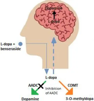
2.5.3. Aromatic amino acid decarboxylase (AADC) inhibitors
The most effective treatment for PD currently are the combinations of benserazide and L-dopa and carbidopa and L-dopa. Carbidopa is tenfold less potent than benserazide as a peripheral AADC inhibitor in both animals and humans. Benserazide inhibits the decarboxylation of L- dopa only in the extracerebral tissues, thus permitting the formation of DA in the striatum and in the hypothalamus. Because benserazide is well tolerated, relatively nontoxic even when used chronically and is the most potent peripheral AADC inhibitor presently available, it appears to be the drug of choice for the development of controlled release formulations in which L-dopa is combined with a peripheral AADC inhibitor. These controlled-release systems may reduce the clinical fluctuations in patients where the “wearing-off” and “on-off”
phenomena occurs (Da Prada et al., 1987).
NH O
N H
3C
H
3C
Figure 2.9: An illustration of the breakdown of L-dopa in the presence of an AADC inhibitor in the body (Satoh et al., 2015).
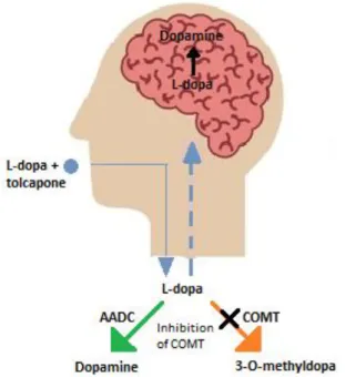
2.5.4. Catechol-O-methyltransferase (COMT) inhibitors
When L-dopa is given as monotherapy, its extensive peripheral metabolism results in very limited access of the amino acid to the CNS. L-Dopa is rapidly decarboxylated by AADC and 3-O-methylated by COMT. The COMT enzyme converts L-dopa and catecholamines to their methoxy derivatives to yield reaction products such as 3-O-methyl-dopa and 3-O-methyl-DA, as well as the 3-O-methylated, deaminated compound homovanillic acid (HVA), the major final metabolite of DA metabolism in humans (Factor et al., 2001; Kuno, S., 1997; Standaert &
Young, 2006; Teräväinen et al., 2001). Reversible COMT inhibitors are currently used clinically in PD therapy and include tolcapone and entacapone. Entacapone has a shorter duration of action (2 hours) and acts mostly in the periphery, where tolcapone has a relatively longer duration of action (8-12 hours) and acts in both the brain and periphery. The common adverse effects of these agents are nausea, vivid dreams, confusion and hallucinations, which can be attributed to an increase in brain DA (Olanow & Watkins, 2007).
Figure 2.10: An illustration of the breakdown of L-dopa in the presence of a COMT inhibitor in the body (Satoh et al., 2015).
2.5.5. Monoamine oxidase (MAO) inhibitors
MAO-A and MAO-B are enzymes that catalyse the oxidation of biogenic and xenobiotic amines (Bortolato & Shih, 2011; Edmondson et al., 2007). These two isoforms of MAO have different substrate preference and inhibitor selectivity. For example, the irreversible inhibitor, clorgyline, inhibits the oxidation of serotonin and norepinephrine that is catalysed by MAO-A.
MAO-B on the other hand uses benzylamine and phenylethylamine as substrates, and is irreversibly inhibited by selegiline, also an irreversible MAO inhibitor. DA is the common substrate for both isoforms (Youdim & Bakhle, 2006). DA is, however, preferentially deaminated by MAO-B in the human nigrostriatal system, and MAO-B inhibitors are thus used to block the MAO-catalysed metabolism of DA and increase DA bioavailability in the PD brain.
MAO-B inhibitors therefore are expected to increase DA levels and thus compensate the nigrostriatal deficit in DA and consequently provide symptomatic relief of the motor symptoms of PD (Finberg, 2014; Riederer & Laux, 2011; Robakis & Fahn, 2015; Youdim & Bakhle, 2006).
Selegiline and the second-generation drug, rasagiline, ar