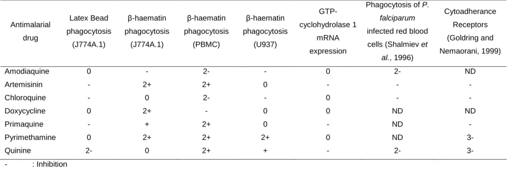
However, the effects of antimalarial drugs on hemozoin phagocytosis and GTP-cyclohydrolase 1 mRNA expression by monocytes are unknown. Effect of temperature and time on phagocytosis of J774A.1 β-hematin determined by counting and OD 400 assays.
Introduction
Malaria
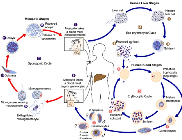
- Life-Cycle of the human malaria parasite
A small percentage of trophozoites develop into male and female gametocytes (7) that are ingested by the mosquito during a blood meal (8). The oocysts rupture (12) and a fraction of the released sporozoites end up in the salivary glands.
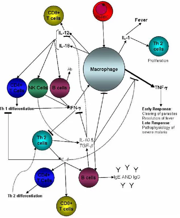
The immune response of monocytes to malaria
- Haemozoin formation by malaria parasites
- The structure of haemozoin
- The mechanism of haemozoin formation
- Biology of the mononuclear phagocyte system
- Morphology of the mononuclear phagocyte
- Composition and metabolism of the mononuclear phagocyte
- Functions of the mononuclear phagocyte
- The role of monocytes/macrophages in the immune response to malaria
- Pro-inflammatory cytokines in malaria infection
- Anti-inflammatory cytokines in the malaria infection
- The role of dendritic cells in the immune response to malaria
- Neopterin, a marker of inflammation
- Biosynthesis of neopterin and 7,8-dihydroneopterin
- Functions of 7,8-dihydroneopterin and neopterin
- Neopterin during malaria infections
Oliveira et al., (2005) showed that hemozoin formation occurs within lipid bodies in the worm's gut. IFN-γ leads to a rapid and sustained increase in plasma neopterin levels (Muller et al., 1991).

Antimalarial drugs
- Quinoline containing drugs
- Antifolates
- Antibiotics
- Artemisinin
- Resistance to antimalarial drugs
- Immunomodulatory effects of antimalarial drugs
- Effect of antimalarial drugs on cytokine production
- Effect of antimalarial drugs on monocyte metabolism
- Effect of antimalarial drugs on monocyte surface receptors
- Effect of antimalarial drugs on the production of monocyte reactive
- Effect of antimalarial drugs on monocyte p hagocytosis
Doxycycline (4-dimethylamino-1, 4, 4a, 5, 5a, 6, 11, 12a-octahydro and pentahydroxy-6-methyl-1,11-dioxo-2-naphthacenecarboxamide) belongs to the tetracycline group and has blood schizontocide. activity (Wernsdorfer, 1997) and tissue schizontocidal activity against pre-erythrocytic forms in hepatocytes (Marussig et al., 1993). Quinine has also been shown to increase phosphatidylserine synthesis in the Jurkat T cell line (Aussel et al., 1990).
Aim of study
This was further confirmed by high levels of inducible nitric oxide synthase gene expression (Prada et al., 1996b). Peripheral blood mononuclear cells isolated from healthy subjects receiving prophylactic doses of chloroquine showed reduced phagocytosis of opsonized sheep red blood cells and unopsonized zymosan particles (Osorio et al., 1992).
Effect of antimalarial drugs on the phagocytosis of haemozoin ( β -haematin)
Introduction
The host immunological response to the parasite may contribute to the pathophysiology of the disease in humans (Malaguarnera and Musumeci, 2002; Miller et al., 2002). These are exported from the food vacuole and finally degraded to amino acids, probably by aminopeptidases in the cytoplasm of the parasite (Gavigan et al., 2001). About 15% of these amino acids are then used in parasite protein synthesis (Krugliak et al., 2002).
Initiation may involve lipids (various unsaturated fatty acids and mono- and dioleoylglycerol) or proteins (histidine-rich protein (HRP) or the heme detoxification protein (HDP) (Jani et al., 2008)) or both (Egan, 2008b). Ingested hemozoin persists unmodified for long periods within the phagocyte and alters a number of monocyte functions (Schwarzer et al., 2008). Chloroquine and pyrimethamine both interfere with IL-2 production and thus inhibit T-cell proliferation (Landewe et al., 1995; Viora et al., 1996).
Quinine, chloroquine and primaquine also inhibit monocyte esterases, but to a lesser extent (Markovic et al., 1988).
Materials and Methods
- Materials
- Culturing of J774A.1 and U937 cell lines
- Isolation of peripheral blood mononuclear cells (PBMC)
- Adhesion of J774A.1, U937 and PBMC (monocytes) onto coverslips
- Characterisation of the cell lines
- Synthesis and analysis of β -haematin
- Preparation of samples for transmission electron microscopy (TEM)
- Treatment of monocytes with antimalarial drugs
- Measuring phagocytosis of β -haematin and latex beads by monocytes
- Statistical analysis
Phagocytosis still occurs in malaria patients being treated with antimalarial drugs, but the effect of antimalarial drugs on hemozoin phagocytosis is unknown. A spectrophotometric assay was developed to determine the effect of the drugs on the phagocytosis of β-hematin by the U937 cell line and PBMC. Aliquots (5 ml) of mononuclear cells were washed with 10 ml of complete media and the suspension spun at 150 g for 10 min.
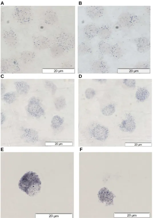
Cell viability and concentration were determined by trypan-blue exclusion (98%) and cells were counted using a hemocytometer at 400x magnification (Strober, 2001). The rest of the cells were fixed overnight at 4ºC in 3% (v/v) glutaraldehyde in 0.05 M sodium cacodylate buffer, pH 7.1. Software was also used to maximize red image contrast and 'touch count' the total number of cells and cells containing latex beads observed in each field.
The maximized contrast blue image was used to “touch count” the total cells and cells containing β-haematin in each field.
Results
- Characterisation of the monocytes
- Detection of peroxidase and alkaline phosphatase activity in monocytes
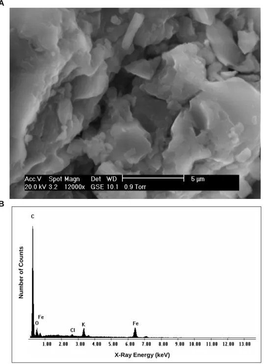
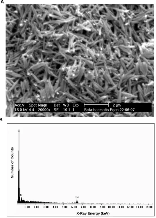
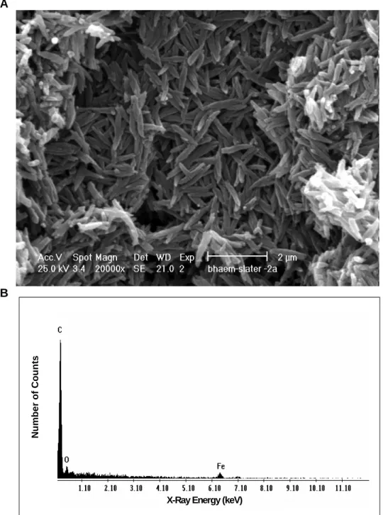
- Synthesis and analysis of β -haematin
- Measuring phagocytosis of latex beads and β -haematin by monocytes
- Comparison of the counting and OD 400 assay methods to determine the
- Effect of febrile temperature on phagocytosis of β -haematin by monocytes
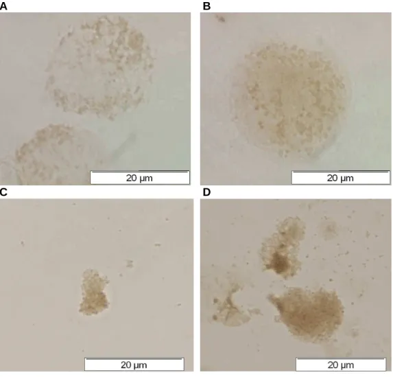
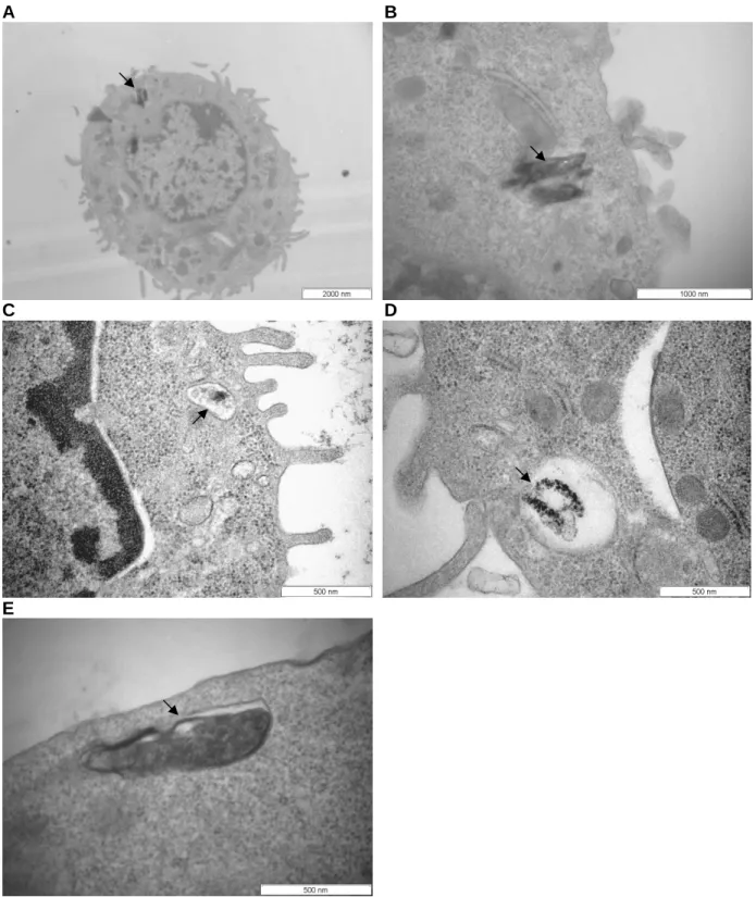
Transmission electron micrographs (TEM) of J774A.1 cells after phagocytosis of ββββ-hematin. B) TEM (A) at higher magnification to illustrate two β-hematin crystals surrounded by a discontinuous membrane; (C, D, E) β-hematin crystals in phagocytic vacuoles after (C) 30 min, (D, E) 2 h of phagocytosis. J774A.1 cells were treated with different concentrations of β-hematin and (A) the percentage of phagocytosis was determined by a counting assay or (B) the amount of heme in cell lysates was measured by the OD 400 assay. Effect of temperature and time on phagocytosis of J774A.1 ββββ- hematin determined by counting and OD 400 assays.
The effects of the drugs on the phagocytosis of β-hematin or latex beads by the J774A.1 cells were determined by the counting test as described in section 2.2.9. In contrast, the phagocytosis of β-hematin by the J774A.1 cells was activated by five out of seven of the antimalarial drugs examined (Figure 2.14B). Both tests showed that artemisinin, pyrimethamine and quinine activated the phagocytosis of β-hematin by the J774A.1 cells.
Effect of antimalarial drugs on PBMC ββββ-hematin phagocytosis measured by OD 400 assay.
Discussion
- Characterisation of the monocytes
- Characterisation of β -haematin
- Phagocytosis of latex beads and β -haematin
- Comparison of the counting and OD 400 assays to monitor phagocytosis of
- Effect of febrile temperature on phagocytosis of β -haematin
- Effects of antimalarial drugs on the phagocytosis of monocytes
Based on the scanning electron micrographs and transmission electron micrographs, the β-haematin crystals produced by both methods (Egan et al., 1994; The method according to Egan et al. 1994), due to the shorter preparation time, were adopted in this study. As the promonocytes develop into monocytes, which then eventually mature into macrophages, there is an increase in the phagocytic capacity of the cells (Cline et al., 1978).
It has been observed that the purity of monocytes/macrophages produced in this way is not very high (Brodersen et al., 1973; Koller et al., 1973). It has recently been demonstrated that the phagocytosis of β-haematin requires cholesterol-rich lipid domains in surface lipid rafts (Tiemi Shio et al., 2009). However, immunosuppression caused by the parasite and its products, such as hemozoin, contributes to the pathogenesis of the disease (Miller et al., 2002).
Hemozoin or β-hematin is known to have inhibitory as well as stimulatory effects on monocyte function (Schwarzer et al., 2008).

Effect of antimalarial drugs, haemozoin ( β -haematin), Plasmodium
Introduction
Neopterin is biosynthetically derived from guanosine triphosphate (GTP) (Murr et al., 2002) by GTP cyclohydrolase 1, which catalyzes the cleavage of the purine to produce 7,8-dihydronoepterin triphosphate (Werner et al., 1990). The main reaction that generates neopterin from 7,8-dihydroneopterin is oxidation by hypochlorous acid (HOCl) (Widner et al., 2000), which is released from neutrophils and macrophages during inflammation (Chisolm et al., 1999; Schraufstatter et al. , 1990) ). Neopterin derivatives are produced by human monocyte-derived macrophages (Huber et al., 1984) and dendritic cells (Wirleitner et al., 2002) when stimulated with the cytokine, interferon gamma (IFN-γ), released from activated T helper (Th) cells subtype 1 (Romagnani, 1991).
Since these cells promote cytotoxic T cell-mediated immune responses, the increased production of neopterin in body fluids can be used to monitor cell-mediated immunity (Wachter et al., 1989). However, many clinical laboratories also use immuno-based methods such as enzyme-linked immunosorbent assay (ELISA) to measure neopterin (Westermann et al., 2000). 7,8-Dihydronopterin inhibited the direct oxidation of plasma membranes of U937 cells by ferrous ions (Gieseg et al., 2001b).
Elevated levels of neopterin have been detected in the urine (Reibnegger et al., 1984) and plasma (Kremsner et al., 1989; Ringwald et al., 1991) of malaria patients infected with Plasmodium falciparum.
Materials and Methods
- Materials
- Coupling of neopterin to albumin carriers
- Preparation of immunogen and immunisation of chickens
- Isolation of Immunoglobulin Y (IgY) from egg yolks
- Monitoring the titre of antibody production in chickens with an ELISA
- Preparation of affinity matrices
- Affinity purification of IgY
- Development of a competitive ELISA to detect neopterin secretion in the
- Culturing of the U937 cell line
- Synthesis and analysis of β -haematin
- Treatment of U937 cells with interferon- γ (IFN- γ ) and antimalarial drugs, β -
- Isolation of Total RNA from U937 cells
- Statistical analysis
An indirect ELISA was performed to determine the optimal layer concentration of the neopterin-ovalbumin conjugate linked at a molar ratio of 200:1. During this incubation, the wells of the Nunc-Immuno™ ELISA plate were blocked as described in section 3.2.5. After the wells were washed, 100 µl of the neopterin-IgY solutions were added and incubated for 1 hour at 37ºC.
One-fiftieth of the resulting cDNA was used as a template for quantitative PCR SYBR® Green I (Molecular Probes®, U.S.A.) with the two-step SensiMix™ kit in the RotorGene 6000 Series real-time PCR cycler (Corbett Life Science Research , Australia ). The change in CT (∆CT) for each experimental sample was then subtracted from the ΔCT of the unstimulated control sample. For the 2−∆∆CT calculation to be valid, the amplification efficiency of the target (GTP-CH1) and reference genes (β-actin or GAPDH) must be approximately equal.
The ∆CT values calculated for each cDNA dilution were plotted against the logarithm of the cDNA input and the dynamic range of the assay was determined by removing outlier dilutions (usually the highest and/or lowest dilution).
Results
- Evaluation of chicken anti-rabbit albumin antibody and anti-neopterin
- Affinity purification of chicken anti-neopterin antibodies
- Development of a competitive ELISA to measure the neopterin secreted into
- Analysis of the relative expression of GTP-cyclohydrolase 1 mRNA
- Effect of phorbol-12-myristate-13-acetate and interferon- γ on the
- Effect of antimalarial drugs on the expression of GTP-cyclohydrolase 1
- Effects of β -haematin, latex beads, non-infected and P. falciparum-
This suggested that the capacity of the affinity matrix coupled to the neopterinovalbumin conjugate (40:1) to bind anti-neopterin antibodies was limited. An indirect ELISA was performed to ascertain the optimal coating concentration of neopterin-ovalbumin conjugate (200:1) to detect anti-neopterin IgY. ELISA to determine the appropriate coating concentration of neopterin-ovalbumin conjugate (200:1) to detect anti-neopterin IgY.
Dilutions of the affinity-purified antibodies (µg/ml) were incubated with 5-fold dilutions of free neopterin for 1 hour in microcentrifuge tubes. Variations in the coating concentrations of the 200:1 neopterin-ovalbumin conjugate from 524 to 16.375 ng/ml neopterin were examined in the competitive ELISA. However, no increase in A405 values with decreasing concentrations of free neopterin was observed at any of the coating concentrations of the neopterin-ovalbumin conjugate (Figure 3.6).
The affinity-purified antibodies did not bind to the neopterin coating at any of the concentrations of neopterin or antibody examined.

Discussion
- Production of anti-neopterin IgY in chickens
- Development of a competitive ELISA
- Competitive ELISA using the neopterin-ovalbumin conjugate to coat the
- Competitive ELISA using microtitre plates coated with neopterin added
- Analysis of relative expression of GTP-cyclohydrolase 1 mRNA transcripts
- Effect of phorbol-12-myristate-13-acetate and interferon- γ on the
- Effect of antimalarial drugs on the expression of GTP-cyclohydrolase 1
- Effect of β -haematin, latex beads, non-infected and P. falciparum-
Furthermore, neopterin is light sensitive and easily degraded, and although all the reactions with neopterin were performed in the dark, degradation of neopterin cannot be eliminated (Laich et al., 2002). In particular, chicken IgY has been bred against antimalarial drugs and used in a competitive ELISA (Goldring et al., 2005). INF-γ has been reported to increase the expression of GTP-cyclohydrolase 1 mRNA in human umbilical vein endothelial cells (Gesierich et al., 2003; Katusic et al., 1998).
Short-term administration of estradiol benzoate resulted in an increase in the GTP-cyclohydrolase 1 mRNA levels in dopaminergic and noradrenergic cell bodies (Serova et al., 2004). The GTP-cyclohydrolase 1 mRNA expression was found to be post-transcriptionally regulated by iron perturbations (Oexle et al., 2003). Kharazmi et al., (1986) also reported that chloroquine, quinine, pyrimethamine and tetracycline had no effect on the phagocytosis of S.
This is in contrast to reports, where iron was found to inhibit the mRNA expression of GTP-cyclohydrolase 1 (Oexle et al., 2003) and P. However, ICAM-1 on the surface of monocytes, which has been shown to be associated with malaria Infected erythrocytes (Ockenhouse et al., 1991), are involved in the sequestration of infected red blood cells and contribute to the pathogenesis of complicated and severe malaria (Chulay and Ockenhouse, 1990). Hemozoin reduces IFN-γ-mediated expression of ICAM-1 on the surface of monocytes ( Schwarzer et al., 1998 ).