EXTRACELLULAR MATRIX FACTORS INFLUENCE MYOBLAST ACTIVATION, DIFFERENTIATION AND FUSION
By
NICHOLAS LEE WALKER MSc (cum laude)
Submitted in fulfillment of the academic requirements for the degree of Doctor of Philosophy in the School of Life Sciences,
Discipline of Biochemistry University of KwaZulu-Natal
Pietermaritzburg
November 2015
As the candidate’s supervisor I have approved this thesis/dissertation for submission.
Signed: _____________ Name: Dr. C. U. Niesler Date: _____________________
ABSTRACT
Satellite cells are muscle stem cells that reside in a niche between the basal lamina and sarcolemma of mature muscle fibers. Upon muscle injury, these cells are activated to myoblasts that subsequently proliferate, migrate and differentiate into myotubes in order to facilitate repair. Extracellular matrix (ECM) and growth factors are known to regulate certain aspects of myogenesis however, a comprehensive study of the direct effects of niche ECM factors on C2C12 myogenesis has not previously been conducted and forms the core of this study.
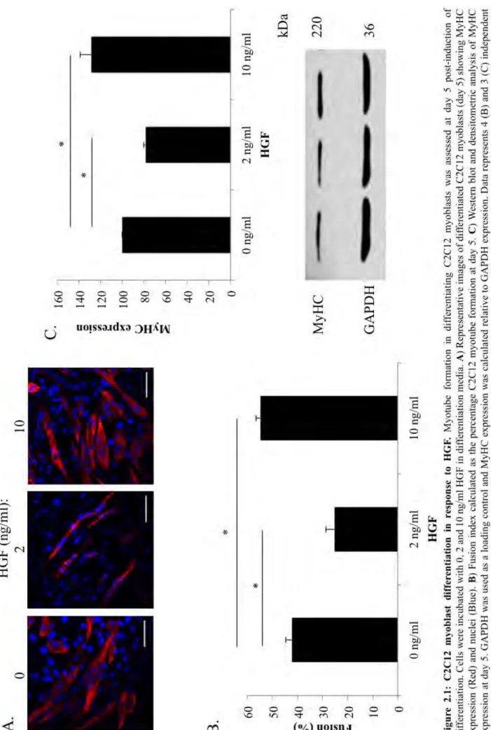
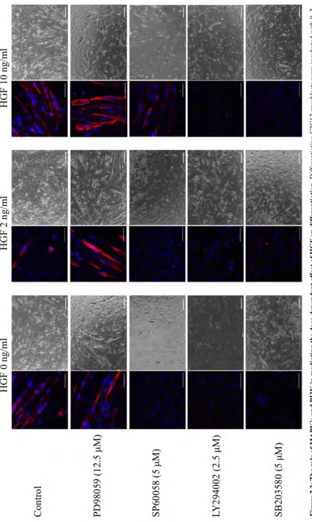
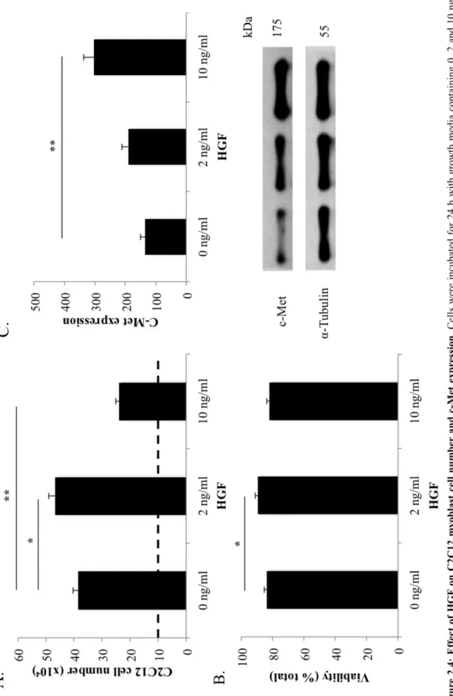
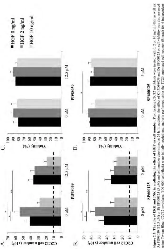
We first examined the role of hepatocyte growth factor (HGF) on C2C12 myogenesis. HGF is known to initiate activation of quiescent satellite cells in the niche and regulate various aspects of differentiation. In this study, we determined that HGF has a dose-dependent dual role in C2C12 myogenesis. HGF (2 ng/ml) significantly promoted cell division, but reduced myogenic commitment and fusion. Conversely, 10 ng/ml HGF reduced proliferative capability, but increased differentiation. This is potentially regulated by changes in c-Met expression; analysis revealed significantly decreased c-Met expression in differentiating cells cultured with 2 ng/ml HGF, but increased expression in proliferating cells with 10 ng/ml HGF. Furthermore, investigation into the mechanisms by which HGF affects myogenesis, revealed that mitogen-activated protein kinase (MAPKs: ERK, JNK or p38K) and phosphatidylinositol-3-kinase (PI3K) inhibition abrogated the HGF-stimulated increase in cell number. Interestingly, PI3K and p38 kinase facilitated the negative effect of HGF on proliferation, while ERK inhibition abrogated the HGF-mediated decrease in differentiation.
Next, we analyzed the effect of the satellite cell niche ECM on C2C12 myogenesis. Collagen IV and laminin, the major components of the basal lamina, bind to and interact with satellite cells via integrins and other cell surface proteins such as the tetraspanin, CD9. Matrigel significantly increased terminal fusion but had no effect on Pax7+ and MyoD+ cell numbers.
Collagen IV, the second largest constituent of Matrigel, was observed to significantly increase MyoD+ cell numbers and terminal fusion without effecting percentage Pax7+ cell numbers. Furthermore collagen IV stimulated an increase in CD9 expression on differentiating cells, such that cells cultured on collagen IV required higher levels of neutralizing anti-CD9 monoclonal antibodies to reduce fusion. These results indicate, for the
first time, that the interaction of collagen IV with CD9 is a critical mediator of skeletal muscle fusion and that the observed pro-myogenic effect is accompanied, on a molecular level, by an increase in the number of committed MyoD positive cells.
Extracellular matrix (ECM) and growth factors are known to have complex interactions that may modulate their activity in vivo. Lastly, in an attempt to more closely mimic in vivo conditions, murine C2C12 myoblasts were cultured on collagen IV in HGF-supplemented media followed by assessment of differentiation and proliferation. Collagen IV was not able to negate the negative effect of HGF (2 ng/ml) on fusion but was able to restore normal MyHC expression. Due to a collagen IV stimulated increase in CD9 expression in differentiating myoblasts, cells cultured on collagen IV required higher levels of neutralizing anti-CD9 monoclonal antibodies to reduce fusion; an effect not observed when cells were differentiated in the presence of HGF alone. HGF (10 ng/ml) treated samples we unable to demonstrate any fusion when CD9 was completely blocked suggesting that CD9 is a crucial co-factor in HGF (10 ng/ml)-induced fusion. These results show, for the first time, that collagen IV is able to modulate certain aspects of the dual role of HGF on myogenesis.
In summary, we identified a novel dose-dependent dual role of HGF in myogenesis and uncovered that these effects are mediated by changes in c-Met expression and downstream MAPK and PI3K signalling. We showed, for the first time, that collagen IV is able to positively mediate C2C12 differentiation via a CD9-dependent pathway. Lastly, we revealed that collagen IV is able to mediate the dose-dependent effects of HGF on C2C12 myogenesis.
PREFACE
The experimental work described in this dissertation was carried out in the Discipline of Biochemistry, School Life Sciences, University of KwaZulu-Natal, Pietermaritzburg, from January 2012 to September 2015, under the supervision of Dr. C. U. Niesler.
These studies represent original work by the author and have not otherwise been submitted in any form for any degree or diploma to any tertiary institution. Where use has been made of the work of others it is duly acknowledged in the text.
Signed:
………
Date:
………...
DECLARATION 1 - PLAGIARISM
I, Nicholas Lee Walker, declare that:
1. The research reported in this thesis, except where otherwise indicated, is my original research.
2. This thesis has not been submitted for any degree or examination at any other university.
3. This thesis does not contain other persons’ data, pictures, graphs or other information, unless specifically acknowledged as being sourced from other persons.
4. This thesis does not contain other persons' writing, unless specifically acknowledged as being sourced from other researchers. Where other written sources have been quoted, then:
a. Their words have been re-written but the general information attributed to them has been referenced
b. Where their exact words have been used, then their writing has been placed in italics and inside quotation marks, and referenced.
5. This thesis does not contain text, graphics or tables copied and pasted from the Internet, unless specifically acknowledged, and the source being detailed in the thesis and in the References sections.
Signed:
………
Date:
………
Declaration Plagiarism 22/05/08 FHDR Approved
DECLARATION 2 – PUBLICATIONS
DETAILS OF CONTRIBUTION TO PUBLICATIONS that form part and/or include research presented in this thesis(include publications in preparation, submitted, in press and published and give details of the contributions of each author to the experimental work and writing of each publication).
Walker NL, Kyle P. Goetsch*, Trish R. Kahamba*, Nicholas J. Woudberg and Carola U.
Niesler#. Hepatocyte growth factor (HGF) modulates myoblast proliferation and differentiation in a dose-dependent manner. Growth Factors, 33, 229-41.
Walker NL and Niesler CU. Collagen IV promotes myoblast fusion in a CD9-dependent manner [Contributed all data and analysis of figures in the paper]. Submitted, Cell and Tissue Research; 30 November 2015.
Walker NL and Niesler CU. Collagen IV modulates the dose-dependent effect of HGF in C2C12 myogenesis. [Contributed all data and analysis of figures in the paper]. In preparation.
Signed:
………..
Date:
………..
Declaration Publications FHDR 22/05/08 Approved
TABLE OF CONTENTS
ABSTRACT ... I PREFACE ... III DECLARATION 1 - PLAGIARISM ... IV DECLARATION 2 - PUBLICATIONS ... V TABLE OF CONTENTS ... VI LIST OF FIGURES AND TABLES ... X ABBREVIATIONS ... XI CONFERENCE CONTRIBUTIONS ... XIII ACKNOWLEDGEMENTS ... XIV
CHAPTER 1: LITERATURE REVIEW ... 1
1.1INTRODUCTION: ... 1
1.2SATELLITE CELL CHARACTERIZATION: ... 2
1.3PAX AND MYOGENIC REGULATORY FACTORS (MRFS): ... 4
1.4SKELETAL MUSCLE REPAIR: ... 7
1.5SATELLITE CELL NICHE: ... 9
1.6 EXTRACELLULAR MATRIX AND GROWTH FACTORS IN THE NICHE AND WOUND: ... 11
1.6.1 Fibronectin ... 11
1.6.2 Collagens ... 12
1.6.3 Laminins ... 13
1.6.4 Decorin ... 14
1.6.5 Matrigel ... 14
1.6.6 HGF ... 15
1.6.6.1 Stability: ... 16
1.6.6.2 c-Met receptor: ... 16
1.6.6.3 Intracellular signaling: ... 17
1.6.6 TGF-β ... 18
1.6.6 FGF ... 19
1.7EXTRACELLULAR MATRIX – GROWTH FACTOR INTERACTIONS: ... 19
1.83D SKELETAL MUSCLE GENERATION: ... 21
1.9SUMMARY, OBJECTIVES AND AIMS: ... 22
CHAPTER 2 ... 24
DOSE-DEPENDENT MODULATION OF MYOGENESIS BY HGF: IMPLICATIONS FOR
C-MET EXPRESSION AND DOWNSTREAM SIGNALLING PATHWAYS ... 24
2.1INTRODUCTION ... 25
2.2MATERIALS AND METHODS ... 28
2.2.1 Cell culture ... 28
2.2.2 HGF addition ... 28
2.2.3 Inhibitors ... 28
2.2.4 Cell counts ... 29
2.2.5 Immunocytochemistry ... 29
2.2.6 Fusion Index ... 29
2.2.7 Western Blotting ... 30
2.2.8 Statistical Analysis ... 30
2.3RESULTS ... 31
2.3.1 C2C12 myoblast differentiation is regulated through HGF ... 31
2.3.2 HGF regulates c-Met receptor expression during myoblast differentiation ... 33
2.3.3 Inhibition of the PI3K, JNK and p38, but not ERK, modulates the dose-dependent effect of HGF on C2C12 myoblast differentiation ... 35
2.3.4 HGF regulates myoblast proliferation in a dose-dependent manner ... 37
2.3.5 ERK and JNK mediate pro-proliferative effects of HGF ... 39
2.3.6 Inhibition of the PI3K and p38 pathways negates the effect of HGF on myoblast proliferation ... 41
2.4DISCUSSION ... 43
CHAPTER 3 ... 48
COLLAGEN IV PROMOTES MYOBLAST FUSION IN A CD9-DEPENDENT MANNER ... 48
3.1INTRODUCTION ... 49
3.2MATERIALS AND METHODS: ... 52
3.2.1 Cell Culture ... 52
3.2.2 Differentiation ... 52
3.2.3 ECM Coating ... 52
3.2.3.1 Collagen IV Coating ... 53
3.2.3.2 Laminin-111 Coating ... 53
3.2.3.3 Matrigel Coating ... 53
3.2.4 Immunocytochemistry and Confocal Microscopy ... 53
3.2.4.1 Assessment of percentage Pax7+ and MyoD+ cells ... 54
3.2.4.2 Assessment of terminal differentiation via a fusion index ... 54
3.2.5 Protein separation by SDS-PAGE ... 55
3.2.6 Western Blot analysis ... 55
3.2.6.1 Densitometry ... 56
3.2.7 Statistical analysis ... 56
3.3RESULTS ... 57
3.3.1 Matrigel increases terminal fusion of C2C12 myoblasts ... 57
3.3.2 C2C12 cells down-regulate Pax7 expression in response to differentiation cues ... 58
3.3.3 Matrigel does not effect Pax7 expression in differentiating C2C12 myoblasts ... 59
3.3.4 Matrigel does not effect MyoD expression in differentiating C2C12 myoblasts ... 60
3.3.5 Laminin does not affect C2C12 activation differentiation ... 61
3.3.6 Collagen IV increases fusion of C2C12 myoblasts ... 62
3.3.7 Collagen IV does not effect Pax7 expression in differentiating C2C12 myoblasts ... 63
3.3.8 Collagen IV increases MyoD expression in differentiating C2C12 myoblasts ... 64
3.3.9 Collagen IV increases CD9 expression in differentiating C2C12 myoblasts ... 65
3.3.10 CD9 mediates C2C12 fusion in response to collagen IV ... 66
3.4DISCUSSION ... 68
CHAPTER 4 ... 72
COLLAGEN IV MODULATES THE DOSE-DEPENDENT EFFECT OF HGF ON C2C12 MYOGENESIS ... 72
4.1INTRODUCTION ... 73
4.2MATERIALS AND METHODS: ... 75
4.2.1 Cell Culture ... 75
4.2.2 Differentiation ... 75
4.2.3 ECM Coating and HGF Addition ... 75
4.2.3.1 Collagen IV coating ... 75
4.2.3.2 HGF addition ... 76
4.2.4 Immunocytochemistry and Confocal Microscopy ... 76
4.2.4.1 Assessment of percentage MyoD+ cells ... 77
4.2.4.2 Assessment of terminal differentiation via a fusion index ... 77
4.2.5 Protein separation by SDS-PAGE ... 77
4.2.6 Western Blot analysis ... 78
4.2.6.1 Densitometry ... 79
4.2.7 Statistical analysis ... 80
4.3RESULTS ... 81
4.3.1 Collagen IV mediated HGF-modulated C2C12 myogenesis ... 81
4.3.2 Collagen IV prevents the dose-dependent effect of HGF on MyoD expression ... 83
4.3.3 HGF treated C2C12 cell differentiation is not more resistant to CD9 blocking than control samples ... 84
4.3.4 Collagen IV does not significantly effect C2C12 myoblast proliferation, viability or CD9 expression ... 86
4.3.5 Collagen IV does not modulate the dose-dependent effect of HGF on proliferation ... 88
4.4DISCUSSION ... 89
CHAPTER 5: DISCUSSION ... 91
APPENDIX I: SUPPLEMENTARY FIGURES ... 95
APPENDIX II: CONFERENCE ATTENDENCE AND PRESENTATIONS ... 96
POSTGRADUATE RESEARCH DAY,FACULTY OF SCIENCE &AGRICULTURE,UKZN,2012 ... 96
INDIAN OCEAN RIM MUSCLE COLLOQUIUM (IORMC)2013,SINGAPORE ... 98
THE SOUTH AFRICAN SOCIETY FOR BIOCHEMISTRY AND MOLECULAR BIOLOGY (SASBMB), CAPE TOWN,SOUTH AFRICA,2014 ... 99
"STEM CELLS:FROM BASIC RESEARCH TO BIOPROCESSING.",LONDON,UK,2015 ... 100
"STEM CELLS IN DRUG DISCOVERY.",CAMBRIDGE,UK,2015 ... 101
INDIAN OCEAN RIM MUSCLE COLLOQUIUM (IORMC)2016 ... 102
APPENDIX III: CLUSTAL W (1.81) MULTIPLE SEQUENCE ALIGNMENTS ... 104
A3.1COLLAGEN IV(HUMAN VS.MOUSE)SCORE 79.2899 ... 104
A3.2HGF(HUMAN VS.MOUSE)SCORE 89.4882 ... 104
A3.3LAMININ 111 VS.LAMININ 211(MOUSE)SCORE 33.8746 ... 104
APPENDIX IV: LYSATE PREP., PROTEIN DETERMINATION AND SDS-PAGE ... 105
A4.1LYSATE PREPARATION ... 105
A4.1.1 Reagents ... 105
A4.1.2 Method ... 105
A4.2BRADFORD ASSAY ... 105
A4.2.1 Reagents ... 105
A4.2.2 Method ... 106
A4.3 SDS-PAGE ... 106
A4.3.1 Reagents ... 106
APPENDIX V: WESTERN BLOT AND DOT BLOT ... 109
A5.1 WESTERN BLOT ... 109
A5.1.1 Reagents ... 109
A5.2DOT BLOT ... 109
REFERENCES ... 111
LIST OF FIGURES AND TABLES
FIGURE1.1:SATELLITECELLLOCALIZATION ... 3
FIGURE1.2:THEPROGRESSIONFROMQUIESCENTSATELLITECELLTO MULTINUCLEATEDMYOFIBERSHOWINGTHEEXPRESSIONOFTRANSCRIPTION FACTORSDURINGTHISPROCESS... ... 4
TABLE1.1:COMMONSATELLITECELLPROTEINSANDTHEIRFUNCTIONS. ... 6
FIGURE1.3:NORMALMUSCLEREGENERATION.... ... 8
FIGURE1.4:SCARTISSUEFORMATIONFOLLOWINGREPEATEDOREXTREMELY SEVEREMUSCLEINJURY.... ... 9
TABLE1.2:COMPARISONOFCOMPONENTSFOUNDWITHINTHESATELLITECELL NICHEANDTHOSEFOUNDINAFIBROTICWOUND. ... 11
TABLE1.3:THEKNOWNEFFECTSOFVARIOUSECMANDGROWTHFACTORSONSTEM CELLDIFFERENTIATION. ... 15
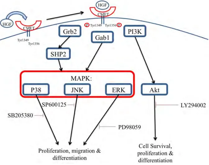
FIGURE1.5:SCHEMATICREPRESENTATIONOFC-METACTIVATEDBYHGF. ... 18
FIGURE2.1:C2C12MYOBLASTDIFFERENTIATIONINRESPONSETOHGF. ... 32
FIGURE2.2:EFFECTOFHGFONC-METEXPRESSIONINDIFFERENTIATINGC2C12 MYOBLASTS. ... 34
FIGURE2.3:THEROLEOFMAPK’SANDPI3KINMEDIATINGTHEDOSE-DEPENDENT EFFECTOFHGFONDIFFERENTIATION ... 36
FIGURE2.4:EFFECTOFHGFONC2C12MYOBLASTCELLNUMBERANDC-MET EXPRESSION. ... 38
FIGURE2.5:THEROLEOFERKANDJNKINMEDIATINGTHEEFFECTOFHGFONCELL NUMBER. ... 40
FIGURE2.6:THEROLEOFPI3KANDP38KINMEDIATINGTHEEFFECTOFHGFONCELL NUMBER. ... 42
FIGURE2.7:SCHEMATICREPRESENTATIONOFOURFINDINGS ... 47
TABLE3.1:ANTIBODYCONCENTRATIONSUSEDINCONFOCALMICROSCOPY ... 54
FIGURE3.1:MATRIGELINCREASESFUSIONOFC2C123MYOBLASTS.. ... 57
FIGURE3.2:PERCENTAGEPAX7+CELLSINPROLIFERATINGVERSUS DIFFERENTIATINGC2C12CELLS.. ... 58
FIGURE3.3:EFFECTOFMATRIGELONPAX7EXPRESSIONOFC2C12MYOBLASTS. ... 59
FIGURE3.4:EFFECTOFMATRIGELONMYOGENICCOMMITMENTOFC2C12 MYOBLASTS.. ... 60
FIGURE3.5:THEEFFECTOFLAMININ-111ONTHEDIFFERENTIATIONOFC2C12 MYOBLASTS ... 61
FIGURE3.6:THEEFFECTOFCOLLAGENIVONTHEDIFFERENTIATIONOFC2C12 MYOBLASTS ... 62
FIGURE3.7:EFFECTOFCOLLAGENIVONPAX7EXPRESSIONOFC2C12MYOBLASTS ... 63
FIGURE3.8:EFFECTOFCOLLAGENONMYOGENICCOMMITMENTOFC2C12
MYOBLASTS.. ... 64
FIGURE3.9:C2C12CD9EXPRESSIONINRESPONSETOCOLLAGENIVAND DIFFERENTIATIONCUES.. ... 65
FIGURE3.10:COLLAGENIVTREATEDC2C12MYOBLASTSAREMORERESISTANTTO DISRUPTIONOFDIFFERENTIATIONBYBLOCKINGOFCD9 ... 67
FIGURE3.11:SCHEMATICREPRESENTATIONOFCD9BLOCKING.. ... 70
TABLE4.1:ANTIBODYCONCENTRATIONSUSEDINCONFOCALMICROSCOPY ... 77
TABLE4.2:ANTIBODYCONCENTRATIONSUSEDINWESTERNBLOT ... 79
FIGURE4.1:COLLAGENIVMODULATESHGF-MEDIATEDMYOGENESIS ... 82
FIGURE4.2:HGF,INTHEPRESENCEOFCOLLAGENIV,DOESNOTMODULATE PERCENTAGEMYOD+C2C12MYOBLASTS ... 83
FIGURE4.3:CD9NEUTRALIZINGANTIBODIESDECREASEBOTHHGFANDCOLLAGEN IVSTIMULATEDFUSION. ... 84
FIGURE4.4:EFFECTOFCOLLAGENIVONC2C12MYOBLASTCELLNUMBERS, VIABILITYANDCD9EXPRESSION ... 87
FIGURE4.5:COLLAGENIVDOESNOTALTERTHEDOSE-DEPENDENTEFFECTOFHGF ONC2C12PROLIFERATION ... 88
FIGUREA1:EFFECTOFDIMETHYLSULFOXIDE(DMSO)ONC2C12CELLVIABILITY ... 95
FIGUREA4.1:BOVINESERUMALBUMIN(BSA)BRADFORDSTANDARDCURVE ... 106
TABLEA4.3.1:PREPARATIONOF10%AND12.5%LAEMMLIRUNNINGANDSTACKING GELSFORSDS-PAGE ... 108
FIGUREA5.2.1:DOTBLOTLAYOUTFORPRIMARYANDSECONDARYANTIBODY OPTIMIZATIONS. ... 110
ABBREVIATIONS
2D two-Dimensional 3D three-Dimensional
BMPs bone morphogenetic proteins BrdU Bromodeoxyuridine
BSA bovine serum albumin BSC biological safety cabinet CD9 cluster of differentiation (9) CD34 cluster of differentiation (34) CD44 cluster of differentiation (44) DDR discoidin domain receptor DM differentiation media
DMEM Dulbecco’s Modified Eagle Serum DMSO Dimethyl sulfoxide
ECM extracellular matrix
ECL enhanced chemiluminescence EGF epidermal growth factor EHS Engelbreth-Holm-Swarm
ERK Extracellular signal-regulated kinases FCS fetal calf serum
FGF fibroblast growth factor FGF-2 fibroblast growth factor-2
GAPDH glyceraldehyde-3-Phosphate Dehydrogenase GDFs growth differentiation factors
GM growth media
HGF hepatocyte growth factor
HS horse serum
HRPO horseradish peroxidase ICC Immunocytochemistry IGF Insulin-like growth factor JNK Jun N-terminal kinases
MAPK mitogen-activated protein kinase MRF(s) myogenic regulatory factor(s) MRF4 myogenic regulatory factor 5 Myf5 myogenic factor 5
MyoD myoblast determination protein MyHC myosin heavy chain
NCAM neural cell adhesion molecule
Pax3 paired-box protein 3 Pax7 paired-box protein 7 PBS phosphate buffered saline PenStrep Penicillin-Streptomycin PDGF platelet-derived growth factor PI3K Phosphoinositide 3-kinase
SDS-PAGE sodium dodecyl sulfate polyacrylamide gel electrophoresis SEM structural equation modeling
TGF-β1 transforming growth factor-β1 TBST tris buffered saline with tween UV ultra-violet
VEGF Vascular endothelial growth factor
CONFERENCE CONTRIBUTIONS
Walker N.L. & Niesler C.U. Effect of Growth and Extracellular Matrix Factors on Differentiation of Myoblasts. Presented at the Postgraduate Research Day, College of Science, Agriculture and Engineering, University of KwaZulu-Natal, Pietermaritzburg, South Africa; 2012.
Walker N.L. & Niesler C.U. Collagen IV is pro-myogenic and also regulates the dose- dependent effect of HGF on myogenesis. Presented at the Indian Ocean Rim Muscle Colloquium (IORMC), Singapore; 2013.
Walker N.L. & Niesler C.U. Collagen IV is pro-myogenic and also regulates the dose- dependent effect of HGF on myogenesis. Presented at SASBMB, South Africa; 2014.
Walker N.L. & Niesler C.U. The extracellular matrix modulates the effect of HGF on myogenesis. Presented at “Stem Cells: From basic research to bioprocessing”, London, UK;
2015.
Walker N.L. & Niesler C.U. The extracellular matrix modulates the effect of HGF on myogenesis. Presented at “Stem Cells in Drug Discovery”, Cambridge, UK; 2015.
ACKNOWLEDGEMENTS
I would like to start off by thanking Dr. Niesler for her constant advice and assistance in making this work possible. Your wisdom is astounding and I feel privileged to have been able to work in your laboratory.
Dr. Kyle Goetsch, you have been a helping hand whenever I have needed it and without your training on the confocal microscope this thesis would not be possible. You always have time to give valuable advice and for this I am extremely grateful.
Dr. Celia Snyman, your lessons in persistence have proven invaluable. Without such a diligent, motivated and determined figure in our laboratory, I would have thought science impossible.
Thanks to Rhys McColl for his assistance in ECL and Western Blotting as well as all the good times in the lab. Wanani Sibanda, Trish Kahamba, Colin Venter and Mtho Nkosi; thank you for your motivation and help with everything.
Rob Krause, your extensive knowledge of SDS-PAGE, Western Blotting and ECL has made you the troubleshooting genius of our department and your friendly demeanor makes you approachable. Thank you for all your help and advise.
Thanks to Prof. Coetzer and Prof. Goldring. You run a tight ship and I feel privileged to be a part of the Department of Biochemistry at UKZN.
Thanks to the Center for Electron Microscopy, for their assistance in sample preparation and use of the confocal microscope. I would especially like to thank Shirley for her assistance and for always being willing to help.
A special thanks to Robyn, Pat, Charmaine and all the wonderful women in the admin department for making our department run so well.
Kate, I would like to thank you for your patience with me as I have been working on this thesis. Your high standards and composure have been valuable examples in times of
frustration. You have been an ear when I needed to talk and your love has been unconditional, thank you.
Finally I would like to thank my family. Rex, you were my hero growing up and everyday your memory serves as an example to me of true character. Dad, your advice over the years has been my compass and I feel privileged to call you my father. Mom, your love, dedication and hard work are inspirational. I truly believe you are the best mother in the world. Ames, you are by far the most loving person I know and I feel privileged to know that you will be there for me whenever I need you. My extended family, of whom there are too many to mention, thank you.
To the NRF and UKZN for their financial support and to everyone else I have not mentioned, thank you.
CHAPTER 1: LITERATURE REVIEW
1.1 Introduction:
Myofibers (skeletal muscle cells) are post-mitotic and therefore unable to divide and repair damaged muscle themselves (Grounds et al., 2002). However, a cellular repair system is present in adult muscle in the form of a stem/progenitor cell known as the satellite cell (De Bari et al., 2003). Satellite cells reside in a niche between the basal lamina and plasma membrane of the adjacent myofiber (Mauro, 1961, Grefte et al., 2012). They are positioned along the entire length of the muscle fiber and ensure the capability of muscle repair regardless of the site of injury (Muir et al., 1965). Morphologically, satellite cells are fusiform in shape with a small nucleus and reduced organelle content (Charge and Rudnicki, 2004).
In healthy, uninjured muscle, satellite cells are metabolically inactive, a state known as quiescence. Upon muscle damage, they are activated from quiescence by hepatocyte growth factor (HGF) and begin to proliferate (Allen et al., 1995). Activated satellite cells, also known as myoblasts, migrate along the basal lamina towards the site of injury where they differentiate and facilitate tissue repair (Allen et al., 1995). The precise composition of the extracellular matrix (ECM) changes as the cell moves from its niche, along the basal lamina and into the wound. The two main protein components of the basal lamina are collagen IV and laminin (Boonen and Post, 2008). However, upon entering a fibrotic wound, myoblasts come into contact with high levels of collagen I, fibronectin and decorin (Vaz et al., 2012).
Previous research has highlighted the importance of the ECM and growth factors in regulating the activation, commitment, migration and differentiation of myoblasts (Borojevic, 1999, Goetsch et al., 2011, Grefte et al., 2012, Melo et al., 1996, Osses and Brandan, 2002, Schenke-Layland et al., 2007, Vaz, 2009) and a comprehensive assessment of the effects of HGF and the niche ECM on myogenesis forms the basis of this study.
1.2 Satellite cell characterization:
Satellite cells were first identified by Mauro (1961) in the hind limb muscles of frogs.
Research by Muir et al. (1965) identified and further characterized these cells in skeletal muscle of mice and fruit bats. Electron microscopy allowed the visualization of mammalian satellite cells residing in their niche between the basal lamina and plasma membrane of a myofiber (Figure 1.1) (Muir et al., 1965). Although satellite cells were suspected to play a role in skeletal muscle repair in 1961, conclusive proof of this was only shown some 17 years later. Snow (1978), using autoradiography, showed that satellite cells were essential for the regeneration of muscle in Sprague-Dawley rats. Here, satellite cells autoradiographically labeled with tritiated thymidine were transplanted into non-radioactive littermates. Following muscle damage, newly repaired myotubes appeared radioactively labeled thus proving that the satellite cells had differentiated into myotubes during fiber repair. In this study, activated satellite cells were also shown to be capable of myotube formation in vitro confirming their stem cell-like nature (Snow, 1978).
Satellite cells are not the only mononucleated cells found in skeletal muscle tissue (Figure 1.1). Pericytes are contractile cells that regulate capillary flow to the myofiber. They have recently been shown to retain a certain level of plasticity allowing them to differentiate into muscle cells and fibroblasts, and are able to assist in skeletal muscle regeneration (Dellavalle et al., 2007).
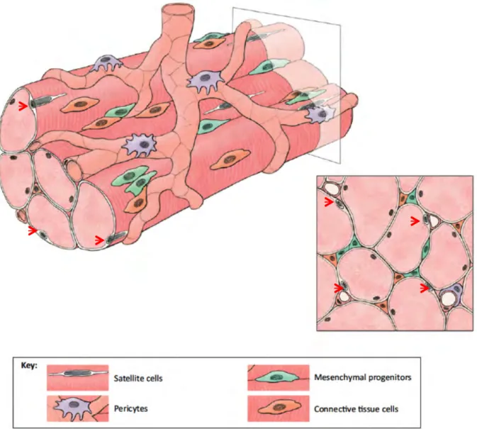
Figure 1.1: Satellite cell localization. Skeletal muscle is made up of densely packed, parallel myofibers. These fibers are multinucleated (containing myonuclei) and are surrounded by pericytes and satellite cells (red arrows indicate satellite cells). Blood vessels supply the fiber with nutrients, growth factors and circulating inflammatory cells in times of muscle injury. Mesenchymal progenitors and connective tissue cells are also found integrated within skeletal muscle tissue (Pannerec et al., 2012).
Mesenchymal progenitors are capable of differentiating into a range of cell types, including adipocytes and muscle (Uezumi et al., 2010). Within skeletal muscle, they secrete hepatocyte growth factor (HGF) which plays a vital role in the activation of satellite cells upon muscle injury (Catlow et al., 2003). Connective tissue cells are one of the most abundant cell types found embedded in skeletal muscle; they form a sheath of connective tissue around each muscle fiber (Rowe, 1981). This not only protects the fiber, but also transfers tension between adjacent muscle fibers and supplies a element of elasticity to the muscle bundle (Trotter and Purslow, 1992). Unlike myofibers, satellite cells are mononuclear and express specific satellite cell proteins such as Pax7 and muscle regulatory factors (MRFs), which allow them to be identified in skeletal muscle tissue (Charge and Rudnicki, 2004)
1.3 Pax and Myogenic Regulatory Factors (MRFs):
Paired-box (Pax) proteins 3/7 and the myogenic regulatory factors (MRFs) are transcription factors specific to muscle cell lineages (Seale et al., 2000). They play distinct, stage-specific roles during myogenesis and their use as molecular markers has allowed great insight into myogenesis. The expression of these transcription factors at different stages of muscle regeneration is outlined in Figure 1.2.
Figure 1.2: The progression from quiescent satellite cell to multinucleated myofiber showing the expression of transcription factors during this process. Quiescent satellite cells express Pax7 with low levels of Myf5. Myoblasts express the transcriptions factors Pax7, MyoD and myogenin. Differentiated myocytes down-regulate expression of Pax7, but continue to express MyoD and myogenin. Mature multinucleated muscle fibers express transcription factors myogenin and MRF4 as well as the structural protein Myosin Heavy Chain (MyHC) (Charge and Rudnicki, 2004, Cornelison and Wold, 1997, Fuchtbauer and Westphal, 1992, Gayraud- Morel et al., 2007, Le Grand and Rudnicki, 2007, Ustanina et al., 2007, Bentzinger et al., 2012).
Pax3 is a major regulator of embryonic muscle development, however, its importance in postnatal skeletal muscle growth and repair is overshadowed by Pax7 (Young and Wagers, 2010). Once activated, proliferating myoblasts increase expression of the paired-box transcription factor Pax7 (Le Grand and Rudnicki, 2007). Pax7 is known to play a role in the self-renewal pathway that is responsible for maintaining the population of satellite cells in muscle. Pax7 is expressed in both quiescent and activated satellite cells, but is down- regulated prior to differentiation (Relaix et al., 2006). After several proliferative cycles, myoblasts exit the cell cycle and enter a state of differentiation which allows subsequent fusion and myotube formation (Dhawan and Rando, 2005). If Pax7 expression is not downregulated, cells do not differentiate, but rather re-enter a quiescent state.
Following down-regulation of Pax7 in differentiating cells, MyoD expression increases, suggesting that Pax7 plays a role in self renewal via direct suppression of MyoD expression (Olguin and Olwin, 2004). MyoD expression is accompanied by up-regulation of Myf5, myogenin and myosin heavy chain (MyHC) expression, before subsequent fusion into multinucleated muscle fibers as reflected in Figure 1.2 (Le Grand and Rudnicki, 2007).
Through various knockout studies it has been shown that MyoD is a crucial regulatory factor of post-natal myogenesis with knockouts unable to effectively differentiate satellite cells.
Triple knockouts of myogenin, MRF4 and Myf5 also show a complete inability to perform successful myogenesis (Gayraud-Morel et al., 2007). This indicates that the regulatory factors work in unison to ensure successful myogenesis.
Satellite cells express a number of cell surface proteins including M-cadherin, neural cell adhesion molecule (N-CAM), c-Met and CD9 (Table 1.1). c-Met and CD9 are of particular relevance to this study as HGF signals through c-Met and collagen IV is known to bind to CD9. These surface proteins will be discussed in more detail in the following sections.
Table 1.1: Common satellite cell proteins and their functions.
Markers Expression Function References
Transcription Factors:
Pax3
Pax7
Q
Q/A
Myogenic specification (embryonic)
Myogenic specification (adult), self renewal
(Horst et al., 2006)
(Seale et al., 2000)
MRFs:
MyoD A Commitment to differentiation (Cornelison et al., 2000) Myf5 A/D Commitment to differentiation (Ustanina et al., 2007)
Myogenin MRF4
D D
Differentiation, fusion Differentiation, fusion
(Fuchtbauer and Westphal, 1992)
(Bentzinger et al., 2012) Cell surface proteins:
c-Met CD9
Q/A Q/A
Receptor for HGF
Role in integrin signaling, fusion
(Cornelison and Wold, 1997) (Beauchamp et al., 2000) (Tachibana and Hemler, 1999, Charrin et al., 2013).
Structural proteins:
MyHC D Terminal differentiation (Bader et al., 1982)
Abbreviations: Q: Quiescent; A: Activated; D: Differentiated; NCAM: neural cell adhesion molecule; Pax3/7:
paired-box transcription factor 3/7; Myf5: myogenic factor 5; MyHC: myosin heavy chain.
C2C12 cells are an immortalized myoblast line used as a research model for myogenesis isolated from C3H mice following a crush injury experiment (Yaffe and Saxel, 1977). Upon stimulation to differentiate, C2C12 cells are observed to downregulate Pax7, upregulate MyoD and begin differentiation (Zammit et al., 2006b). This is followed by the expression of the abovementioned MRFs and followed by fusion into actin and myosin positive myotubes (Burattini et al., 2004, Olguin et al., 2007). This pattern of expression of transcription factors as differentiation progresses closely mirrors that of primary culture myoblast differentiation and in vivo myogenesis (Olguin et al., 2007). This outlines the value in quantifying C2C12 transcription factor and MRF levels as a primary step in understanding specific stages of myogenesis.
1.4 Skeletal muscle repair:
Adult skeletal muscle is considered an extremely stable tissue type. Schmalbruch and Lewis (2000) estimate that approximately 2% of mononuclei are replaced each week in healthy adult mice (Schmalbruch and Lewis, 2000). Skeletal muscle also possesses the ability to rapidly regenerate following injury.
Muscle tissue repair can be classified into two distinct stages, namely degradation and regeneration. Degeneration begins with necrosis and partial or full autolysis of the fiber ensues (Schmalbruch and Lewis, 2000). This degradation releases factors which act as chemotactic signals to inflammatory cells in the bloodstream. Neutrophils are present at the site of injury within 6 hours of injury followed by macrophages approximately 48 hours post-injury (Figure 1.3A) (Tidball, 1995). Although fibroblasts are recruited to the site of injury, deposition of large amounts of fibrotic scar tissue is not characteristic for these types of injuries (Schmalbruch and Lewis, 2000). The released factors from the damaged muscle fiber also stimulate activation of the regeneration process. This is characterized by mitotic cell division of satellite cells followed by migration into the injury site and later, fusion of myoblasts to existing myofibers or to one another forming new myotubes (Figure 1.3B) (Schmalbruch and Lewis, 2000). Late in the regeneration stage, remodeling occurs and myotubes increase in size due to hypertrophy. Following complete regeneration, repaired myofibers are morphologically indistinguishable from undamaged fibers and exhibit complete contractile functionality (Figure 1.3C) (Schmalbruch and Lewis, 2000).
Figure 1.3: Normal muscle regeneration. Neutrophils and macrophages enter injury site. Activated myoblasts differentiate into multinucleated myotubes and fuse to existing myofibers thereby bridging the injury without scar tissue formation. Full muscle repair ensues (Constructed from (Jarvinen et al., 2000, Occleston et al., 2010, Tidball and Wehling-Henricks, 2007)).
However, in cases of extremely severe or frequent injury (Figure 1.4A), excessive ECM deposition by infiltrating fibroblasts may result in the formation of a fibrotic scar (Figure 1.4B) (Jarvinen et al., 2000). The fibrotic scar, composed of ECM factors including fibronectin and collagen III (which is later remodeled to collagen I), prevents myoblasts from fusing and bridging the original muscle fiber (Figure 1.4C) (Tidball and Wehling-Henricks, 2007). This, in turn, results in a loss of contractile ability (Tidball and Wehling-Henricks, 2007). This type of scarring also occurs as a result of repetitive injuries characteristic of myopathies such as Duchenne muscular dystrophy, where increasing fibrosis causes transdifferentiation of myoblasts into fibroblasts (Grounds, 2014).
Figure 1.4: Scar tissue formation following repeated or extremely severe muscle injury. Fibroblasts enter the site of injury and deposit ECM proteins such as collagen I and fibronectin. Myotubes then attach to either side of the fibrotic scar and fuse with the existing muscle fibers. This results in fibrotic scar tissue formation and impeded muscle function (Constructed from (Jarvinen et al., 2000, Occleston et al., 2010, Tidball and Wehling-Henricks, 2007)).
1.5 Satellite cell niche:
The stem cell niche is defined as the local microenvironment that supports, maintains and regulates stem cell identity and function. In particular, the stem cell niche has been shown to regulate self-renewal via a mechanism known as asymmetric cell division (Kuang et al., 2008). Asymmetric cell division is characterized by mitosis resulting in daughter cells of different cellular fates (Morrison and Kimble, 2006) as opposed to symmetric cell division which results in identical daughter cells. With regard to myogenesis, asymmetric cell division results in one daughter cell that will migrate to the site of injury and differentiate,
facilitating regeneration. The other daughter cell will return to the niche and re-enter quiescence (Kuang et al., 2007). This process is extremely important in maintaining a population of satellite cells within the muscle and thus ensuring its regenerative potential.
The satellite cell niche is found along the surface of muscle fibers between the sarcolemma and the basal lamina. The satellite cell is in contact with the muscle fiber, components of the basal lamina and the vascular system that supplies the niche (Fuchs et al., 2004). The muscle fiber has been shown to supply the satellite cell with mechanical, electrical, and chemical signals that play a role in the regulation of its function (Pallafacchina et al., 2010). The basal lamina surrounding myofibers consists of various ECM components such as laminin-211, collagen IV and various proteoglycans such as syndecans, glypican-1, perlecan, decorin and biglycan. These factors contribute to maintaining quiescence of the satellite cells while still in their niche (Pallafacchina et al., 2010). The vascular system supplies nutrients and oxygen to the muscle fiber. This network supplies extrinsic signals from the circulatory system, which along with factors released by macrophages and fibroblasts, regulate the quiescence, activation and proliferation of satellite cells (Pallafacchina et al., 2010).
Growth and ECM factors of the satellite cell niche and wound (Table 1.2) regulate the process of muscle repair and regeneration (Allen et al., 1995, Amento and Beck, 1991, Catlow et al., 2003, De Bari et al., 2003, Ewton et al., 1988, Fuchs et al., 2004, Goetsch et al., 2011, Grinnell, 1984, Lehto et al., 1985, Lin et al., 2010, Podleski et al., 1979, Schabort et al., 2009, Schenke-Layland et al., 2007, Schonherr et al., 1995, Tatsumi et al., 1998, Vaz, 2009, von der Mark and Ocalan, 1989, Yao et al., 1996). An understanding of how these factors affect myogenesis may provide clues as how to improve the restoration of muscle function post-injury.
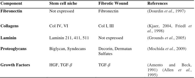
Table 1.2: Comparison of components found within the satellite cell niche and those found in a fibrotic wound.
Component Stem cell niche Fibrotic Wound References
Fibronectin Not expressed Fibronectin (Dourdin et al., 1997)
Collagens Col IV, VI Col I, III (Kjaer, 2004, Friedl et
al., 1998)
Laminin Laminin 211, 411, 511 Not expressed (Grounds et al., 2005) Proteoglycans Biglycan, Syndecans Decorin, Dermatan
Sulfates
(Mochida et al., 2009)
Growth Factors HGF, TGF-β TGF-β (Amento and Beck,
1991) (Allen et al., 1995)
1.6 Extracellular matrix and growth factors in the niche and wound:
The extracellular matrix (ECM) surrounding skeletal muscle was initially thought to act solely as a scaffold to support and maintain the structure of the tissue, but has since been shown to regulate many cellular processes (Grounds et al., 2005). These include cell survival, activation, proliferation, migration and differentiation (Friedl and Brocker, 2000). A vast array of proteins, proteoglycans and polysaccharides form the lattice-like meshwork of the ECM (Wu et al., 2005). Individual components of the ECM such as laminin, fibronectin, collagen I, tenacin and decorin have been shown to influence stem cell activation, migration and differentiation in various tissues (Lin et al., 2010, Schenke-Layland et al., 2007, Friedl and Brocker, 2000, Wehrle-Haller and Chiquet, 1993). Growth factors control many aspects of myogenesis including activation, proliferation, migration, differentiation and fusion of myoblasts into myotubes. In this respect, important growth factor families include the hepatocyte growth factor (HGF) family, the fibroblast growth factor (FGF) family and the transforming growth factor beta (TFG-β) family.
1.6.1 Fibronectin: Fibronectin has been shown to be crucial in embryogenesis with fibronectin knockout mice rarely developing beyond day 11 of embryogenesis (George et al., 1993). This is due to the vital role that fibronectin plays in guiding the migration of various cell types during early embryogenesis (Darribere and Schwarzbauer, 2000). Fibronectin, in its insoluble form, is a glycoprotein that forms fibrils as components of the ECM. This protein exists in two other forms: cell surface fibronectin oligomers and a soluble dimeric form located in the blood (Grinnell, 1984). The insoluble form of fibronectin has been shown
to be able to bind various ECM components including collagen and tenacin (Hocking et al., 2008). Along with tenacin, fibronectin is among the first ECM factors to be produced by fibroblasts in severely injured muscle tissue (Grinnell, 1984). Fibronectin forms multimeric cross-linked structures with fibrin which act as a scaffold for invading inflammatory cells and myoblasts (Dourdin et al., 1997). Fibronectin has been shown to improve the myogenic differentiation of C2C12 cells in vitro (Table 1.3) (Garcia et al., 1999, Lin et al., 2010).
Garcia et al (1999) showed that blocking with monoclonal antibodies against fibronectin itself, or the integrin with which it interacts (α5β1), abrogates the observed increase in C2C12 differentiation.
1.6.2 Collagens: Collagens are a family of structural proteins known to perform numerous functions in vivo. Fibrillar collagens include collagen I, II, III, IV, V, VI and XI and are all composed of three polypeptide α-chains which arrange into a triple helix (Ricard- Blum and Ruggiero, 2005). The basal lamina has been shown to be rich in collagen IV, which forms the basic scaffold into which laminin networks are integrated (Timpl and Brown, 1996). Collagen IV binds to cell surface receptors knows as integrins as well as tetraspanins (Leitinger and Hohenester, 2007). Integrins are the major transmembrane receptors involved in cell adhesion to the ECM (Humphries et al., 2006).
Although little is known regarding the effect of collagen IV on myoblast differentiation, collagen IV is known to interact with the CD9 receptor (Castro-Sanchez et al., 2010). This cell surface glycoprotein is expressed on the surface of C2C12 cells and has been shown to be vital in the normal development of skeletal muscle. CD9 expression is upregulated in the early stages of C2C12 differentiation and blocking CD9 using monoclonal antibodies substantially inhibits and delays conversion of C2C12 cells to elongated myotubes (Tachibana and Hemler, 1999, Charrin et al., 2013). This suggests that collagen IV may positively regulate myogenesis via CD9. Knockout studies have shown that collagen IV is not essential for early embryo development, but is essential for the correct structural assembly of various basement membranes during late development (Poschl et al., 2004). At day 10.5 to 11.5 lethality occurs in collagen IV null mice due to structural abnormalities in the basement membrane between parietal endoderm cells and trophoblast cells known as the Reichert’s membrane. Collagen IV has been shown to increase the differentiation of human and mouse embryonic stem cell types into mesodermal cell lineages including hematopoietic,
endothelial, and smooth muscle cells (Table 1.3) (Ali et al., 1998, Schenke-Layland et al., 2007, Taru Sharma et al., 2012).
Collagen I is the most abundant collagen subtype within mammalian tissues and is the final product of collagen remodeling during scar tissue formation (Ricard-Blum and Ruggiero, 2005). TGF-β promotes fibrosis by stimulating collagen I deposition by fibroblasts following severe muscle injury (Tidball and Wehling-Henricks, 2007). Additionally, the interstitial ECM surrounding skeletal muscle fibers has been shown to be rich in collagen I, which increases integrin α1β1 expression on fibroblasts (Kjaer, 2004). Increased expression of integrin α1β1 causes the clumping of fibroblasts on collagen fibers via a critical GFOGER (O denotes hydroxyproline) motif within the collagen’s I domain (Emsley et al., 2000).
Collagen I also binds to discoidin domain receptor 1 and 2 (DDR1 and DDR2) (Leitinger and Hohenester, 2007). DDRs are receptor tyrosine kinases (RTKs) involved in the regulation of cell growth, differentiation and metabolism (Mohan et al., 2001). Collagen I has been shown to inhibit the differentiation of rat primary culture satellite cells (Table 1.3) (Kjaer, 2004, Grefte et al., 2012).
1.6.3 Laminins: Laminins, composed of multiple heterodimers consisting of α, β and γ polypeptide chains, are a major protein component of the basal lamina. They are key bridge molecules, connecting the myofiber to the basal lamina; their absence results in congenital muscle dystrophies (Grounds et al., 2005). These conditions arise as, without the connection of the myofiber to the basal lamina, the contractile force generated by the myofiber cannot be transferred effectively to the interstitial connective tissue (Grounds et al., 2005). Laminin- 211, found around the sarcolemma of muscle fibers, binds to collagen IV in the basal lamina (Grounds et al., 2005). Laminin-111 and laminin-211 (merosin) differ in their alpha domains, however laminin-111 has been shown to improve the repair of skeletal muscle in merosin deficient mice (Van Ry et al., 2013). This suggests a similar functional nature of these two isoforms. Although the effect of laminin deficiency is well documented in congenital muscular dystrophies, its effect on myoblast differentiation remains a topic for debate.
Laminin-111 has been shown by some to increase C2C12 differentiation (Grossi et al., 2007) while others have found that laminin had no effect on C2C12 differentiation (Vaz, 2009) (Table 1.3).
1.6.4 Decorin: Decorin, a proteoglycan, is composed of a leucine-rich core protein containing 12 subunits with each subunit containing a 24 amino acid polypeptide (Mochida et al., 2009). Decorin plays a vital role in collagen fibrillogenesis by binding to collagen and causing a delay in fibril assembly. This results in the reduction of the fibril diameter and ensures correct fibril assembly (Mochida et al., 2009). Irregular collagen contours and a loosely packed collagen network are the result of decorin scarcity during myogenesis (Mochida et al., 2009). Goetsch et al., 2011 found that decorin increases collagen I- stimulated, but not fibronectin-stimulated migration of mouse myoblasts. Riquelme et al.
(2001) found that by minimizing the expression of decorin using antisense mRNA, they enhanced terminal differentiation of C2C12 cells (Table 1.3). In contrast, recent studies have shown decorin to aid in the differentiation of C2C12 cells (Table 1.3) through the suppression of myostatin activity (Kishioka et al., 2008). Li et al (2007) showed that decorin inhibits transforming growth factor TGF-β1 and can reduce the formation of fibrous scar tissue in vivo. This resulted in improved muscle healing after injury using decorin gene transfer (Li et al., 2007). The knockout of decorin expression performed by Riquelme et al.
(2001) may possibly have activated a compensatory mechanism that is responsible for their observed improvement in C2C12 terminal differentiation. The findings of Li et al (2007) were observed using an animal model as opposed to the C2C12 in vitro model used in the two other studies mentioned. These differences in the models used may contribute to the observed contrasting results.
1.6.5 Matrigel: Matrigel, as it is known by its trade name, is an exogenous mixture of mainly collagen IV, laminin-111 and various proteoglycans (Hughes et al., 2010). These components closely mimic the ECM of the satellite cell niche, providing a valuable tool in the study of niche ECM factors. Matrigel is commonly utilized to improve the proliferative and myogenic potential of primary isolated myoblasts (Grefte et al., 2012). Grefte et al showed that Matrigel did indeed improve the fusion of C2C12 cells in vitro. Melo et al.
(1996) demonstrated that the ECM is essential for skeletal muscle differentiation by culturing C2C12 myoblasts in the presence or absence of Matrigel. Cells plated in the absence of Matrigel were unable to form myotubes by day 3 while those differentiated on Matrigel had fused at this time point. Interestingly, when cells were plated in the absence of Matrigel, the production of the differentiation-stage specific transcription factor myogenin was not prevented, however later muscle-specific gene products such as MyHC were not produced by day 3 of differentiation (Melo et al., 1996, Osses and Brandan, 2002). This is
compelling evidence that the components of Matrigel speed up C2C12 terminal differentiation.
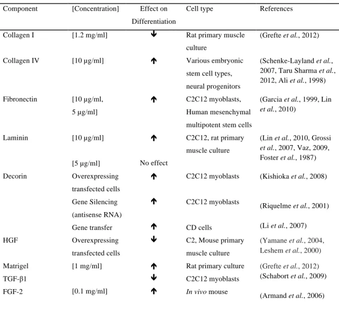
Table 1.3: The known effects of various ECM and growth factors on stem cell differentiation.
Component [Concentration] Effect on Differentiation
Cell type References
Collagen I [1.2 mg/ml] ê Rat primary muscle
culture
(Grefte et al., 2012)
Collagen IV [10 µg/ml] é Various embryonic
stem cell types, neural progenitors
(Schenke-Layland et al., 2007, Taru Sharma et al., 2012, Ali et al., 1998) Fibronectin [10 µg/ml,
5 µg/ml]
é C2C12 myoblasts,
Human mesenchymal multipotent stem cells
(Garcia et al., 1999, Lin et al., 2010)
Laminin [10 µg/ml]
[5 µg/ml]
é
No effect
C2C12, rat primary muscle culture
(Lin et al., 2010, Grossi et al., 2007, Vaz, 2009, Foster et al., 1987)
Decorin Overexpressing
transfected cells Gene Silencing (antisense RNA) Gene transfer
é é é
C2C12 myoblasts
C2C12 myoblasts
CD cells
(Kishioka et al., 2008)
(Riquelme et al., 2001) (Li et al., 2007)
HGF Overexpressing
transfected cells
ê C2, Mouse primary muscle culture
(Yamane et al., 2004, Leshem et al., 2000) Matrigel
TGF-β1 FGF-2
[1 mg/ml]
[0.1 mg/ml]
é ê é
Rat primary culture C2C12 myoblasts In vivo mouse
(Grefte et al., 2012) (Schabort et al., 2009) (Armand et al., 2006)
1.6.6 HGF: Hepatocyte growth factor (HGF) exists in two forms, pro-HGF and mature HGF.
Pro-HGF is secreted by mesenchymal cells as a single chain and remains bound in the ECM in an inactive form. Upon muscle injury, it is cleaved and activated by a serine protease (Catlow et al., 2003). Mature (active) HGF is dimeric and exists as a heavy α-chain (69 kDa) and light β-chain (34 kDa) heterodimer. This growth factor is known to play a role in development and regeneration of a range of tissues including the endothelium, kidney, lung and skeletal muscle (Nakamura and Mizuno, 2010). HGF has been shown to impede the differentiation of embryonic mouse tongue cells and increase the proliferation and migration of C2C12 myoblasts in vitro (Yamane et al., 2004, Barbero et al., 2001). With regards to
skeletal muscle, HGF is of vital importance in the initial stages of muscle regeneration (Matsumoto and Nakamura, 1997).
1.6.6.1 Stability:
In vivo studies investigating the effect of HGF on liver regeneration have shown that HGF has a blood half-life of approximately 3 minutes (Appasamy et al., 1993, Ido et al., 2004, Xue et al., 2003). This is mainly due to HGF uptake by the liver and does not suggest that HGF is inherently unstable (Appasamy et al., 1993). In fact, two studies have demonstrated that HGF is stable at a range of temperatures, pH’s and ionic strengths (Nayeri et al., 2002, Nayeri et al., 2004). HGF has however been observed to degrade significantly over a 3 hour period when placed in culture flow environments. However, HGF levels remained at over 90% 3 hours post addition to static media at 37 °C and 5% CO2 (Meneghello et al., 2014).
1.6.6.2 c-Met receptor:
The c-Met receptor is a high affinity receptor for HGF. It exists as a 190 kDa transmembrane protein composed of an α-chain (50 kDa) and a β-chain (140 kDa) (Tam et al., 2000). Upon binding of HGF to the c-Met receptor, Met kinase becomes active (Bottaro et al., 1991). This results in 2 tyrosine residues (Tyr1349 and Tyr1356) in the carboxy-terminal tail becoming phosphorylated (Figure 1.5). These residues become docking sites for a range of adaptor proteins including phosphatidylinositol-3-kinase (PI3K), Grb2-associated adaptor protein (Gab1) and growth factor receptor-bound protein 2 (Grb2) (Figure 1.5). These pathways proceed to mediate Met-dependent cell proliferation, migration, survival and differentiation (Faria et al., 2011). These effects will be further discussed in section 1.6.6.3. Blocking the binding of HGF to the c-Met receptor has been shown to prevent the activation